1. Induced Disease Models Products GPCR/G Protein Neuronal Signaling Autophagy Immunology/Inflammation MAPK/ERK Pathway Apoptosis
  2. Nervous System Disease Models Dopamine Receptor Autophagy Mitophagy COX PGE synthase Interleukin Related p38 MAPK Apoptosis Caspase
  3. Parkinson's Disease Models
  4. Oxidopamine hydrobromide

Oxidopamine hydrobromide  (Synonyms: 6-Hydroxydopamine hydrobromide; 6-OHDA hydrobromide)

Cat. No.: HY-B1081A Purity: 99.95%
SDS COA Handling Instructions

Oxidopamine (6-OHDA) hydrobromide is an antagonist of the neurotransmitter dopamine. Oxidopamine hydrobromide is a widely used neurotoxin and selectively destroys dopaminergic neurons. Oxidopamine hydrobromide promotes COX-2 activation, leading to PGE2 synthesis and pro-inflammatory cytokine IL-1β secretion. Oxidopamine hydrobromide can be used for the research of Parkinson’s disease (PD), attention-deficit hyperactivity disorder (ADHD), and Lesch-Nyhan syndrome.

For research use only. We do not sell to patients.

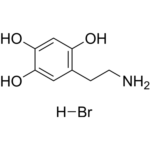
Oxidopamine hydrobromide Chemical Structure

Oxidopamine hydrobromide Chemical Structure

CAS No. : 636-00-0

Size Price Stock Quantity
Free Sample (0.1 - 0.5 mg)   Apply Now  
50 mg USD 66 In-stock
100 mg USD 79 In-stock
200 mg USD 119 In-stock
500 mg USD 238 In-stock
1 g USD 370 In-stock
5 g   Get quote  
10 g   Get quote  

* Please select Quantity before adding items.

This product is a controlled substance and not for sale in your territory.

Customer Review

Based on 19 publication(s) in Google Scholar

Other Forms of Oxidopamine hydrobromide:

Top Publications Citing Use of Products
  • Biological Activity

  • Purity & Documentation

  • References

  • Customer Review

Description

Oxidopamine (6-OHDA) hydrobromide is an antagonist of the neurotransmitter dopamine. Oxidopamine hydrobromide is a widely used neurotoxin and selectively destroys dopaminergic neurons. Oxidopamine hydrobromide promotes COX-2 activation, leading to PGE2 synthesis and pro-inflammatory cytokine IL-1β secretion. Oxidopamine hydrobromide can be used for the research of Parkinson’s disease (PD), attention-deficit hyperactivity disorder (ADHD), and Lesch-Nyhan syndrome[1][2][3][4].

IC50 & Target

COX-2


IL-1β


Caspase-3


Caspase-8


Caspase-9


In Vitro

Oxidopamine hydrobromide (0-500 μM, 24 h) decreases the viability of both Neuro-2a cells and SH-SY5Y cells in a concentration-dependent manner[1].
Oxidopamine hydrobromide (75-150 μM, 0-24 h) induces COX-2 expression and nuclear translocation[1].
Oxidopamine hydrobromide (75-150 μM, 0-24 h) causes PGE2 biosynthesis and pro-inflammatory cytokine IL-1β production[1].
Oxidopamine hydrobromide (0-150 μM, 12 h) induces apoptosis and mitochondrial membrane depolarization of pheochromocytoma PC12 cells[3].
Oxidopamine hydrobromide (75 μM, 0-12 h) induces p38 phosphorylation[3].

MedChemExpress (MCE) has not independently confirmed the accuracy of these methods. They are for reference only.

Cell Viability Assay[1]

Cell Line: Neuro-2a cells and SH-SY5Y cells
Concentration: 0-500 µM
Incubation Time: 24 or 48 h
Result: Induced neurotoxicity, caused cytotoxicity in both Neuro-2a cells and SH-SY5Y cells in a concentration dependent manner. EC50=111 µM for 24 h incubation and 109 µM for 48 h incubation in the Neuro-2a cells; EC50=118 µM for 24 h incubation and 107 µM for 48 h incubation in the SH-SY5Y cells.

RT-PCR[1]

Cell Line: Neuro-2a cells and SH-SY5Y cells
Concentration: 75 or 150 µM
Incubation Time: 0, 6 or 24 h
Result: Quickly and robustly induced COX-2 in a time-dependent manner. Induced COX-2 activation characterized by expression induction and nuclear translocation. Substantially increased PGE2 in the culture medium by nearly 5-fold in Neuro-2a cells (at 75 µM) and 3-fold in SH-SY5Y cells (at 150 µM). Significantly upregulated the pro-inflammatory cytokine interleukin-1β (IL-1β) within Neuro-2a cells and SH-SY5Y cells.

Apoptosis Analysis[3]

Cell Line: PC12 cells
Concentration: 0, 25, 50, 75, and 150 μM
Incubation Time: 0, 2, 4, 6, 12, and 20 h
Result: Induced apoptosis of PC12 cells. Increased the activities of caspase-3, -8 and -9 in PC12 cells in a time- and concentration-dependent manner. Increased these caspase activities at 2-4 h and reached a maximum at 12 h. Decreased cells with high mitochondrial membrane potential (JC-1 aggregate) in a time- and concentration-dependent manner.

Western Blot Analysis[3]

Cell Line: PC12 cells
Concentration: 75 μM
Incubation Time: 0, 3, 5, 6, 8, 10, and 12 h
Result: Increased the level of p-p38 in a time-dependent manner.
In Vivo

Oxidopamine (6-OHDA) hydrobromide can induce Parkinson's disease models[5][6].

Induction of Parkinson's disease model[5][6]
Background
The chemical structure of Oxidopamine (6-OHDA) hydrobromide is similar to dopamine (DA), enabling it to compete with DA for uptake sites and be subsequently taken into cells. Once inside the cells, oxidopamine hydrochloride can be oxidized and decomposed, generating reactive oxygen species, which further produce oxygen free radicals through MAO (monoamine oxidase) or directly cause mitochondrial dysfunction, leading to the death of dopaminergic neurons.
Specific Mmodeling Methods
Rat: Sprague-Dawley (SD) • Male • 200-250 g •
Administration: 5μg/2μL/site • stereotaxically injected in the fight striatum • single dose.
Note
(1) Lesions were made by the unilateral injection of Oxidopamine hydrochloride (5 μg in 2 μl/site) into the right striatum at the two coordinates:
① AP, ?0.7; L, ?3.0; DV, ?5.5 and 4.5 mm from Bregma.
② AP, ?0.2; L, ?2.6; DV, ?5.5 and 4.5 mm from Bregma.
The two coordinates were injected Oxidopamine hydrochloride 10 μg in 4 μl/2 sites.
(2) Oxidopamine hydrochloride was prepared freshly in dark to avoid autooxidation, and was administered using a 5 μl microinjector at a rate of 0.5 μl/min. The syringe was left in place for 5 min before slowly retracting it to allow for toxin diffusion and prevent the toxin reflux.
(3) On the 56th day after the injury, the animals were decapitated under deep halothane anesthesia. Their brains were quickly removed from the skull, rinsed with chilled saline, and tissue samples containing the caudate-putamen head were dissected from both the lesioned and unlesioned striata on ice.
(4) The animals were housed in an environment with a 12-hour light/dark cycle, with the temperature maintained at 22-23°C. They were allowed free access to food and tap water.
Modeling Indicators
Behavioral monitoring: Rats exhibit rotation with a rotation count greater than 210 r/30 min. Molecular changes: Elevated levels of COX-2, TNF-α mRNA, and COX-2 protein. Histopathological changes: Chromatin condensation into clumps around the nucleus, along with evident mitochondrial swelling and vacuolation. Induced nigrostriatal nerve terminal lesions. Decreased striatal dopamine levels and reduced number of tyrosine hydroxylase immunoreactive cells in the ipsilateral substantia nigra, accompanied by long-term significant atrophy of remaining dopaminergic neurons.
Correlated Product(s): /
Opposite Product(s): Resveratrol (HY-16561)

MedChemExpress (MCE) has not independently confirmed the accuracy of these methods. They are for reference only.

Molecular Weight

250.09

Formula

C8H12BrNO3

CAS No.
Appearance

Solid

Color

Light brown to gray

SMILES

OC1=CC(CCN)=C(O)C=C1O.[H]Br

Shipping

Room temperature in continental US; may vary elsewhere.

Storage

4°C, stored under nitrogen

*The compound is unstable in solutions, freshly prepared is recommended.

Solvent & Solubility
In Vitro: 

H2O : ≥ 100 mg/mL (399.86 mM)

DMSO : 50 mg/mL (199.93 mM; ultrasonic and warming and heat to 60°C; Hygroscopic DMSO has a significant impact on the solubility of product, please use newly opened DMSO)

*"≥" means soluble, but saturation unknown.

Preparing
Stock Solutions
Concentration Solvent Mass 1 mg 5 mg 10 mg
1 mM 3.9986 mL 19.9928 mL 39.9856 mL
5 mM 0.7997 mL 3.9986 mL 7.9971 mL
View the Complete Stock Solution Preparation Table

* Please refer to the solubility information to select the appropriate solvent. The compound is unstable in solutions, freshly prepared is recommended.

* Note: If you choose water as the stock solution, please dilute it to the working solution, then filter and sterilize it with a 0.22 μm filter before use.

  • Molarity Calculator

  • Dilution Calculator

Mass (g) = Concentration (mol/L) × Volume (L) × Molecular Weight (g/mol)

Mass
=
Concentration
×
Volume
×
Molecular Weight *

Concentration (start) × Volume (start) = Concentration (final) × Volume (final)

This equation is commonly abbreviated as: C1V1 = C2V2

Concentration (start)

C1

×
Volume (start)

V1

=
Concentration (final)

C2

×
Volume (final)

V2

In Vivo:

Select the appropriate dissolution method based on your experimental animal and administration route.

For the following dissolution methods, please ensure to first prepare a clear stock solution using an In Vitro approach and then sequentially add co-solvents:
To ensure reliable experimental results, the clarified stock solution can be appropriately stored based on storage conditions. As for the working solution for in vivo experiments, it is recommended to prepare freshly and use it on the same day.
The percentages shown for the solvents indicate their volumetric ratio in the final prepared solution. If precipitation or phase separation occurs during preparation, heat and/or sonication can be used to aid dissolution.

  • Protocol 1

    Add each solvent one by one:  10% DMSO    40% PEG300    5% Tween-80    45% Saline

    Solubility: ≥ 2.5 mg/mL (10.00 mM); Clear solution

    This protocol yields a clear solution of ≥ 2.5 mg/mL (saturation unknown).

    Taking 1 mL working solution as an example, add 100 μL DMSO stock solution (25.0 mg/mL) to 400 μL PEG300, and mix evenly; then add 50 μL Tween-80 and mix evenly; then add 450 μL Saline to adjust the volume to 1 mL.

    Preparation of Saline: Dissolve 0.9 g sodium chloride in ddH₂O and dilute to 100 mL to obtain a clear Saline solution.
  • Protocol 2

    Add each solvent one by one:  10% DMSO    90% (20% SBE-β-CD in Saline)

    Solubility: ≥ 2.08 mg/mL (8.32 mM); Clear solution

    This protocol yields a clear solution of ≥ 2.08 mg/mL (saturation unknown).

    Taking 1 mL working solution as an example, add 100 μL DMSO stock solution (20.8 mg/mL) to 900 μL 20% SBE-β-CD in Saline, and mix evenly.

    Preparation of 20% SBE-β-CD in Saline (4°C, storage for one week): 2 g SBE-β-CD powder is dissolved in 10 mL Saline, completely dissolve until clear.

For the following dissolution methods, please prepare the working solution directly. It is recommended to prepare fresh solutions and use them promptly within a short period of time.
The percentages shown for the solvents indicate their volumetric ratio in the final prepared solution. If precipitation or phase separation occurs during preparation, heat and/or sonication can be used to aid dissolution.

  • Protocol 1

    Add each solvent one by one:  PBS

    Solubility: 50 mg/mL (199.93 mM); Clear solution; Need ultrasonic

In Vivo Dissolution Calculator
Please enter the basic information of animal experiments:

Dosage

mg/kg

Animal weight
(per animal)

g

Dosing volume
(per animal)

μL

Number of animals

Recommended: Prepare an additional quantity of animals to account for potential losses during experiments.
Calculation results:
Working solution concentration: mg/mL
This product has good water solubility, please refer to the measured solubility data in water/PBS/Saline for details.
The concentration of the stock solution you require exceeds the measured solubility. The following solution is for reference only.If necessary, please contact MedChemExpress (MCE).
Purity & Documentation

Purity: 99.95%

References

Complete Stock Solution Preparation Table

* Please refer to the solubility information to select the appropriate solvent. The compound is unstable in solutions, freshly prepared is recommended.

Optional Solvent Concentration Solvent Mass 1 mg 5 mg 10 mg 25 mg
DMSO / H2O 1 mM 3.9986 mL 19.9928 mL 39.9856 mL 99.9640 mL
5 mM 0.7997 mL 3.9986 mL 7.9971 mL 19.9928 mL
10 mM 0.3999 mL 1.9993 mL 3.9986 mL 9.9964 mL
15 mM 0.2666 mL 1.3329 mL 2.6657 mL 6.6643 mL
20 mM 0.1999 mL 0.9996 mL 1.9993 mL 4.9982 mL
25 mM 0.1599 mL 0.7997 mL 1.5994 mL 3.9986 mL
30 mM 0.1333 mL 0.6664 mL 1.3329 mL 3.3321 mL
40 mM 0.1000 mL 0.4998 mL 0.9996 mL 2.4991 mL
50 mM 0.0800 mL 0.3999 mL 0.7997 mL 1.9993 mL
60 mM 0.0666 mL 0.3332 mL 0.6664 mL 1.6661 mL
80 mM 0.0500 mL 0.2499 mL 0.4998 mL 1.2496 mL
100 mM 0.0400 mL 0.1999 mL 0.3999 mL 0.9996 mL

* Note: If you choose water as the stock solution, please dilute it to the working solution, then filter and sterilize it with a 0.22 μm filter before use.

  • No file chosen (Maximum size is: 1024 Kb)
  • If you have published this work, please enter the PubMed ID.
  • Your name will appear on the site.
Help & FAQs
  • Do most proteins show cross-species activity?

    Species cross-reactivity must be investigated individually for each product. Many human cytokines will produce a nice response in mouse cell lines, and many mouse proteins will show activity on human cells. Other proteins may have a lower specific activity when used in the opposite species.

Your Recently Viewed Products:

Inquiry Online

Your information is safe with us. * Required Fields.

Product Name


Salutation

Applicant Name *


Email Address *

Phone Number *


Organization Name *

Department *


Requested quantity *

Country or Region *


Remarks

Bulk Inquiry

Inquiry Information

Product Name:
Oxidopamine hydrobromide
Cat. No.:
HY-B1081A
Quantity:
MCE Japan Authorized Agent: