From 11:00 pm to 12:00 pm EST ( 8:00 pm to 9:00 pm PST ) on January 6th, the website will be under maintenance. We are sorry for the inconvenience. Please arrange your schedule properly.
Astegolimab (MSTT 1041A; RG 6149) is a human IgG2 monoclonal antibody. Astegolimab blocks IL-33 signaling by targeting the IL-33 receptor ST2. Astegolimab reduces p53 expression, mitigates IL33-upregulated SASP factors such as IL1α, IL6 and MCP1. Astegolimab mitigates IL33-increased p-p65/p65 ratio. Astegolimab blocks CM-induced neutrophil extracellular trap (NET) formation. Astegolimab is used in chronic obstructive pulmonary disease (COPD) and myocardial research [1] .
Subtilisin (EC 3.4.21.14) is a bacterial serine protease. Subtilisin induces Apoptosis. Subtilisin stimulates the expression of pro-allergic cytokines (IL-1α,IL-33). Subtilisin induces prototypic allergic lung inflammation. Subtilisin exhibits anticancer activity against breast and colon cancer. Subtilisin shows antifouling activity. Subtilisin can be used as a detergent additive [1] .
Candidalysin is a cytolytic peptide toxin secreted by the fungus Candida albicans. Candidalysin drives epithelial immune responses by activating the EGFR-MAPK signaling pathway, inducing MMP expression and calcium influx, and regulating the c-Fos transcription factor and MKP1 via p38 MAPK and ERK1/2 respectively. Candidalysin is essential for mucosal and systemic infections, activating NLRP3 to promote inflammatory responses, neutrophil recruitment, and Th17 immunity. Candidalysin activates LDH causing membrane damage and exhibiting cytotoxicity [1]
Lutikizumab (ABT-981) is an anti-IL-1α and IL-1β dual variable domain immunoglobulin. Lutikizumab binds and inhibits IL-1α and IL-1β. Lutikizumab can be used for the research of osteoarthritis [1].
Nidanilimab (CAN04) is a fully humanized monoclonal anti-IL1RAP antibody with a Kd value of 1.10 pM. Nidanilimab blocks IL1α and IL1β signaling and stimulates the immune system to destroy tumour cells. Nidanilimab can be used in research of non-small lung cancer (NSCLC) and pancreatic ductal adenocarcinoma (PDAC) [1] .
Bermekimab (MABp1) is a human monoclonal antibody targeting interleukin-1α(IL-1α) . Bermekimab can prevent tumor-related inflammation. Bermekimab can be used in the research of atopic dermatitis [1].
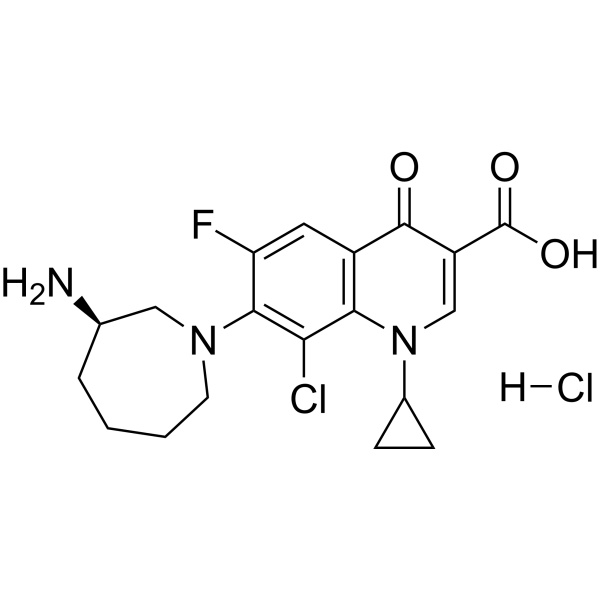
Besifloxacin Hydrochloride is a fourth generation fluoroquinolone antibiotic. Besifloxacin Hydrochloride is a DNA gyrase and topoisomerase IV inhibitor. Besifloxacin Hydrochloride has broad-spectrum antibacterial activity, it is effective against Gram-negative and Gram-positive aerobic and anaerobic strains and reduces the incidence of drug resistance. Besifloxacin Hydrochloride has anti-inflammatory activity. Besifloxacin Hydrochloride can be used in bacterial conjunctivitis research [1] .
Nadifloxacin (OPC7251) is a broad-spectrum quinolone antibiotic. Nadifloxacin inhibits bacterial DNA gyrase and topoisomerase IV, interfering with DNA replication. It also suppresses the production of proinflammatory cytokines (such as IL-1α, IL-6, and IL-8). Nadifloxacin exhibits antibacterial activity against various pathogens, including Propionibacterium acnes and Staphylococcus aureus. Nadifloxacin also exhibits anti-inflammatory activity. Nadifloxacin can be used in the research of skin infections such as acne vulgaris, folliculitis, and impetigo [1] .
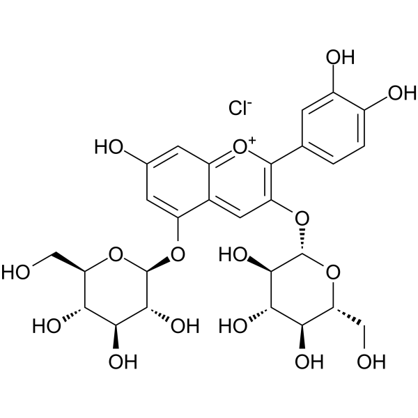
Cyanidin 3,5-diglucoside chloride is an anthocyanin. Cyanidin 3,5-diglucoside chloride inhibits inflammatory cytokines (IL-1α,IL-1β, and IL-6) expression and NO production. Cyanidin 3,5-diglucoside chloride inhibits the phosphorylation of STAT3, IκB, ERK, p38, and AKT. Cyanidin 3,5-diglucoside chloride inhibits high pressure-induced decrease in GLAST. Cyanidin 3,5-diglucoside chloride exerts anti-inflammatory and skin barrier modulating effects. Cyanidin 3,5-diglucoside chloride can be used in retinal research [1] .
Ascorbyl tetra-2-hexyldecanoate (tetra-isopalmitoyl Ascorbic acid; IPAA) is a lipophilic derivative of vitamin C (L-ascorbic acid). Ascorbyl tetra-2-hexyldecanoate (100 μM ) can prevent the decrease in viability of HaCaT keratinocytes induced by UVB, hydrogen peroxide, or tert-butyl hydroperoxide, and reduce the production of IL-1α and prostaglandin E2 (PGE2). Topical application of ascorbyl tetra-2-hexyldecanoate (1%) increases epidermal viability thickness, stratum corneum water content, and skin smoothness, and reduces skin roughness in hairless mice. Ascorbyl tetra-2-hexyldecanoate can be used to develop skin whitening agents in the beauty industry.
JT002 is an orally active and selective NLRP3 inflammasome assembly inhibitor. JT002 reduces NLRP3-dependent proinflammatory cytokine (such as IL-1β, IL-1α, IL-18) production and pyroptosis. JT002 blocks NLRP3 inflammasome complex formation. JT002 reduces airway hyperresponsiveness and airway neutrophilia in mice. JT002 can be used for the study of a variety of inflammatory diseases, such as Muckle-Wells syndrome (MWS) [1].
ATUX-1215 is an activator of protein phosphatase 2A (PP2A). ATUX-1215 reduced the phosphorylation of ERK, p38, JNK, and Akt and the secretion of IL-12p70, GM-CSF, and IL1α in Bleomycin hydrochloride (HY-17565A)-treated animals. ATUX-1215 can slow the progression of lung fibrosis [1].
IQ-3 is a specific inhibitor of the c-Jun N-terminal kinase (JNK) family, with preference for JNK3. IQ-3 exhibits Kd values of 0.24 μM, 0.29 μM and 0.066 μM for JNK1, JNK2 and JNK3, respectively [1].
Goflikicept (RPH 104) is a fusion protein that selectively binds and inactivates both circulating IL-1β and IL-1α. Goflikicept has the potential for the research of ST-segment elevation myocardial infarction (STEMI) [1].
Vilamakitug (XB-2001) is an anti-IL-1α human IgG4 κ monoclonal antibody [1]. Recommend Isotype Controls: Human IgG4 (S228P) kappa, Isotype Control (HY-P99003).
PG-901 is a full, selective MC5R agonist (EC50 = 0.072 nM for hMC5R). PG-901 is also a full antagonist at the hMC3R and the hMC4R (Kb: 1.0 nM and 0.53 nM, respectively). PG-901 significantly diminishes Cytokines (IL-1α,IL-1β, IL-6). PG-901 increases glucose tolerance, reduces blood glucose, decreases retinal damage [1] .
Oleaside A is a polar cardenolide monoglycoside isolated from Nerium oleander, inhibits the induction of ICAM-1 induced by IL-1α and TNF-α, and has anti-tumor activity [1].
DA-E 5090 is an orally effective inhibitor of IL-1 production that can be converted into a pharmacologically active deacetylated form (DA-E5090) in vivo. In this study, the effects of DA-E5090 on IL-1 production in vitro were examined by LPS-stimulated human monocytes. The results showed that DA-E5090 could dose-dependently inhibit the production of IL-1α and IL-1β (1-10 μM) by LPS-stimulated human monocytes, as determined by LAF assay and ELISA. Northern blotting analysis showed that DA-E5090 inhibited the transcription of IL-1α and IL-1β mRNA.
(S,S)-Z-FA-FMK is a cell-permeable, irreversible cathepsin B inhibitor. (S,S)-Z-FA-FMK blocks LPS-induced production of IL-1α and IL-1β. (S,S)-Z-FA-FMK can be used as a negative control for caspase-1 and caspase-2 inhibitors because it lacks an aspartic acid residue at the P1 position [1] .
Ac-LEVD-CHO (TFA) is an inhibitor of caspase-4. Ac-LEVD-CHO (TFA) can inhibit the expression and secretion of IL-1α expression as well as the activation of caspase-4 induced by the T. denticola periodontal pathogen surface protein Td92 in human gingival fibroblasts. Ac-LEVD-CHO (TFA) can also reduce the apoptosis due to the expression of the dominant negative adenoviral RNA-dependent protein kinase in A549 and PC3 cancer cell lines [1] .
PDE1-IN-5 (Compound 10c) is a selective PDE1C inhibitor (IC50: 15 nM). PDE1-IN-5 has anti- inflammatory activity, and inhibits expression of iNOS, TNF-α, IL-1α, IL-1β, and IL-6 induced by LPS. PDE1-IN-5 has anti-inflammatory bowel disease (IBD) effects in the dextran sodium sulfate (DSS)-Induced colitis mice model. PDE1-IN-5 can be used for research of IBD [1].
Nadifloxacin (Standard) is the analytical standard of Nadifloxacin (HY-B0506). This product is intended for research and analytical applications. Nadifloxacin (OPC7251) is a broad-spectrum quinolone antibiotic. Nadifloxacin inhibits bacterial DNA gyrase and topoisomerase IV, interfering with DNA replication. It also suppresses the production of proinflammatory cytokines (such as IL-1α, IL-6, and IL-8). Nadifloxacin exhibits antibacterial activity against various pathogens, including Propionibacterium acnes and Staphylococcus aureus. Nadifloxacin also exhibits anti-inflammatory activity. Nadifloxacin can be used in the research of skin infections such as acne vulgaris, folliculitis, and impetigo [1] .
Nadifloxacin-d5 (OPC7251-d5) is deuterium labeled Nadifloxacin (HY-B0506). Nadifloxacin (OPC7251) is a broad-spectrum quinolone antibiotic. Nadifloxacin inhibits bacterial DNA gyrase and topoisomerase IV, interfering with DNA replication. It also suppresses the production of proinflammatory cytokines (such as IL-1α, IL-6, and IL-8). Nadifloxacin exhibits antibacterial activity against various pathogens, including Propionibacterium acnes and Staphylococcus aureus. Nadifloxacin also exhibits anti-inflammatory activity. Nadifloxacin can be used in the research of skin infections such as acne vulgaris, folliculitis, and impetigo [1] .
Cyanidin 3,5-diglucoside (chloride) (Standard) is the analytical standard of Cyanidin 3,5-diglucoside (chloride) (HY-129138). This product is intended for research and analytical applications. Cyanidin 3,5-diglucoside chloride is an anthocyanin. Cyanidin 3,5-diglucoside chloride inhibits inflammatory cytokines (IL-1α,IL-1β, and IL-6) expression and NO production. Cyanidin 3,5-diglucoside chloride inhibits the phosphorylation of STAT3, IκB, ERK, p38, and AKT. Cyanidin 3,5-diglucoside chloride inhibits high pressure-induced decrease in GLAST. Cyanidin 3,5-diglucoside chloride exerts anti-inflammatory and skin barrier modulating effects. Cyanidin 3,5-diglucoside chloride can be used in retinal research [1] .
Octyl decyldimethyl ammonium chloride, a quaternary ammonium salt, is an antibacterial agent. Octyl decyldimethyl ammonium chloride disrupts cell membranes, leading to cytotoxicity. Octyl decyldimethyl ammonium chloride has risk of skin irritation and increases pro-inflammatory IL-1α secretion [1].
(Rac)-Besifloxacin Hydrochloride is a fourth generation fluoroquinolone antibiotic. (Rac)-Besifloxacin Hydrochloride is a DNA gyrase and topoisomerase IV inhibitor. (Rac)-Besifloxacin Hydrochloride has broad-spectrum antibacterial activity, it is effective against Gram-negative and Gram-positive aerobic and anaerobic strains and reduces the incidence of drug resistance. (Rac)-Besifloxacin Hydrochloride has anti-inflammatory activity. Besifloxacin Hydrochloride can be used in bacterial conjunctivitis research [1] .
NEK7 degrader-3 is an orally active and brain-penetrant NEK7molecular glue degrader with a DC50 of 33.1 nM. NEK7 degrader-3 mediates interaction between NEK7 and E3 ligase cereblon, promoting proteasomal degradation of NEK7 and attenuating NLRP3 inflammasome-mediated inflammatory responses. NEK7 degrader-3 inhibits caspase-1 activity, cytokine release of IL-1β, IL-1α, and IL-18, and pyroptosis-related plasma membrane permeabilization. NEK7 degrader-3 shows antiinflammatory activity in an LPS (HY-D1056)-induced neuroinflammation mouse model. NEK7 degrader-3 can be used for the research of neuroinflammation [1].
DB-310 is a selective immunoproteasome LMP2 inhibitor with an IC50 value of 80.62 nM. DB-310 inhibits the production of IL-1α in microglia. DB-310 improves cognitive function in the Tg2576 transgenic mouse model of Alzheimer's disease. DB-310 can be used for research related to Alzheimer's disease [1].
Lithocholic amide-C2-N(didecane) (Compound LC10) is a Lithocholic acid (HY-B0172) analogue. Lithocholic amide-C2-N(didecane) can form lipid nanoparticles spontaneously in the aqueous milieu, permeate through the skin, penetrate the deeper dermal layers, and exert anti-inflammatory effects against psoriasis-like chronic skin inflammations. Lithocholic amide-C2-N(didecane) can inhibit abnormal proliferation of keratinocytes, downregulate the mRNA expression of the psoriasis-associated receptor EphA2 and reduce serum levels of multiple pro-inflammatory factors such as IL-1α, IL-1β, and IFN-γ by inhibiting activation of the Th17/Th2 inflammatory pathway [1].
IQ-3 (Standard) is the analytical standard of IQ-3 (HY-107600). This product is intended for research and analytical applications. IQ-3 is a specific inhibitor of the c-Jun N-terminal Kinase (JNK) family, with preference for JNK3. IQ-3 exhibits Kd values of 0.24 μM, 0.29 μM and 0.066 μM for JNK1, JNK2 and JNK3, respectively [1].
IL-1β-IN-4 is a selective IL-1β competitive binder. IL-1β-IN-4 interferes with the interaction between IL-1β and its cognate receptor IL-1R1, with an IC50 of 8.3 μM. IL-1β-IN-4 can be used in studies related to inflammatory and immune diseases [1].
Tyrosinase-IN-50 (Compound 14) is a Tyrosinase inhibitor (IC50 is 0.06 μM or 0.16 μM). Tyrosinase-IN-50 inhibits melanogenesis in multiple cell types. Tyrosinase-IN-50 can be used for the research of hyperpigmentation-related diseases and inflammatory diseases [1].
Candidalysin is a cytolytic peptide toxin secreted by the fungus Candida albicans. Candidalysin drives epithelial immune responses by activating the EGFR-MAPK signaling pathway, inducing MMP expression and calcium influx, and regulating the c-Fos transcription factor and MKP1 via p38 MAPK and ERK1/2 respectively. Candidalysin is essential for mucosal and systemic infections, activating NLRP3 to promote inflammatory responses, neutrophil recruitment, and Th17 immunity. Candidalysin activates LDH causing membrane damage and exhibiting cytotoxicity [1]
PG-901 is a full, selective MC5R agonist (EC50 = 0.072 nM for hMC5R). PG-901 is also a full antagonist at the hMC3R and the hMC4R (Kb: 1.0 nM and 0.53 nM, respectively). PG-901 significantly diminishes Cytokines (IL-1α,IL-1β, IL-6). PG-901 increases glucose tolerance, reduces blood glucose, decreases retinal damage [1] .
(S,S)-Z-FA-FMK is a cell-permeable, irreversible cathepsin B inhibitor. (S,S)-Z-FA-FMK blocks LPS-induced production of IL-1α and IL-1β. (S,S)-Z-FA-FMK can be used as a negative control for caspase-1 and caspase-2 inhibitors because it lacks an aspartic acid residue at the P1 position [1] .
Ac-LEVD-CHO (TFA) is an inhibitor of caspase-4. Ac-LEVD-CHO (TFA) can inhibit the expression and secretion of IL-1α expression as well as the activation of caspase-4 induced by the T. denticola periodontal pathogen surface protein Td92 in human gingival fibroblasts. Ac-LEVD-CHO (TFA) can also reduce the apoptosis due to the expression of the dominant negative adenoviral RNA-dependent protein kinase in A549 and PC3 cancer cell lines [1] .
Astegolimab (MSTT 1041A; RG 6149) is a human IgG2 monoclonal antibody. Astegolimab blocks IL-33 signaling by targeting the IL-33 receptor ST2. Astegolimab reduces p53 expression, mitigates IL33-upregulated SASP factors such as IL1α, IL6 and MCP1. Astegolimab mitigates IL33-increased p-p65/p65 ratio. Astegolimab blocks CM-induced neutrophil extracellular trap (NET) formation. Astegolimab is used in chronic obstructive pulmonary disease (COPD) and myocardial research [1] .
Lutikizumab (ABT-981) is an anti-IL-1α and IL-1β dual variable domain immunoglobulin. Lutikizumab binds and inhibits IL-1α and IL-1β. Lutikizumab can be used for the research of osteoarthritis [1].
Nidanilimab (CAN04) is a fully humanized monoclonal anti-IL1RAP antibody with a Kd value of 1.10 pM. Nidanilimab blocks IL1α and IL1β signaling and stimulates the immune system to destroy tumour cells. Nidanilimab can be used in research of non-small lung cancer (NSCLC) and pancreatic ductal adenocarcinoma (PDAC) [1] .
Bermekimab (MABp1) is a human monoclonal antibody targeting interleukin-1α(IL-1α) . Bermekimab can prevent tumor-related inflammation. Bermekimab can be used in the research of atopic dermatitis [1].
Goflikicept (RPH 104) is a fusion protein that selectively binds and inactivates both circulating IL-1β and IL-1α. Goflikicept has the potential for the research of ST-segment elevation myocardial infarction (STEMI) [1].
Vilamakitug (XB-2001) is an anti-IL-1α human IgG4 κ monoclonal antibody [1]. Recommend Isotype Controls: Human IgG4 (S228P) kappa, Isotype Control (HY-P99003).
Cyanidin 3,5-diglucoside chloride is an anthocyanin. Cyanidin 3,5-diglucoside chloride inhibits inflammatory cytokines (IL-1α,IL-1β, and IL-6) expression and NO production. Cyanidin 3,5-diglucoside chloride inhibits the phosphorylation of STAT3, IκB, ERK, p38, and AKT. Cyanidin 3,5-diglucoside chloride inhibits high pressure-induced decrease in GLAST. Cyanidin 3,5-diglucoside chloride exerts anti-inflammatory and skin barrier modulating effects. Cyanidin 3,5-diglucoside chloride can be used in retinal research [1] .
Oleaside A is a polar cardenolide monoglycoside isolated from Nerium oleander, inhibits the induction of ICAM-1 induced by IL-1α and TNF-α, and has anti-tumor activity [1].
Cyanidin 3,5-diglucoside (chloride) (Standard) is the analytical standard of Cyanidin 3,5-diglucoside (chloride) (HY-129138). This product is intended for research and analytical applications. Cyanidin 3,5-diglucoside chloride is an anthocyanin. Cyanidin 3,5-diglucoside chloride inhibits inflammatory cytokines (IL-1α,IL-1β, and IL-6) expression and NO production. Cyanidin 3,5-diglucoside chloride inhibits the phosphorylation of STAT3, IκB, ERK, p38, and AKT. Cyanidin 3,5-diglucoside chloride inhibits high pressure-induced decrease in GLAST. Cyanidin 3,5-diglucoside chloride exerts anti-inflammatory and skin barrier modulating effects. Cyanidin 3,5-diglucoside chloride can be used in retinal research [1] .
IL-1 alpha, found in non-hematopoietic cells, connects innate and adaptive immunity. Binding to IL1R1, it activates signaling pathways involving MYD88, IRAK1, and IRAK4, leading to NF-kappa-B and MAPK pathway activation. Released during cell death, IL-1 alpha induces inflammation, senses DNA damage, and interacts with TMED10, IL1R1, and S100A13 for secretion and plasma membrane translocation. IL-1 alpha Protein, Cynomolgus (HEK293, His) is the recombinant cynomolgus-derived IL-1 alpha protein, expressed by HEK293 , with C-His labeled tag.
IL-1 alpha is a ubiquitous and pivotal pro-inflammatory cytokine. IL-1 alpha is produced by monocytes and macrophages as a proprotein, which is proteolytically processed and released in response to cell injury, and thus induces apoptosis. IL-1 alpha plays an important role in inflammation and bridges the innate and adaptive immune systems. IL-1 alpha mediates the activation of NF-kappa-B and the three MAPK pathways p38, p42/p44 and JNK pathways. IL-1 alpha protein, Human (HEK293) is a recombinant protein consisting of 159 amino acids (S113-A271) and is produced by HEK293 cells.
IL-1 alpha is a ubiquitous and pivotal pro-inflammatory cytokine. IL-1 alpha is produced by monocytes and macrophages as a proprotein, which is proteolytically processed and released in response to cell injury, and thus induces apoptosis. IL-1 alpha plays an important role in inflammation and bridges the innate and adaptive immune systems. IL-1 alpha mediates the activation of NF-kappa-B and the three MAPK pathways p38, p42/p44 and JNK pathways.
IL-1 alpha is a ubiquitous and pivotal pro-inflammatory cytokine. IL-1 alpha is produced by monocytes and macrophages as a proprotein, which is proteolytically processed and released in response to cell injury, and thus induces apoptosis. IL-1 alpha plays an important role in inflammation and bridges the innate and adaptive immune systems. IL-1 alpha mediates the activation of NF-kappa-B and the three MAPK pathways p38, p42/p44 and JNK pathways. IL-1 alpha protein, Mouse is a recombinant protein consisting of 156 amino acids (S115-A270) and is produced by E. coli.
IL-1 alpha is a ubiquitous and pivotal pro-inflammatory cytokine. IL-1 alpha is produced by monocytes and macrophages as a proprotein, which is proteolytically processed and released in response to cell injury, and thus induces apoptosis. IL-1 alpha plays an important role in inflammation and bridges the innate and adaptive immune systems. IL-1 alpha mediates the activation of NF-kappa-B and the three MAPK pathways p38, p42/p44 and JNK pathways. IL-1 alpha protein, Human is a recombinant protein consisting of 159 amino acids (S113-A271) and is produced by E. coli.
IL-1 alpha protein is an important cytokine found intracellularly in non-hematopoietic cells that bridges innate and adaptive immunity. Binds to IL1R1 and IL1RAP to form a high-affinity receptor complex, initiates signaling through MYD88, IRAK1 or IRAK4, and activates the NF-kappa-B and MAPK pathways. Animal-Free IL-1 alpha Protein, Mouse (His) is the recombinant mouse-derived animal-FreeIL-1 alpha protein, expressed by E. coli , with C-His labeled tag. This product is for cell culture use only.
IL-1 alpha protein is located in cells and is a key cytokine connecting innate immunity and adaptive immunity. After binding to IL1R1 through IL1RAP, it forms a high-affinity receptor complex, initiates a signaling cascade with adapter molecules such as MYD88, IRAK1 or IRAK4, and activates the NF-kappa-B and MAPK pathways. Animal-Free IL-1 alpha Protein, Human (His) is the recombinant human-derived animal-FreeIL-1 alpha protein, expressed by E. coli , with C-His labeled tag. This product is for cell culture use only.
IL-1 alpha is a ubiquitous and pivotal pro-inflammatory cytokine. IL-1 alpha is produced by monocytes and macrophages as a proprotein, which is proteolytically processed and released in response to cell injury, and thus induces apoptosis. IL-1 alpha plays an important role in inflammation and bridges the innate and adaptive immune systems. IL-1 alpha mediates the activation of NF-kappa-B and the three MAPK pathways p38, p42/p44 and JNK pathways. IL-1 alpha protein, Rat is a recombinant protein consisting of 156 amino acids (S115-S270) and is produced by E. coli.
IL-1 alpha, a cytokine present in non-hematopoietic cells, links innate and adaptive immunity. Binding to its receptor IL1R1, IL-1 alpha forms a receptor complex, activating signaling pathways involving MYD88, IRAK1, and IRAK4. It activates NF-kappa-B and MAPK pathways. Released during cell death, IL-1 alpha induces inflammation, senses DNA damage, and interacts with TMED10, IL1R1, and S100A13 for secretion and plasma membrane translocation. IL-1 alpha Protein, Rhesus macaque is the recombinant Rhesus Macaque-derived IL-1 alpha protein, expressed by E. coli , with tag free.
IL-1 alpha is a ubiquitous and pivotal pro-inflammatory cytokine. IL-1 alpha is produced by monocytes and macrophages as a proprotein, which is proteolytically processed and released in response to cell injury, and thus induces apoptosis. IL-1 alpha plays an important role in inflammation and bridges the innate and adaptive immune systems. IL-1 alpha mediates the activation of NF-kappa-B and the three MAPK pathways p38, p42/p44 and JNK pathways. GMP IL-1 alpha Protein, Human is a recombinant protein consisting of 153 amino acids (A117-S269) and is produced by E. coli.
IL-1 alpha protein is located in cells and is a key cytokine connecting innate immunity and adaptive immunity. After binding to IL1R1 through IL1RAP, it forms a high-affinity receptor complex, initiates a signaling cascade with adapter molecules such as MYD88, IRAK1 or IRAK4, and activates the NF-kappa-B and MAPK pathways. IL-1 alpha Protein, Human (HEK293, Fc) is the recombinant human-derived IL-1 alpha protein, expressed by HEK293 , with C-hFc labeled tag.
IL-1 alpha is a ubiquitous and pivotal pro-inflammatory cytokine. IL-1 alpha is produced by monocytes and macrophages as a proprotein, which is proteolytically processed and released in response to cell injury, and thus induces apoptosis. IL-1 alpha plays an important role in inflammation and bridges the innate and adaptive immune systems. IL-1 alpha mediates the activation of NF-kappa-B and the three MAPK pathways p38, p42/p44 and JNK pathways. IL-1 alpha protein, Human (Biotinylated, HEK293, Avi) is a biotinylated recombinant protein with Avi label that consists of 159 amino acids (S113-A271) and is produced by E. coli.
IL-1 alpha protein is an important cytokine found intracellularly in non-hematopoietic cells that bridges innate and adaptive immunity. Binds to IL1R1 and IL1RAP to form a high-affinity receptor complex, initiates signaling through MYD88, IRAK1 or IRAK4, and activates the NF-kappa-B and MAPK pathways. IL-1 alpha Protein, Mouse (HEK293, His) is the recombinant mouse-derived IL-1 alpha protein, expressed by HEK293, with C-His labeled tag.
The IL-1α/IL-1F1 protein is found intracellularly in most non-hematopoietic cells and plays a crucial role in mediating inflammation and linking innate and adaptive immunity. IL1RAP binds to IL1R1 to form a high-affinity receptor complex that activates cascades and pathways.IL-1 alpha/IL-1F1 Protein, Pig is the recombinant Porcine-derived IL-1 alpha/IL-1F1 protein, expressed by E. coli , with tag free.
IL-1 alpha protein is located in cells and is a key cytokine connecting innate immunity and adaptive immunity. After binding to IL1R1 through IL1RAP, it forms a high-affinity receptor complex, initiates a signaling cascade with adapter molecules such as MYD88, IRAK1 or IRAK4, and activates the NF-kappa-B and MAPK pathways. IL-1 alpha Protein, Human (HEK293, His) is the recombinant human-derived IL-1 alpha protein, expressed by HEK293, with C-His labeled tag.
The IL-1α/IL-1F1 protein is found intracellularly in most non-hematopoietic cells and plays a crucial role in mediating inflammation and linking innate and adaptive immunity. IL1RAP binds to IL1R1 to form a high-affinity receptor complex that activates cascades and pathways. Animal-Free IL-1 alpha/IL-1F1 Protein, Pig (His) is the recombinant pig-derived animal-FreeIL-1 alpha/IL-1F1 protein, expressed by E. coli , with C-His labeled tag. This product is for cell culture use only.
Nadifloxacin-d5 (OPC7251-d5) is deuterium labeled Nadifloxacin (HY-B0506). Nadifloxacin (OPC7251) is a broad-spectrum quinolone antibiotic. Nadifloxacin inhibits bacterial DNA gyrase and topoisomerase IV, interfering with DNA replication. It also suppresses the production of proinflammatory cytokines (such as IL-1α, IL-6, and IL-8). Nadifloxacin exhibits antibacterial activity against various pathogens, including Propionibacterium acnes and Staphylococcus aureus. Nadifloxacin also exhibits anti-inflammatory activity. Nadifloxacin can be used in the research of skin infections such as acne vulgaris, folliculitis, and impetigo [1] .
Western blot analysis of extracts from THP-1(lane 2(20μg), Jurkat (lane 3(20μg) and NIH3T3(lane 4(20μg) using FOXO1A (HY-P80132) Rabbit mAb. Proteins were transferred
to a PVDF membrane and blocked with 5% non-fat milk in TBST for 2 hour at room temperature. The primary antibody (1/1000) and Loading control antibody (Beta Actin, HY-P80438, 1/10000) was
used in 5% non-fat milk in TBST at 4°C overnight. Goat Anti-Mouse/Rabbit IgG-HRP Secondary Antibody (1/10000) was used for 1 hour at room temperature.
Western blot analysis of extracts from THP-1(lane 2(20μg), Jurkat (lane 3(20μg) and NIH3T3(lane 4(20μg) using FOXO1A (HY-P80132) Rabbit mAb. Proteins were transferred
to a PVDF membrane and blocked with 5% non-fat milk in TBST for 2 hour at room temperature. The primary antibody (1/1000) and Loading control antibody (Beta Actin, HY-P80438, 1/10000) was
MedchemExpress Validation 03
Western blot analysis of extracts from THP-1(lane 2(20μg), Jurkat (lane 3(20μg) and NIH3T3(lane 4(20μg) using FOXO1A (HY-P80132) Rabbit mAb. Proteins were transferred
MedchemExpress Validation 04
Western blot analysis of extracts from THP-1(lane 2(20μg), Jurkat (lane 3(20μg) and NIH3T3(lane 4(20μg) using FOXO1A (HY-P80132) Rabbit mAb. Proteins were transferred
to a PVDF membrane and blocked with 5% non-fat milk in TBST for 2 hour at room temperature. The primary antibody (1/1000) and Loading control antibody (Beta Actin, HY-P80438, 1/10000) was
used in 5% non-fat milk in TBST at 4°C overnight. Goat Anti-Mouse/Rabbit IgG-HRP Secondary Antibody (1/10000) was used for 1 hour at room temperature.
MedchemExpress Validation
Western blot analysis of extracts from THP-1(lane 2(20μg), Jurkat (lane 3(20μg) and NIH3T3(lane 4(20μg) using FOXO1A (HY-P80132) Rabbit mAb. Proteins were transferred
to a PVDF membrane and blocked with 5% non-fat milk in TBST for 2 hour at room temperature. The primary antibody (1/1000) and Loading control antibody (Beta Actin, HY-P80438, 1/10000) was
used in 5% non-fat milk in TBST at 4°C overnight. Goat Anti-Mouse/Rabbit IgG-HRP Secondary Antibody (1/10000) was used for 1 hour at room temperature.
MedchemExpress Validation
Western blot analysis of extracts from THP-1(lane 2(20μg), Jurkat (lane 3(20μg) and NIH3T3(lane 4(20μg) using FOXO1A (HY-P80132) Rabbit mAb. Proteins were transferred
to a PVDF membrane and blocked with 5% non-fat milk in TBST for 2 hour at room temperature. The primary antibody (1/1000) and Loading control antibody (Beta Actin, HY-P80438, 1/10000) was
used in 5% non-fat milk in TBST at 4°C overnight. Goat Anti-Mouse/Rabbit IgG-HRP Secondary Antibody (1/10000) was used for 1 hour at room temperature.
MedChemExpress values your privacy and your trust is important to us. We use cookies to enhance your website experience. Some cookies are necessary to run the website.
Privacy and Cookie Policy