



Candidalysin

Based on 1 Customer Validation
Candidalysin is a cytolytic peptide toxin secreted by the fungus Candida albicans. Candidalysin drives epithelial immune responses by activating the EGFR-MAPK signaling pathway, inducing MMP expression and calcium influx, and regulating the c-Fos transcription factor and MKP1 via p38 MAPK and ERK1/2 respectively. Candidalysin is essential for mucosal and systemic infections, activating NLRP3 to promote inflammatory responses, neutrophil recruitment, and Th17 immunity. Candidalysin activates LDH causing membrane damage and exhibiting cytotoxicity
For research use only. We do not sell to patients.
- Purity : 99.81%
- CAS No.: 1906866-53-2
- Formula: C153H266N38O38S2
- Molecular Weight:3310.11
-
Storage:
Sealed storage, away from moisture.
Powder -80°C, 2 years , -20°C, 1 year* In solvent : -80°C, 6 months; -20°C, 1 month (sealed storage, away from moisture)


To place orders, for customer services and technical support, please contact: MedChemExpress USA
Tel: 609-228-6898 E-mail: [email protected] [email protected]


-
 Biological Activity
-
Chemical Information
-
Solvent & Solubility
- Protocols
- Purity & Documentation
- References
-
 Help & FAQs


-

HY-B0215
AcetylcysteineMucolytic Agent -
HY-13740
ResiquimodTLR7/8 Agonist -
HY-17031
SBE-β-CDExcipient -

HY-10583
Y-27632 dihydrochlorideROCK Inhibitor -

HY-15763
ErastinFerroptosis Inducer -
HY-B0180
ImiquimodTLR7 Agonist -
HY-114277
SotorasibKRAS G12C Inhibitor -
HY-K0011
Protease Inhibitor Cocktail, mini-Tablet (EDTA-Free) -
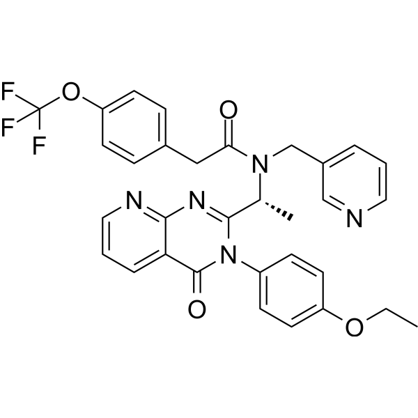
HY-15319
AMG 487CXCR3 Antagonist -
HY-K0010
Protease Inhibitor Cocktail (EDTA-Free, 100× in DMSO)


Biological Activity
Description
In Vitro
Candidalysin (3-70 μM, 2 h) induces phosphorylation of EGFR at Y1068 and Y845 in TR146 cells in a dose-dependent manner[1].
Candidalysin (3-70 μM, 2 h-24 h) induces immune response in TR146 cells that are mediated by MMP and calcium influx[1].
Candidalysin (1.5-70 μM, 24 h) induces the release of LDH in A431 vaginal epithelial cells in a dose-dependent manner[2].
Candidalysin (1.5-70 μM, 24 h) upregulates the expression levels of proinflammatory cytokines IL-1α, IL-1β, G-CSF, GM-CSF, IL-6 and IL-8 secreted by A431 vaginal epithelial cells[2].
Candidalysin (10-50 μM, 2 h) activates NLRP3 inflammasome and specifically stimulates bone marrow-derived macrophages (BMDM) to secrete IL-1β[3].
MedChemExpress (MCE) has not independently confirmed the accuracy of these methods. They are for reference only. Further protocols information, click here.
-
Cell Line:TR146 cells
-
Concentration:3, 15, 70 μM
-
Incubation Time:2 h
-
Result:Up-regulated the protein levels of pEGFR Y1068 and pEGFR Y845.
-
Cell Line:BMDM
-
Concentration:10, 50 μM
-
Incubation Time:2 h
-
Result:Up-regulated the protein levels of IL-1β.
In Vivo
Candidalysin (wild-type C. albicans infection) (5×10⁸ cfu/ml, Intravaginally inoculated, once) induces neutrophil recruitment and mucosal damage in the vulvovaginal candidiasis (VVC) mouse model[2].
MedChemExpress (MCE) has not independently confirmed the accuracy of these methods. They are for reference only.
-
Animal Model:Murine OPC model (female Balb/c mice 22-25 g)[1]
-
Dosage:107 cfu/ml C. albicans yeast culture
-
Administration:Sub-lingually for 75 min, once
-
Result:Compared with those lacking the candidalysin parent gene ECE1 (ece1Δ/Δ) or the candidalysin-encoding region (ece1Δ/Δ+ECE1Δ184-279), wild-type (WT) C. albicans infection induced lower levels of pEGFR Y1068 in tongue tissues.
-
Animal Model:Murine VVC model (female 6-8 weeks old C57BL/6 mice)[2]
-
Dosage:5×10⁸ cfu/ml C. albicans, 10 μL of the standardized blastoconidial cell suspension, generating an inoculum size of 5×10⁶ blastoconidia.
-
Administration:Intravaginally inoculated, once
-
Result:Increased fungal burden.
Increased LDH activity.


Chemical Information
-
CAS No. 1906866-53-2
-
Appearance Solid
-
Molecular Weight 3310.11
-
Formula C153H266N38O38S2
-
Color White to off-white
-
Sequence
Ser-Ile-Ile-Gly-Ile-Ile-Met-Gly-Ile-Leu-Gly-Asn-Ile-Pro-Gln-Val-Ile-Gln-Ile-Ile-Met-Ser-Ile-Val-Lys-Ala-Phe-Lys-Gly-Asn-Lys
-
Sequence Shortening
SIIGIIMGILGNIPQVIQIIMSIVKAFKGNK
-
Shipping
Room temperature in continental US; may vary elsewhere.
-
Storage
Sealed storage, away from moisture
Powder -80°C 2 years -20°C 1 year * In solvent : -80°C, 6 months; -20°C, 1 month (sealed storage, away from moisture)


Solvent & Solubility
In Vitro:
DMSO : 25 mg/mL (7.55 mM; Need ultrasonic; Hygroscopic DMSO has a significant impact on the solubility of product, please use newly opened DMSO)
H2O : 25 mg/mL (7.55 mM; Need ultrasonic)
Please refer to the solubility information to select the appropriate solvent. Once prepared, please aliquot and store the solution to prevent product inactivation from repeated freeze-thaw cycles.
Storage method and period of stock solution: -80°C, 6 months; -20°C, 1 month (sealed storage, away from moisture). When stored at -80°C, please use it within 6 months. When stored at -20°C, please use it within 1 month.
* Note: If you choose water as the stock solution, please dilute it to the working solution, then filter and sterilize it with a 0.22 μm filter before use.
Please refer to the solubility information to select the appropriate solvent. Once prepared, please aliquot and store the solution to prevent product inactivation from repeated freeze-thaw cycles.
Storage method and period of stock solution: -80°C, 6 months; -20°C, 1 month (sealed storage, away from moisture). When stored at -80°C, please use it within 6 months. When stored at -20°C, please use it within 1 month.
* Note: If you choose water as the stock solution, please dilute it to the working solution, then filter and sterilize it with a 0.22 μm filter before use.
Concentration (start) × Volume (start) = Concentration (final) × Volume (final)
In Vivo:
Select the appropriate dissolution method based on your experimental animal and administration route.
- For the following dissolution methods, please ensure to first prepare a clear stock solution using an In Vitro approach and then sequentially add co-solvents:
- To ensure reliable experimental results, the clarified stock solution can be appropriately stored based on storage conditions. As for the working solution for In Vivo experiments, it is recommended to prepare freshly and use it on the same day.
- The percentages shown for the solvents indicate their volumetric ratio in the final prepared solution. If precipitation or phase separation occurs during preparation, heat and/or sonication can be used to aid dissolution.
Add each solvent one by one: 10% DMSO 40% PEG300 5% Tween-80 45% Saline
Solubility: ≥ 2.5 mg/mL (0.76 mM); Clear solution
This protocol yields a clear solution of ≥ 2.5 mg/mL (saturation unknown).
Taking 1 mL working solution as an example, add 100 μL DMSO stock solution (25.0 mg/mL) to 400 μL PEG300, and mix evenly; then add 50 μL Tween-80 and mix evenly; then add 450 μL Saline to adjust the volume to 1 mL.
Preparation of Saline: Dissolve 0.9 g sodium chloride in ddH₂O and dilute to 100 mL to obtain a clear Saline solution.
In Vivo Dissolution Calculator
Please enter the basic information of animal experiments:
-
-
-
-
Recommended: Prepare an additional quantity of animals to account for potential losses during experiments.
Working solution concentration: 0.22 mg/mL
This product has good water solubility, please refer to the measured solubility data in water/PBS/Saline for details.

Protocols
-
Nuclear Protein Extraction (High-Salt/Hypotonic Fractionation)
The high-salt/hypotonic fractionation method for nuclear protein extraction is based on the differential solubility of cellular components. Cytoplasmic proteins are extracted first using a hypotonic buffer that causes cell swelling and membrane rupture, followed by centrifugation to separate the cytoplasmic supernatant from the nuclear pellet. The nuclear pellet is then subjected to high-salt extraction (e. g. , 0. 4 M (NH4)2SO4 or 1 M NaCl) to solubilize tightly bound nuclear matrix proteins, including transcription factors, histones, and structural proteins associated with chromatin and the nuclear scaffold. This approach allows for the isolation of both soluble cytoplasmic proteins and salt-resistant nuclear proteins while minimizing cross-contamination.
-
Phagocytosis Functional Assay
A phagocytosis functional assay measures the ability of phagocytic cells, such as neutrophils, macrophages, monocytes, or microglia/macrophages, to bind and internalize particulate targets including bacteria, yeast particles, beads, or myelin particles. Fluorescent flow-cytometry assays detect target uptake as fluorescence associated with gated phagocytes, while pH-sensitive dyes such as pHrodo increase signal in acidic phagosomal compartments and therefore preferentially report internalized particles rather than particles remaining outside the cell. Microscopy or high-content imaging can be used to confirm intracellular localization and, in some protocols, to follow uptake kinetics.
-
RNA extraction experimental
By lysing cells, releasing RNA, and removing impurities such as proteins and DNA, high-purity RNA products are finally obtained. The commonly used traditional method is the guanidine isothiocyanate/phenol/chloroform method (Trizol), which is suitable for a variety of animal materials including animal tissues, microorganisms, cultured cells, etc., and most plant materials.
-
Mitochondrial membrane-potential fluorescent assay
Mitochondrial membrane potential fluorescent assays estimate ΔΨm in living cells using lipophilic cationic dyes such as TMRM, TMRE, rhodamine 123, and JC-1, which accumulate in mitochondria according to membrane polarization; loss of signal after FCCP or CCCP treatment is interpreted as mitochondrial depolarization. TMRM/TMRE and rhodamine 123 are commonly used for semi-quantitative live-cell microscopy or flow cytometry, while JC-1 can report a shift from red aggregate fluorescence to green monomer fluorescence during depolarization; interpretation requires controls because dye concentration, quenching mode, cell type, dye efflux, and mitochondrial mass can affect fluorescence independently of ΔΨm.
-
Cell Cytotoxicity Assay
Cytotoxicity assays are usually based on the assessment of cell membrane damage, which can also be indirectly detected by measuring cell viability. Detection methods include MTT assay, CKK-8 assay, LDH assay and ATP assay, etc.
-
Mammalian live/dead viability and cytotoxicity staining
Live/dead viability and cytotoxicity staining assays are based on the simultaneous detection of intracellular esterase activity in metabolically active (viable) cells and membrane integrity loss in non-viable cells. In commonly used dual-staining approaches, membrane-permeant fluorogenic substrates are converted by intracellular esterases into fluorescent products in live cells, while impermeant DNA-binding dyes selectively enter cells with compromised plasma membranes and label nucleic acids in dead or dying cells, enabling discrimination between viable and non-viable populations by fluorescence microscopy or flow cytometry.
-
Naïve CD4+ T-cell subset differentiation/polarization
Naïve CD4+ T-cell subset differentiation/polarization is an in vitro assay in which purified naïve CD4+ T cells are activated through TCR and CD28 costimulation and cultured with defined cytokines and neutralizing antibodies to generate Th0, Th1, Th2, Th17, or induced Treg-like populations. Differentiation is detected by subset-associated cytokines and transcription factors: IFN-γ/T-bet for Th1, IL-4/GATA3 for Th2, IL-17A/RORγt for Th17, and Foxp3 for induced Treg cells. The assay readout is usually generated by intracellular cytokine staining after restimulation, transcription-factor staining by flow cytometry, ELISA of secreted cytokines, or gene-expression analysis. The result reflects cytokine-directed lineage commitment or polarization rather than antigen-specific immune protection by itself.
-
Mitochondrial membrane-potential and mitochondrial mass staining
Mitochondrial membrane potential staining measures the electrochemical polarization across the mitochondrial inner membrane in live cells using lipophilic cationic fluorescent probes; early rhodamine-based work showed that selective mitochondrial dye accumulation is lost when the mitochondrial transmembrane potential is dissipated. JC-1 reports mitochondrial polarization by shifting from green monomer fluorescence to red J-aggregate fluorescence as dye concentration increases within energized mitochondria; therefore, the red/green fluorescence ratio is used as a relative readout of mitochondrial membrane potential. TMRE or TMRM staining provides a single-channel relative readout because these cationic rhodamine esters accumulate in polarized mitochondria, and lower fluorescence indicates reduced mitochondrial polarization when acquisition and dye-loading conditions are controlled. Mitochondrial mass staining is commonly performed with MitoTracker Green FM or related MitoTracker dyes as
-
Zymosan-Induced Peritonitis
Zymosan-induced peritonitis is a sterile acute-inflammation model produced by intraperitoneal injection of zymosan, a yeast cell-wall particle preparation, followed by quantification of leukocyte recruitment and soluble inflammatory mediators in peritoneal lavage fluid. Low-dose zymosan peritonitis is commonly used as a self-resolving acute inflammation model in which neutrophil recruitment occurs early and monocyte/macrophage accumulation follows later. The assay readouts include total peritoneal leukocyte number, differential neutrophil and monocyte/macrophage counts, peritoneal cytokines and chemokines, plasma or peritoneal exudation, and optional lipidomic or metabolomic changes during inflammation and resolution. Early neutrophil recruitment after zymosan depends strongly on complement and mast-cell C5a receptor signaling, whereas later monocyte recruitment is linked to MCP-1/CCL2 production.
-
Ca2+ Staining Technique
Ca2+ staining is an experimental technique that utilizes specific fluorescent probes (such as Fluo-4 AM, Fura-2, etc.) to qualitatively or quantitatively detect dynamic changes in intracellular Ca2+ concentrations; this is achieved by monitoring the changes in fluorescent signals generated when these probes bind to free intracellular calcium ions. The underlying principle relies primarily on the presence of chelating groups within the probe's molecular structure that possess high affinity for calcium ions.
-
Cell Viability Determination by MTT Colorimetric Assay
The following protocol uses the MTT colorimetric assay as a classic literature-established method for assessing cell viability/metabolic activity in cultured mammalian cells. MTT[3-(4,5-dimethylthiazol-2-yl)-2,5-diphenyltetrazolium bromide] is reduced by metabolically active cells to a colored formazan product; the amount of formazan is quantified spectrophotometrically and provides an indirect measure of metabolically active viable cells. Importantly, MTT reduction reflects cellular oxidoreductase/metabolic activity rather than an absolute direct count of living cells, so changes in cellular metabolism can alter the signal independently of cell number.


Purity & Documentation
-
Data Sheet (287 KB)
-
SDS (254 KB)
- English - EN (254 KB)
- Français - FR (254 KB)
- Deutsch - DE (254 KB)
- Norwegian - NO (254 KB)
- Español - ES (254 KB)
- Swedish - SV (254 KB)
- Italian - IT (254 KB)
- Korean - KR (254 KB)
- Portuguese - PT (254 KB)
-
Handling Instructions (2659 KB)
References
[1]. Ho J, et al. Candidalysin activates innate epithelial immune responses via epidermal growth factor receptor. Nat Commun. 2019 May 24;10(1):2297. [Content Brief]
[2]. Richardson JP, et al. Candidalysin Drives Epithelial Signaling, Neutrophil Recruitment, and Immunopathology at the Vaginal Mucosa. Infect Immun. 2018 Jan 22;86(2):e00645-17. [Content Brief]
[3]. Rogiers O, et al. Candidalysin Crucially Contributes to Nlrp3 Inflammasome Activation by Candida albicans Hyphae. mBio. 2019 Jan 8;10(1):e02221-18. [Content Brief]
[4]. Naglik JR, et al. Candidalysin: discovery and function in Candida albicans infections. Curr Opin Microbiol. 2019 Dec;52:100-109. [Content Brief]


Complete Stock Solution Preparation Table
Please refer to the solubility information to select the appropriate solvent. Once prepared, please aliquot and store the solution to prevent product inactivation from repeated freeze-thaw cycles.
Storage method and period of stock solution: -80°C, 6 months; -20°C, 1 month (sealed storage, away from moisture). When stored at -80°C, please use it within 6 months. When stored at -20°C, please use it within 1 month.
| Optional Solvent | Concentration Solvent Mass | 1 mg | 5 mg | 10 mg | 25 mg |
|---|---|---|---|---|---|
| DMSO / H2O | 1 mM | 0.3021 mL | 1.5105 mL | 3.0210 mL | 7.5526 mL |
| 5 mM | 0.0604 mL | 0.3021 mL | 0.6042 mL | 1.5105 mL |
* Note: If you choose water as the stock solution, please dilute it to the working solution, then filter and sterilize it with a 0.22 μm filter before use.


Candidalysin Related Classifications
HY-P10408 Related Classifications
 Powered by Bioz