



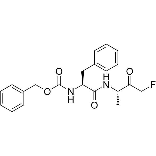
(S,S)-Z-FA-FMK
Based on 1 publication(s) in Google Scholar
(S,S)-Z-FA-FMK is a cell-permeable, irreversible cathepsin B inhibitor. (S,S)-Z-FA-FMK blocks LPS-induced production of IL-1α and IL-1β. (S,S)-Z-FA-FMK can be used as a negative control for caspase-1 and caspase-2 inhibitors because it lacks an aspartic acid residue at the P1 position.
For research use only. We do not sell to patients.
- CAS No.: 105637-38-5
- Formula: C21H23FN2O4
- Molecular Weight:386.42
-
Storage:
Please store the product under the recommended conditions in the Certificate of Analysis.

To place orders, for customer services and technical support, please contact: MedChemExpress USA
Tel: 609-228-6898 E-mail: [email protected] [email protected]


-
 Biological Activity
-
Chemical Information
- Protocols
- Purity & Documentation
- References
-
 Help & FAQs


-
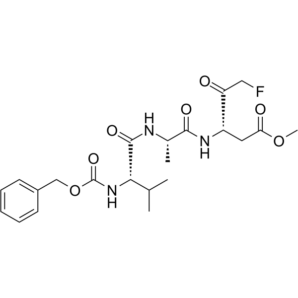
HY-16658
Z-VAD(OMe)-FMKPan-caspase Inhibitor -
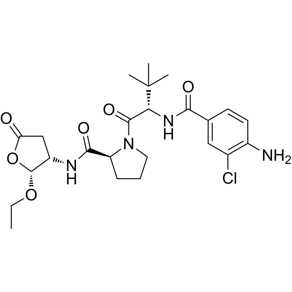
HY-13205
BelnacasanCaspase-1 Inhibitor -
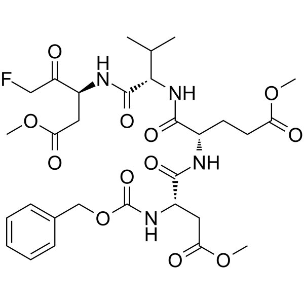
HY-12466
Z-DEVD-FMKCaspase-3 Inhibitor -
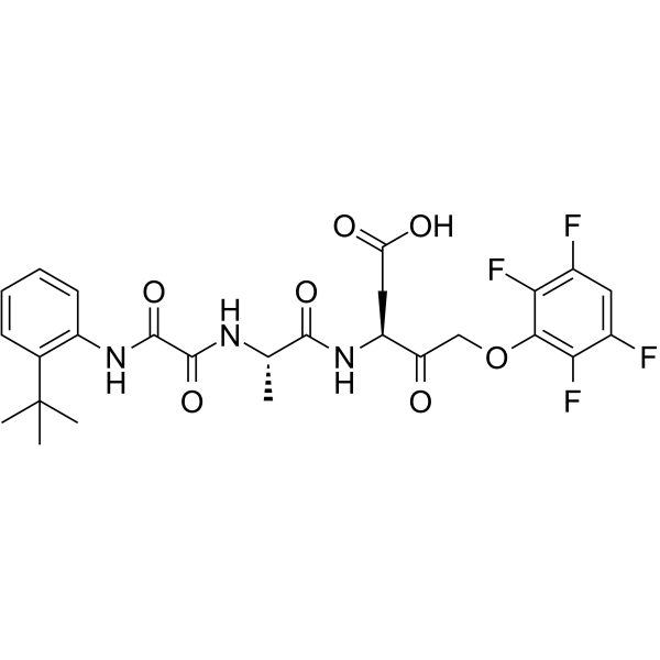
HY-10396
Emricasanpan-Caspase Inhibitor -
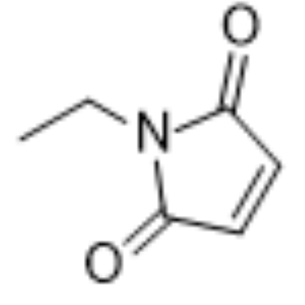
HY-D0843
N-EthylmaleimideCysteine Residues Modifier -
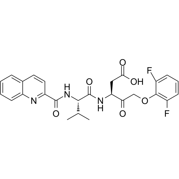
HY-12305
Q-VD-OPhPan-caspase Inhibitor -
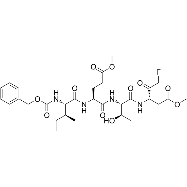
HY-101297
Z-IETD-FMKCaspase-8 Inhibitor -
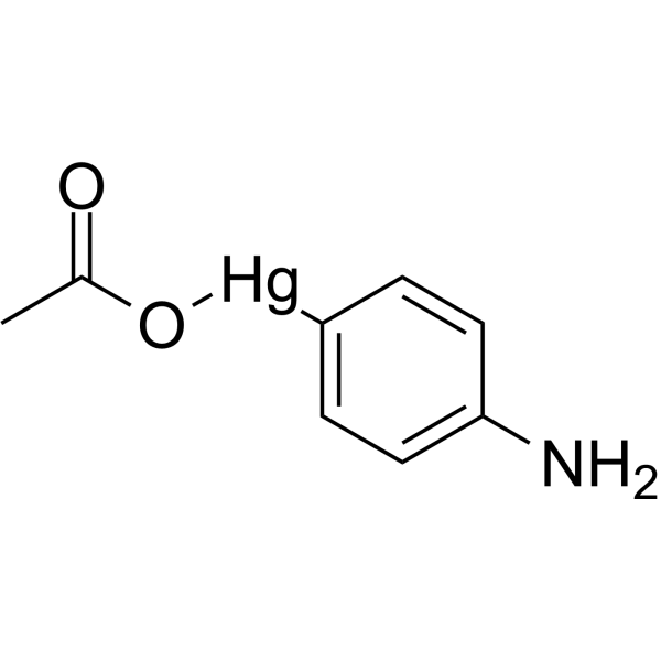
HY-148905
p-Aminophenylmercuric acetateMatrix Metalloproteinases Activator -
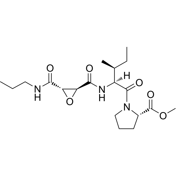
HY-100350
CA-074 methyl esterCathepsin B Inhibitor -
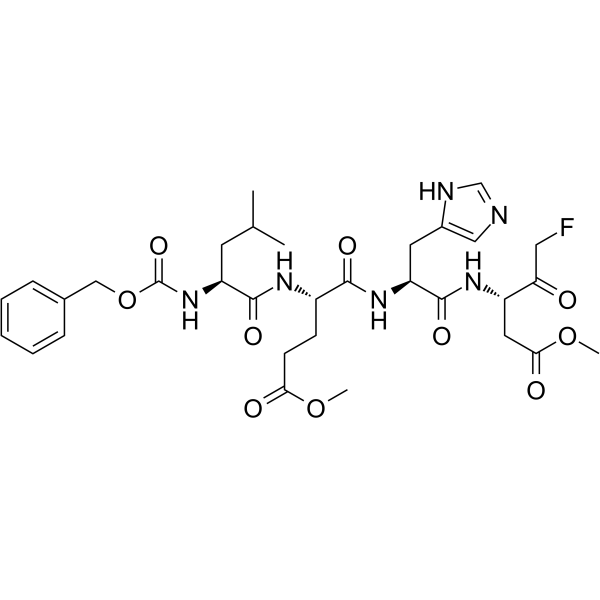
HY-P1010
Z-LEHD-FMKCaspase-9 Inhibitor -
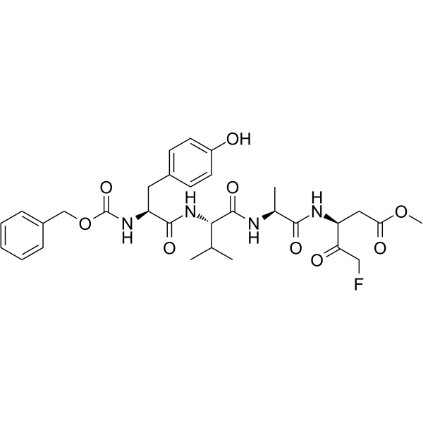
HY-P1009
Z-YVAD-FMKCaspase 1/4 Inhibitor -
HY-13523
PAC-1Caspase Activator
Publications Citing Use of MedChemExpress (MCE) (S,S)-Z-FA-FMK
MoreAll Cathepsin Isoforms
More

Biological Activity
Description
Cellular Effect
|
Cell Line

|
Type | Value | Description | References |
|---|---|---|---|---|
| CHO | IC50 |
>100 μM
Compound: 1; Z-FA-FMK
|
Inhibition of recombinant human cathepsin D (400 residues) expressed in CHO cells using 5-FAM/QXL 520 FRET peptide as substrate preincubated for 20 mins followed by substrate addition by fluorescence plate reader analysis
Inhibition of recombinant human cathepsin D (400 residues) expressed in CHO cells using 5-FAM/QXL 520 FRET peptide as substrate preincubated for 20 mins followed by substrate addition by fluorescence plate reader analysis
|
[PMID: 27563406] |
| CHO | IC50 |
0.3 μM
Compound: 1; Z-FA-FMK
|
Inhibition of recombinant human cathepsin B (332 residues) expressed in CHO cells using Z-FR-AMC as substrate preincubated for 20 mins followed by substrate addition by fluorescence plate reader analysis
Inhibition of recombinant human cathepsin B (332 residues) expressed in CHO cells using Z-FR-AMC as substrate preincubated for 20 mins followed by substrate addition by fluorescence plate reader analysis
|
[PMID: 27563406] |
| HEK-293T | CC50 |
>100 μM
Compound: 1; Z-FA-FMK
|
Cytotoxicity against HEK293T cells after 24 hrs
Cytotoxicity against HEK293T cells after 24 hrs
|
[PMID: 27563406] |


Chemical Information
-
CAS No. 105637-38-5
-
Molecular Weight 386.42
-
Formula C21H23FN2O4
-
Shipping
Room temperature in continental US; may vary elsewhere.
-
Storage
Please store the product under the recommended conditions in the Certificate of Analysis.


Publications (1)
-
Journal Impact Factor
-
Most Recent

Protocols
-
Apoptosis Solutions
Apoptosis is a regulated, generally non-lytic cell-death pathway that removes unwanted, damaged, infected, or abnormal cells through coordinated morphological changes, caspase activation, DNA fragmentation, and membrane remodeling. The intrinsic apoptosis pathway is controlled mainly by mitochondrial outer membrane permeabilization, BCL-2 family proteins, cytochrome c release, apoptosome formation, caspase-9 activation, and downstream executioner caspase-3/7 activation. The extrinsic apoptosis pathway is initiated by death receptors such as Fas, TNFR, and TRAIL receptors, which recruit adaptor proteins and activate caspase-8 before engaging executioner caspases or mitochondrial amplification through BID cleavage. Apoptosis is linked to many phenotypes, including cancer cell killing, tissue homeostasis, immune regulation, neurodegeneration, infection response, and treatment-induced cytotoxicity; unresolved questions include how apoptosis interacts with necroptosis, pyroptosis, ferroptos
-
Pyroptosis Solutions
Pyroptosis is a lytic inflammatory cell-death pathway executed by gasdermin pores, most classically through inflammasome-mediated activation of caspase-1, cleavage of gasdermin D, membrane pore formation, LDH release, and secretion of IL-1β and IL-18. The canonical pathway is commonly modeled by priming cells with an inflammatory signal such as LPS to induce pro-IL-1β and inflammasome components, followed by an activation signal such as ATP or nigericin to activate NLRP3, ASC speck formation, caspase-1 cleavage, GSDMD cleavage, cytokine release, and pyroptotic membrane rupture. The non-canonical pathway is triggered when cytosolic LPS activates mouse caspase-11 or human caspase-4/5, leading to GSDMD cleavage and pyroptosis, and this can secondarily activate NLRP3-dependent IL-1β release. Pyroptosis is linked to inflammatory injury, infection, cancer, liver disease, ocular disease, placental inflammation, and other disease phenotypes, but unresolved questions include which gasdermin fam


Purity & Documentation
References
[1]. Koike S, et al. Advanced Glycation End-Products Induce Apoptosis of Vascular Smooth Muscle Cells: A Mechanism for Vascular Calcification. Int J Mol Sci. 2016;17(9):1567. Published 2016 Sep 16. [Content Brief]
[2]. Harrison JS, et al. The role of VDR and BIM in potentiation of cytarabine-induced cell death in human AML blasts. Oncotarget. 2016;7(24):36447-36460. [Content Brief]

Calculators
Concentration (start) × Volume (start) = Concentration (final) × Volume (final)


(S,S)-Z-FA-FMK Related Classifications
HY-P0109 Related Classifications
 Powered by Bioz