



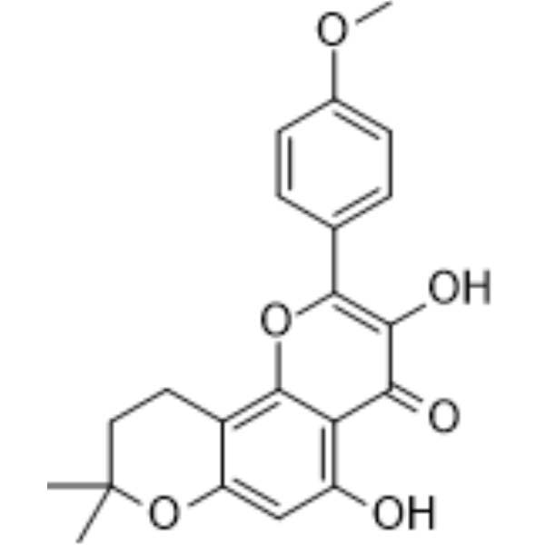
β-Anhydroicaritin
Based on 3 publication(s) in Google Scholar
β-Anhydroicaritin is isolated from Boswellia carterii Birdware, has important biological and pharmacological effects, such as antiosteoporosis, estrogen regulation and antitumor properties. β-Anhydroicaritin ameliorates the degradation of periodontal tissue and inhibits the synthesis and secretion of TNF-α and MMP-3 in diabetic rats. β-Anhydroicaritin decreases the overproduction of NO, IL-10, TNF-α, MCP-1 and IL-6 in inperitonitis mice. β-Anhydroicaritin inhibits the elevation of intracellular Ca2+, and markedly decreases iNOS protein expression.
For research use only. We do not sell to patients.
- Purity : 99.69%
- CAS No.: 38226-86-7
- Formula: C21H20O6
- Molecular Weight:368.38
-
Storage:
4°C, protect from light
* In solvent : -80°C, 6 months; -20°C, 1 month (protect from light)

To place orders, for customer services and technical support, please contact: MedChemExpress USA
Tel: 609-228-6898 E-mail: [email protected] [email protected]


-
 Biological Activity
-
Chemical Information
-
Solvent & Solubility
- Protocols
- Purity & Documentation
- References
-
 Help & FAQs


-
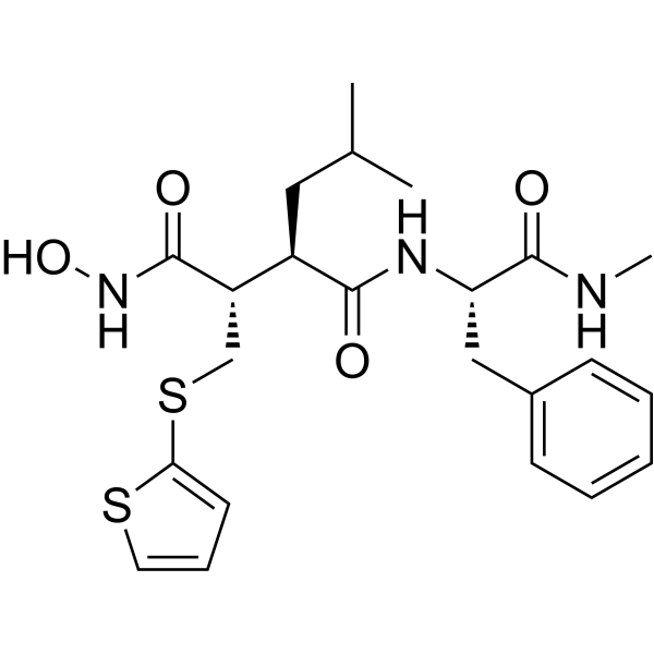
HY-13564
BatimastatMMP Inhibitor -

HY-108847
EtanerceptTNF Inhibitor -
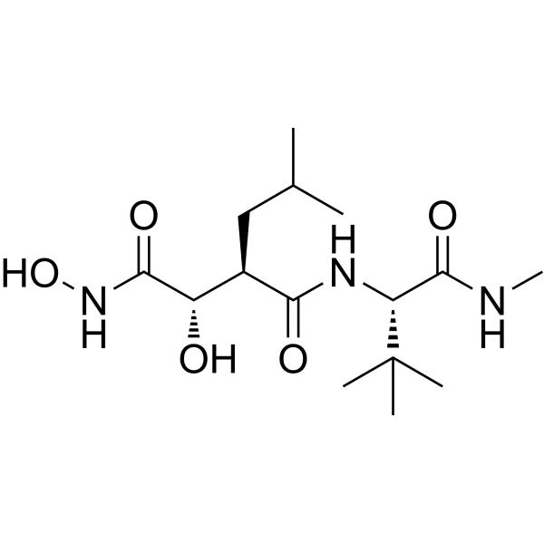
HY-12169
MarimastatMMP Inhibitor -
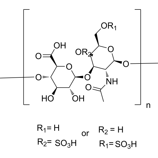
HY-B2162
Chondroitin sulfateGlycosaminoglycan -
HY-19956
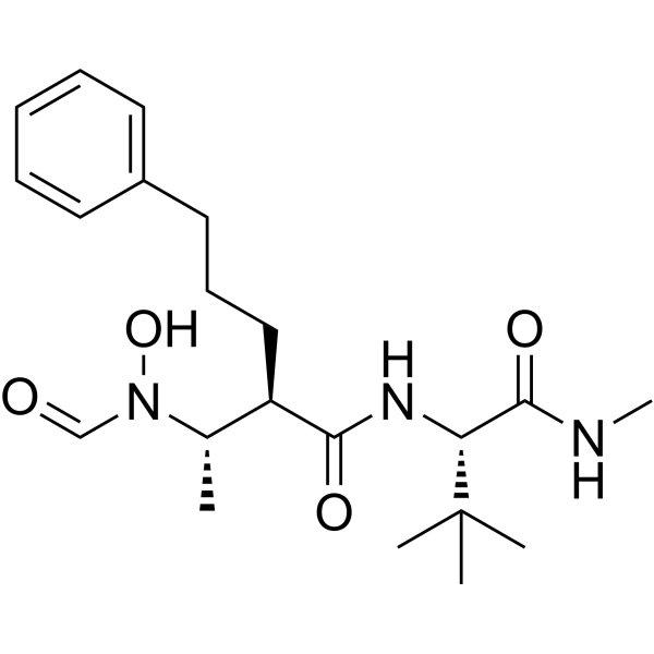
GI254023XADAM10/MMP9 Inhibitor -
HY-115670
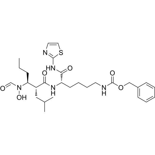
GW280264XADAM10/17 Inhibitor -
HY-B2162D
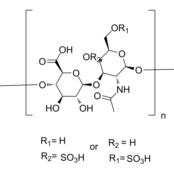
Chondroitin sulfate (from chicken)Sulfated Linear Polysaccharide -
HY-N0532
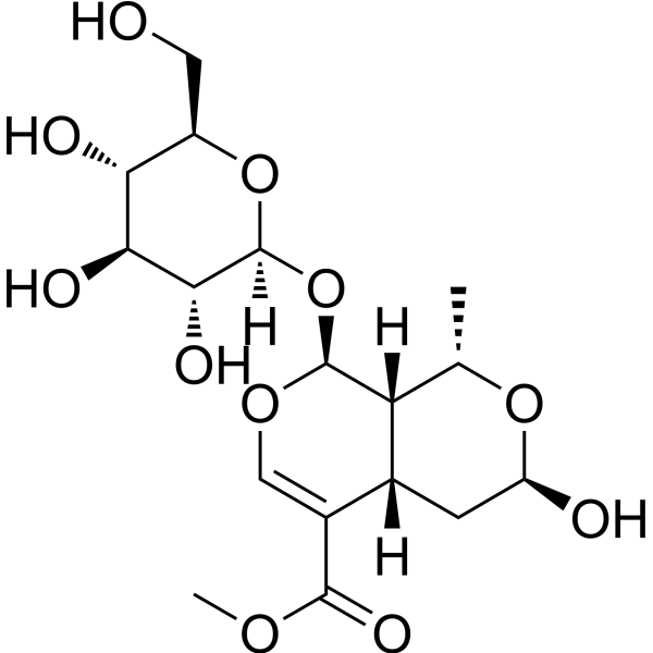
MorronisidePyroptosis Inhibitor -
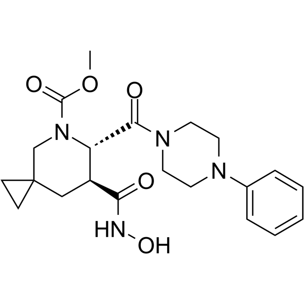
HY-10293
AderbasibADAM10/17 Inhibitor -
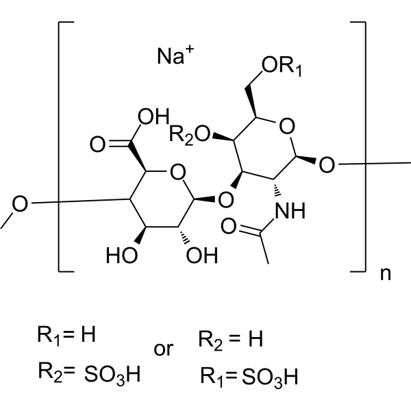
HY-B2162A
Chondroitin sulfate sodium (from shark cartilage)Glycosaminoglycan -
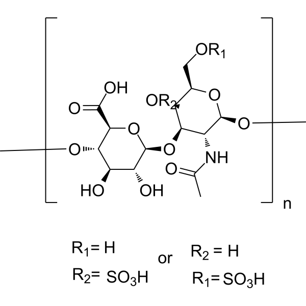
HY-B2162C
Chondroitin sulfate (from pig)Sulfated Linear Polysaccharide -
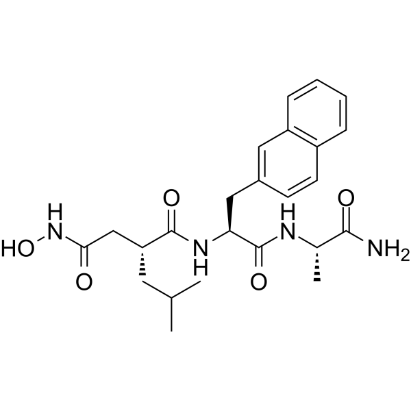
HY-118694
TAPI-0TACE/MMP Inhibitor
Publications Citing Use of MedChemExpress (MCE) β-Anhydroicaritin
More

Biological Activity
Description
|
IL-10 |
IL-6 |
MMP-3 |
Cellular Effect
|
Cell Line

|
Type | Value | Description | References |
|---|---|---|---|---|
| COS-7 | IC50 |
45.5 μM
Compound: 6
|
Inhibition of human recombinant PDE5A1 expressed in COS7 cells
Inhibition of human recombinant PDE5A1 expressed in COS7 cells
|
[PMID: 18778098] |
| NCI/ADR-RES | IC50 |
>100 μM
Compound: 5
|
Cytotoxicity against human MCF7/ADR cells after 72 hrs by MTT assay
Cytotoxicity against human MCF7/ADR cells after 72 hrs by MTT assay
|
[PMID: 19523827] |
| NIH3T3 | IC50 |
>50 μM
Compound: CICT
|
Cytotoxicity against mouse 3T3 cells assessed as reduction in cell viability incubated for 72 hrs by MTT assay
Cytotoxicity against mouse 3T3 cells assessed as reduction in cell viability incubated for 72 hrs by MTT assay
|
[PMID: 38972080] |


Chemical Information
-
CAS No. 38226-86-7
-
Appearance Solid
-
Molecular Weight 368.38
-
Formula C21H20O6
-
Color Light yellow to yellow
-
SMILES
O=C1C2=C(O)C=C3C(CCC(C)(C)O3)=C2OC(C4=CC=C(OC)C=C4)=C1O
-
Synonyms
Cycloicaritin
-
Structure Classification
-
Initial Source
-
Shipping
Room temperature in continental US; may vary elsewhere.
-
Storage
4°C, protect from light
* In solvent : -80°C, 6 months; -20°C, 1 month (protect from light)


Publications (3)
-
Journal Impact Factor
-
Most Recent
-
Food Chem
Effects of sun drying combined with baking processes on the flavor quality of Chongqing Tuocha raw tea. [Abstract]2025 Dec 30:497:146992. PMID: 41285060 -
Food Chem
Flavonoid-mediated metabolic underpinning quality variation in red bud-sport pear mutants. [Abstract]2025 Oct 15:489:144992. PMID: 40466530 -
J Ethnopharmacol
Isoxanthohumol, a component of Sophora flavescens, promotes the activation of the NLRP3 inflammasome and induces idiosyncratic hepatotoxicity. [Abstract]2022 Mar 1:285:114796. PMID: 34740771


Solvent & Solubility
In Vitro:
DMSO : 8.33 mg/mL (22.61 mM; Need ultrasonic; Hygroscopic DMSO has a significant impact on the solubility of product, please use newly opened DMSO)
Please refer to the solubility information to select the appropriate solvent. Once prepared, please aliquot and store the solution to prevent product inactivation from repeated freeze-thaw cycles.
Storage method and period of stock solution: -80°C, 6 months; -20°C, 1 month (protect from light). When stored at -80°C, please use it within 6 months. When stored at -20°C, please use it within 1 month.
Please refer to the solubility information to select the appropriate solvent. Once prepared, please aliquot and store the solution to prevent product inactivation from repeated freeze-thaw cycles.
Storage method and period of stock solution: -80°C, 6 months; -20°C, 1 month (protect from light). When stored at -80°C, please use it within 6 months. When stored at -20°C, please use it within 1 month.
Concentration (start) × Volume (start) = Concentration (final) × Volume (final)

Protocols
-
RNA extraction experimental
By lysing cells, releasing RNA, and removing impurities such as proteins and DNA, high-purity RNA products are finally obtained. The commonly used traditional method is the guanidine isothiocyanate/phenol/chloroform method (Trizol), which is suitable for a variety of animal materials including animal tissues, microorganisms, cultured cells, etc., and most plant materials.
-
Western Blot
Western blotting (WB) is a commonly used experimental method in molecular biology, biochemistry, and immunogenetics for identifying and quantifying target proteins. It combines gel electrophoresis with immunoassay, enabling researchers to analyze protein expression, post-translational modifications, and molecular weight.
-
Mitochondrial membrane-potential fluorescent assay
Mitochondrial membrane potential fluorescent assays estimate ΔΨm in living cells using lipophilic cationic dyes such as TMRM, TMRE, rhodamine 123, and JC-1, which accumulate in mitochondria according to membrane polarization; loss of signal after FCCP or CCCP treatment is interpreted as mitochondrial depolarization. TMRM/TMRE and rhodamine 123 are commonly used for semi-quantitative live-cell microscopy or flow cytometry, while JC-1 can report a shift from red aggregate fluorescence to green monomer fluorescence during depolarization; interpretation requires controls because dye concentration, quenching mode, cell type, dye efflux, and mitochondrial mass can affect fluorescence independently of ΔΨm.
-
Research Protocol for Endocrine Diseases
Endocrine diseases often arise from disrupted hormone production, hormone signaling, or target-tissue responsiveness; for diabetes-focused endocrine disease models, insulin signaling regulates glucose uptake, hepatic glucose output, lipid metabolism, and β-cell compensation. Type 2 diabetes develops through interacting defects in insulin resistance, β-cell dysfunction, adipose inflammation, hepatic glucose overproduction, altered incretin signaling, and ectopic lipid metabolism. A major unresolved question is whether endocrine dysfunction is driven primarily by target-tissue insulin resistance, intrinsic β-cell failure, immune/inflammatory stress, or combined multi-organ failure that differs by disease stage.
-
Mitochondrial membrane-potential and mitochondrial mass staining
Mitochondrial membrane potential staining measures the electrochemical polarization across the mitochondrial inner membrane in live cells using lipophilic cationic fluorescent probes; early rhodamine-based work showed that selective mitochondrial dye accumulation is lost when the mitochondrial transmembrane potential is dissipated. JC-1 reports mitochondrial polarization by shifting from green monomer fluorescence to red J-aggregate fluorescence as dye concentration increases within energized mitochondria; therefore, the red/green fluorescence ratio is used as a relative readout of mitochondrial membrane potential. TMRE or TMRM staining provides a single-channel relative readout because these cationic rhodamine esters accumulate in polarized mitochondria, and lower fluorescence indicates reduced mitochondrial polarization when acquisition and dye-loading conditions are controlled. Mitochondrial mass staining is commonly performed with MitoTracker Green FM or related MitoTracker dyes as
-
Ca2+ Staining Technique
Ca2+ staining is an experimental technique that utilizes specific fluorescent probes (such as Fluo-4 AM, Fura-2, etc.) to qualitatively or quantitatively detect dynamic changes in intracellular Ca2+ concentrations; this is achieved by monitoring the changes in fluorescent signals generated when these probes bind to free intracellular calcium ions. The underlying principle relies primarily on the presence of chelating groups within the probe's molecular structure that possess high affinity for calcium ions.
-
Protocol For Protein Expression And Purification
Recombinant protein expression in Escherichia coli followed by purification of a His-tagged soluble protein by immobilized metal affinity chromatography (IMAC), with optional MBP fusion and TEV tag removal when the construct includes these elements. The biological readout is production of the encoded target protein, detected as an inducible band at the expected molecular mass by SDS-PAGE and quantified by total protein assay or chromatographic absorbance; the purification readout is enrichment of the target protein in elution fractions after selective binding of polyhistidine residues to immobilized Ni2+/metal-chelate resin and elution by imidazole-containing buffer. Expression is driven by an inducible bacterial expression system, commonly T7/lac-based, in which IPTG or lactose/auto-induction activates transcription and translation of the cloned gene; lower induction temperature, lower inducer concentration, induction timing, and solubility-enhancing fusion tags can influence the frac


Purity & Documentation
-
Data Sheet (278 KB)
-
SDS (393 KB)
- English - EN (393 KB)
- Français - FR (393 KB)
- Deutsch - DE (393 KB)
- Norwegian - NO (393 KB)
- Español - ES (393 KB)
- Swedish - SV (393 KB)
- Italian - IT (393 KB)
- Korean - KR (393 KB)
- Portuguese - PT (393 KB)
-
Handling Instructions (2659 KB)
References
[1]. Nguyen VS, et al. Synthesis of Icaritin and β-anhydroicaritin Mannich Base Derivatives and Their Cytotoxic Activities on Three Human Cancer Cell Lines. Anticancer Agents Med Chem. 2017;17(1):137-142 [Content Brief]
[2]. Wu Y, et al. Effect of β-anhydroicaritin on the expression levels of tumor necrosis factor-α and matrix metalloproteinase-3 in periodontal tissue of diabetic rats. Mol Med Rep. 2015 Aug;12(2):1829-37. [Content Brief]
[3]. Lai X, et al. Protective effect of anhydroicaritin against peritonitis in mice. Xi Bao Yu Fen Zi Mian Yi Xue Za Zhi. 2013 Oct;29(10):1036-9. [Content Brief]


Complete Stock Solution Preparation Table
Please refer to the solubility information to select the appropriate solvent. Once prepared, please aliquot and store the solution to prevent product inactivation from repeated freeze-thaw cycles.
Storage method and period of stock solution: -80°C, 6 months; -20°C, 1 month (protect from light). When stored at -80°C, please use it within 6 months. When stored at -20°C, please use it within 1 month.
| Optional Solvent | Concentration Solvent Mass | 1 mg | 5 mg | 10 mg | 25 mg |
|---|---|---|---|---|---|
| DMSO | 1 mM | 2.7146 mL | 13.5729 mL | 27.1459 mL | 67.8647 mL |
| 5 mM | 0.5429 mL | 2.7146 mL | 5.4292 mL | 13.5729 mL | |
| 10 mM | 0.2715 mL | 1.3573 mL | 2.7146 mL | 6.7865 mL | |
| 15 mM | 0.1810 mL | 0.9049 mL | 1.8097 mL | 4.5243 mL | |
| 20 mM | 0.1357 mL | 0.6786 mL | 1.3573 mL | 3.3932 mL |


β-Anhydroicaritin Related Classifications
HY-N1940 Related Classifications
 Powered by Bioz