From 11:00 pm to 12:00 pm EST ( 8:00 pm to 9:00 pm PST ) on January 6th, the website will be under maintenance. We are sorry for the inconvenience. Please arrange your schedule properly.
MOG (35-55) mouse, rat (Myelin Oligodendrocyte Glycoprotein Peptide (35-55), mouse, rat) is a minor component of CNS myelin. MOG (35-55) mouse, rat has encephalitogenic activity and induces T cell proliferative. MOG (35-55) mouse, rat induces Th1 cytokine response as well as relatively high levels of IgG antibodies. MOG (35-55) mouse, rat produces a relapsing-remitting neurological disease with extensive plaque-like demyelination. MOG (35-55) mouse, rat (MOG (35-55)) can be used for experimental autoimmune encephalomyelitis (EAE) modeling .
MOG (35-55) amide, mouse, rat the terminal amidation form of the 35-55 fragment of the myelin oligodendrocyte glycoprotein (MOG) immunogenic peptide (MOG (35-55) (HY-P1240)). MOG(35-55) amide, mouse, rat can be used for experimental autoimmune encephalomyelitis (EAE) modeling .
Cuprizone is a copper chelating agent that forms a deep blue copper ketone complex with copper (II). The copper ketone reaction can be used in colorimetric tests for the presence of trace copper. Cuprizone can be used to induce some schizophrenia-like behavior in mice. Cuprizone acts on copper enzymes, including SOD1, cytochrome oxidase, and DβH, thereby causing oxidative stress and increasing DA levels in certain brain regions such as the medial prefrontal cortex (PFC) .
4-Ethylphenyl sulfate is an orally active and brain-penetrant gut microbial metabolite. 4-Ethylphenyl sulfate downregulates Bcl2 expression, upregulates Bax expression, and induces cancer cell apoptosis via the endogenous apoptotic pathway. 4-Ethylphenyl sulfate induces G2/M cell cycle arrest and reactive oxygen species (ROS) production. 4-Ethylphenyl sulfate impairs oligodendrocyte maturation, reduces oligodendrocyte-neuron interactions, decreases axonal myelination levels, and shifts the oligodendrocyte population toward immature precursor cells. 4-Ethylphenyl sulfate alters brain region-specific neural activity and functional connectivity in mice, and correlates with anxiety-like behaviors in mice .
Poly-L-ornithine hydrobromide (MW 30000-70000) is a poly-lysine derivative with a molecular weight of 30000-70000. Poly-L-ornithine hydrobromide (MW 30000-70000) binds to the surface of cell culture vessels through positively charged amino acid residues to form a coating that promotes cell adhesion and provides cells with a matrix environment required for growth. Poly-L-ornithine hydrobromide (MW 30000-70000) is used as a coating agent in cell culture and can be used for the study of primary culture of neurons (such as dopaminergic neurons and oligodendrocytes) .
MOG (35-55) (MOG (35-55)) TFA is a minor component of CNS myelin. MOG (35-55) (TFA) has encephalitogenic activity and induces T cell proliferative. MOG (35-55) (TFA) induces Th1 cytokine response as well as relatively high levels of IgG antibodies. MOG (35-55) (TFA) produces a relapsing-remitting neurological disease with extensive plaque-like demyelination. MOG (35-55) (MOG (35-55)) TFA can be used for experimental autoimmune encephalomyelitis (EAE) modeling .
Clemastine (HS-592; Meclastine) is an orally active, blood-brain barrier-permeable H1 histamine receptor (H1 histamine receptor) antagonist with potent antiallergic effects. Clemastine also antagonizes muscarinic acetylcholine receptors (mAChR), particularly the M1 and M4 subtypes. In addition to antihistamine effects, Clemastine exhibits multiple pharmacological activities, especially in promoting central nervous system remyelination, activating autophagy and pyroptosis, exerting anti-apoptotic and neuroprotective effects, and suppressing inflammation .
EBP-IN-1 is an orally active, blood-brain barrier-permeable inhibitor of emopamil binding protein (EBP). EBP-IN-1 has an IC50 of 8.2 μM against human ERG potassium channels (in CHO background). By inhibiting the sterol isomerase activity of EBP, EBP-IN-1 causes the accumulation of Zymostenol (HY-113345), and exhibits strong target binding in the brain after repeated administration in rodents. EBP-IN-1 also promotes oligodendrocyte formation in human cortical organoids and can be used in research related to multiple sclerosis .
Cholera toxin B subunit, from vibrio cholerae (CTB, from vibrio cholerae) is non-toxic to cells and possesses no intrinsic adenylate cyclase activity. Cholera toxin B subunit, from vibrio cholerae attaches to cells by binding to ganglioside GM1.8 CTB has been shown to be a good label for microglial cells (due to the enrichment of ganglioside GM1 on their cell surface), but not for oligodendrocytes or astrocytes. Cholera toxin B subunit, from vibrio cholerae has been reported to be an excellent tracer for the study of axonal transport using immunohistochemical methods. Cholera toxin B subunit, from vibrio cholerae has been widely used as a marker of membrane lipid rafts .
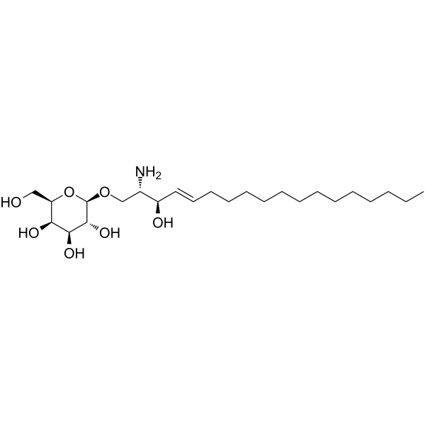
Psychosine (Galactosylsphingosine), a substrate of the galactocerebrosidase (GALC) enzyme, is a potential biomarker for Krabbe disease . Psychosine is a highly cytotoxic lipid, capable of inducing cell death in a wide variety of cell types including, most relevantly to globoid cell leukodystrophy (GLD), oligodendrocytes. Psychosine causes cell death at least in part via apoptosis. Psychosine also is an inhibitor of PKC .
BMS-561392 formate (DPC 333 formate) is a selective ADAM17(TACE) inhibitor. BMS-561392 formate inhibits TNF-α secretion by regulating signaling pathways such as p44 MAPK and NF-κB. BMS-561392 formate also affects the survival of central nervous system-related cells including oligodendrocytes and microglia. BMS-561392 formate promotes microglial apoptosis, enlarges the injury area and exacerbates astrogliosis in a mouse spinal cord injury model. BMS-561392 formate can be used in research related to spinal cord injury and inflammatory diseases .
BN201 promotes neuronal differentiation, the differentiation of precursor cells to mature oligodendrocytes (EC50 of 6.3 μM) in vitro, and the myelination of new axons (EC50 of 16.6 μM). BN201 is able to cross the blood-brain barrier by active transport and activate pathways (IGF-1 pathway) associated with the response to stress and neuron survival. BN201 has potently neuroprotective effects .
7-Dehydro desmosterol is a sterol and an intermediate in cholesterol biosynthesis, which is found in the marine diatom Pseudo-nitzschia multistriata and the nervous system of rodents .
VP3.15 dihydrobromide is a highly potent, orally bioavailable, and CNS-penetrant PDE7-GSK3 dual inhibitor, with IC50 values of 1.59 μM and 0.88 μM against PDE7 and GSK3, respectively . VP3.15 dihydrobromide elevates intracellular cAMP levels, suppresses immune responses, enhances remyelination, limits excessive tau phosphorylation, and alleviates neuroinflammation and neuronal loss. VP3.15 dihydrobromide promotes oligodendrocyte precursor cell differentiation, improves in vivo remyelination, inhibits autoimmune encephalomyelitis, and mitigates germinal matrix-intraventricular hemorrhage-related brain injury, cerebral atrophy, ventricular enlargement, and cognitive impairment. VP3.15 dihydrobromide can be used in research related to multiple sclerosis and germinal matrix-intraventricular hemorrhage .
Halcinonide (SQ-18566) is an orally active Smoothened (Smo) agonist. Halcinonide activates the Hedgehog signaling pathway by binding to Smo and promoting its internalization and expression, thereby activating Gli transcription factors. Halcinonide not only stimulates cell proliferation, increases the expression of cyclin D2/CDK6 and inhibits the degradation of caspase-3, but also suppresses Bcl-2/Bax-mediated apoptosis, oxidative stress and inflammatory responses. Halcinonide activates RxRγ to upregulate the expression of myelin genes, thereby reducing cerebral infarction and improving behavioral deficits. Halcinonide has been used in studies related to multiple sclerosis and ischemic stroke .
T0080 is a central nervous system-penetrant FPR1 inhibitor. By functionally blocking the FPR1 signaling pathway, T0080 effectively reduces neutrophil infiltration into ischemic brain tissue and maintains the integrity of the blood-brain barrier. T0080 alleviates tPA-associated hemorrhagic transformation, inhibits demyelination responses and the expression of NOX2. T0080 also possesses anti-apoptotic (apoptosis) and anti-inflammatory properties, thereby protecting myelin and reducing neurological deficits. T0080 is widely used in studies related to ischemic stroke complicated by hemorrhagic transformation after tPA thrombolysis, as well as multiple sclerosis .
MitoPeDPP is a mitochondrial-targeted fluorescent probe that is sensitive to LPO. MitoPeDPP is synthesized from diphenylpyrenephosphine. MitoPeDPP can be used to study the occurrence of mitochondrial LPO in RSL3-induced oligodendrocyte ferroptosis .
KM91104 is a cell-permeable V-ATPase a3-b2 inhibitor (IC50 = 2.3 µM). KM91104 reduces the metabolic activity, cell proliferation capacity and V-ATPase subunit protein expression levels of primary human hepatic stellate cells, increases intracellular ATP levels and decreases cytoplasmic pH. KM91104 reduces TLR4 expression on the surface of oligodendrocyte precursor cells, blocks the ENV-TLR4 interaction, and reverses oligodendrocyte myelination defects induced by ENV protein .
Geissoschizine methyl ether is an orally active, blood-brain barrier permeable alkaloid, and a partial agonist of the 5-HT1A receptor. It can be isolated from Uncaria hook. Geissoschizine methyl ether potently inhibits the binding of [ 3H]8-OH-DPAT to the 5-HT1A receptor in a concentration-dependent manner, with an IC50 of 0.904 μM. It ameliorates isolation-induced increased aggression and reduced sociability in mice. Geissoschizine methyl ether promotes oligodendrocyte differentiation and remyelination in the medial prefrontal cortex of adult mice .
PT109 is an orally active, blood-brain barrier permeable multi-kinase inhibitor. By inhibiting PTBP1, PT109 promotes the switch of pyruvate kinase isoform from PKM2 to PKM1, thereby effectively inhibiting the proliferation and migration of glioblastoma multiforme and inducing its reprogramming into oligodendrocytes. PT109 also targets and regulates key signaling molecules such as JNK, SGK1, GSK3β to exert neuroprotective effects including promoting neurogenesis, inducing synapse formation and alleviating neuroinflammation. In Alzheimer's disease models, PT109 exhibits significant efficacy in improving spatial learning ability, along with excellent in vivo pharmacokinetic properties. PT109 can be used to investigate metabolic reprogramming of glioblastoma multiforme and neuroprotective mechanisms of Alzheimer's disease .
PD 174265 is a highly selective, reversible EGFR/ErbB2 tyrosine kinase inhibitor (IC50=0.45 nM) and cell differentiation inducer. By blocking receptor autophosphorylation and the downstream ERK signaling pathway (with an IC50 of 0.45 μM for full-length ERK), PD 174265 effectively inhibits tumor growth and exhibits antitumor activity without obvious toxicity in in vivo models. PD 174265 drives oligodendrocyte precursor cells to switch from a proliferative state to a differentiated state, significantly upregulates the expression of myelin proteins such as CNP, PLP and MBP, and induces neurite branching. PD 174265 shows no inhibitory effect on other kinases including insulin, PDGF and basic FGF receptors, and serves as a crucial tool molecule for investigating the treatment of human epidermoid carcinoma and the mechanism of myelin repair in multiple sclerosis .
BMS-561392 (BMS-561392) is a selective ADAM17(TACE) inhibitor. BMS-561392 inhibits TNF-α secretion by regulating signaling pathways such as p44 MAPK and NF-κB. BMS-561392 also affects the survival of central nervous system-related cells including oligodendrocytes and microglia. BMS-561392 promotes microglial apoptosis, enlarges the injury area and exacerbates astrogliosis in a mouse spinal cord injury model. BMS-561392 can be used in research related to spinal cord injury and inflammatory diseases .
NEP(1-40) is a Nogo-66 receptor (NgR) antagonist peptide, reversing the injury-induced shift in distribution of microglia morphologies by limiting myelin-based inhibition .
PTPσ Inhibitor, ISP can bind to recombinant human PTPs and inhibits PTPσ signaling. PTPσ Inhibitor, ISP can penetrate the membrane and relieves the chondroitin sulfate proteoglycan (CSPG)-mediated axonal sprouting inhibition in spinal cord injury model. PTPσ Inhibitor, ISP enhances remyelination in LPC-induced demyelinated spinal cord. PTPσ Inhibitor, ISP also promotes oligodendrocyte progenitor cells (OPCs) migration, maturation, remyelination, and functional recovery in animal models of Multiple Sclerosis (MS) .
4-Ethylphenyl sulfate sodium is an orally active and brain-penetrant gut microbial metabolite. 4-Ethylphenyl sulfate sodium downregulates Bcl2 expression, upregulates Bax expression, and induces cancer cell apoptosis via the endogenous apoptotic pathway. 4-Ethylphenyl sulfate sodium induces G2/M cell cycle arrest and reactive oxygen species (ROS) production. 4-Ethylphenyl sulfate sodium impairs oligodendrocyte maturation, reduces oligodendrocyte-neuron interactions, decreases axonal myelination levels, and shifts the oligodendrocyte population toward immature precursor cells. 4-Ethylphenyl sulfate sodium alters brain region-specific neural activity and functional connectivity in mice, and correlates with anxiety-like behaviors in mice .
Biotin-myelin basic protein (94-102) is a peptide fragemt. Myelin basic protein is responsible for adhesion of the cytosolic surfaces of multilayered compact myelin, it plays an important role in the process of myelination of nerves in the nervous system. Myelin basic protein also acts as a membrane actin-binding protein, which might allow it to participate in transmission of extracellular signals to the cytoskeleton in oligodendrocytes and tight junctions in myelin .
Myelin Oligodendrocyte Glycoprotein (40-54), Rat, Mouse (MOG (40-54)) is a CD8-related self-antigenic epitope of the myelin oligodendrocyte glycoprotein (MOG) protein and is presented in association with H-2Db .
Tasronetide (FTX-101) is a highly selective inhibitor toward the NRP1/Plexin-A1 receptor system, and displays no significant activity on other targets. Tasronetide intercalates within the transmembrane domains of Plexin-A1 and NRP1 of oligodendrocytes, interferes with the heterodimerization of the co-receptor system, effectively disrupts the NRP1/Plexin-A1 receptor complex and mitigates the inhibitory influence of Sema3A on oligodendrocyte migration and differentiation, thereby facilitating increased myelin sheathing around axons. Tasronetide is designed to enhance the recruitment and maturation of oligodendrocyte precursors and can be used for Chronic Op c Neuropathy research .
4α-Hydroxycholesterol is an isomer of 4β-Hydroxycholesterol (HY-124265). 4α-Hydroxycholesterol has a slight effect on the integrity of lysosomal membranes in mouse primary oligodendrocyte cultures .
Cuprizone is a copper chelating agent that forms a deep blue copper ketone complex with copper (II). The copper ketone reaction can be used in colorimetric tests for the presence of trace copper. Cuprizone can be used to induce some schizophrenia-like behavior in mice. Cuprizone acts on copper enzymes, including SOD1, cytochrome oxidase, and DβH, thereby causing oxidative stress and increasing DA levels in certain brain regions such as the medial prefrontal cortex (PFC) .
OLIG2-IN-1 is a potent and selective oligodendrocyte transcription factor 2(OLIG2) inhibitor. OLIG2-IN-1 directly and dose-dependently downregulates nuclear OLIG2 levels with an IC50 value of 0.88 μM. OLIG2-IN-1 exhibits strong anti-proliferative activity in U87 and U251 cells with IC50 values of 7.02 μM and 6.43 μM, respectively. OLIG2-IN-1 can be used for the research of cancer, such as glioblastoma multiforme .
Anti-Monkey/Human CD20 Antibody (2H7) is a mouse-derived IgG2b κ type antibody inhibitor, targeting to monkey/human CD20. Anti-Monkey/Human CD20 Antibody (2H7) specifically depletes B cells. Anti-Monkey/Human CD20 Antibody (2H7) can be used for the researches of inflammation and metabolic disease, such as diabetes and experimental autoimmune encephalomyelitis .
ASN04421891 is a GPR17 agonist with nanomolar EC50 and high specificity. ASN04421891 promotes oligodendrocyte precursor cell maturation to mature myelinating oligodendrocytes. ASN04421891 can be used for the research of cerebral ischaemia, cardiac ischaemia, renal ischaemia, cerebral trauma, multiple sclerosis, schizophrenia, depression, alzheimer's disease, alzheimer-like dementia, parkinson's disease, huntington's chorea, amyotrophic lateral sclerosis (ALS), neuroinflammatory disorders .
Human MOG-specifying DNA is located at chromosome 6 within the human leukocyte antigen (HLA) gene locus. Human MOG-specifying DNA is exclusively expressed in the central nervous system (CNS) on the surface of myelin sheaths and oligodendrocytes (ODCs) processes, with unique methylation patterns in ODCs. Human MOG-specifying DNA can be used for inflammatory demyelinating diseases such as multiple sclerosis (MS) research .
Oxyphenbutazone (monohydrate) (Standard) is the analytical standard of Oxyphenbutazone (monohydrate). This product is intended for research and analytical applications. Oxyphenbutazone monohydrate is a Phenylbutazone (HY-B0230) metabolite, with anti-inflammatory effect. Oxyphenbutazone monohydrate is an orally active non-selective COX inhibitor. Oxyphenbutazone monohydrate selectively kills non-replicating Mycobaterium tuberculosis .
H3R antagonist 1 is a histamine receptor 3 (H3R) inverse agonist. H3R antagonist 1 increases the expression levels of myelin-associated glycoprotein (MAG) and myeline basic protein (MBP) in differentiating oligodendrocytes. H3R antagonist 1 can be used for the study of multiple sclerosis .
H3R antagonist 1 hydrochloride is a histamine receptor 3 (H3R) inverse agonist. H3R antagonist 1 hydrochloride increases the expression levels of myelin-associated glycoprotein (MAG) and myeline basic protein (MBP) in differentiating oligodendrocytes. H3R antagonist 1 hydrochloride can be used for the study of multiple sclerosis .
NEP(1-40) TFA is a Nogo-66 receptor (NgR) antagonist peptide, reversing the injury-induced shift in distribution of microglia morphologies by limiting myelin-based inhibition .
PI3Kδ-IN-21 (Compound 31) is a selective inhibitor for phosphoinositide 3-kinases δ (PI3Kδ), with an IC50 of 13.6 nM. PI3Kδ-IN-21 inhibits proliferation and differentation of T cells through PI3K/AKT/mTOR signaling pathway. PI3Kδ-IN-21 exhibits good pharmacokinetic characters in rat model, and attenuates the experimental autoimmune encephalomyelitis in myelin oligodendrocyte glycoprotein (MOG)-induced EAE model .
KLK6-IN-1 is a reversible small‑molecule inhibitor of KLK6, KLK1, and plasmin. KLK6-IN-1 shows IC50 values of 1.57 μM (KLK6), 5.1 μM (KLK1), 7.4 μM (plasmin), and Ki values of 0.8 μM (KLK6), 2.4 μM (KLK1), 1.3 μM (plasmin). KLK6-IN-1 is highly selective for KLK6 and its proteolytic network. KLK6-IN-1 induces oligodendrocyte differentiation by promoting oligodendrocyte precursor cell maturation. KLK6-IN-1 can be used for the research of multiple sclerosis .
RWT9996 is a balanced GPR17 antagonist. RWT9996 has an inhibitory effect on G protein activation and β-arrestin-2 recruitment induced by MDL-29951. RWT9996 inhibits the phosphorylation of ERK/CREB and the accumulation of inositol phosphate (b IP1) induced by MDL-29951. RWT9996 can be used for the study of neurological diseases .
MitoPeDPP is a mitochondrial-targeted fluorescent probe that is sensitive to LPO. MitoPeDPP is synthesized from diphenylpyrenephosphine. MitoPeDPP can be used to study the occurrence of mitochondrial LPO in RSL3-induced oligodendrocyte ferroptosis .
Poly-L-ornithine hydrobromide (MW 30000-70000) is a poly-lysine derivative with a molecular weight of 30000-70000. Poly-L-ornithine hydrobromide (MW 30000-70000) binds to the surface of cell culture vessels through positively charged amino acid residues to form a coating that promotes cell adhesion and provides cells with a matrix environment required for growth. Poly-L-ornithine hydrobromide (MW 30000-70000) is used as a coating agent in cell culture and can be used for the study of primary culture of neurons (such as dopaminergic neurons and oligodendrocytes) .
Cholera toxin B subunit, from vibrio cholerae (CTB, from vibrio cholerae) is non-toxic to cells and possesses no intrinsic adenylate cyclase activity. Cholera toxin B subunit, from vibrio cholerae attaches to cells by binding to ganglioside GM1.8 CTB has been shown to be a good label for microglial cells (due to the enrichment of ganglioside GM1 on their cell surface), but not for oligodendrocytes or astrocytes. Cholera toxin B subunit, from vibrio cholerae has been reported to be an excellent tracer for the study of axonal transport using immunohistochemical methods. Cholera toxin B subunit, from vibrio cholerae has been widely used as a marker of membrane lipid rafts .
MOG (35-55) mouse, rat (Myelin Oligodendrocyte Glycoprotein Peptide (35-55), mouse, rat) is a minor component of CNS myelin. MOG (35-55) mouse, rat has encephalitogenic activity and induces T cell proliferative. MOG (35-55) mouse, rat induces Th1 cytokine response as well as relatively high levels of IgG antibodies. MOG (35-55) mouse, rat produces a relapsing-remitting neurological disease with extensive plaque-like demyelination. MOG (35-55) mouse, rat (MOG (35-55)) can be used for experimental autoimmune encephalomyelitis (EAE) modeling .
MOG (35-55) amide, mouse, rat the terminal amidation form of the 35-55 fragment of the myelin oligodendrocyte glycoprotein (MOG) immunogenic peptide (MOG (35-55) (HY-P1240)). MOG(35-55) amide, mouse, rat can be used for experimental autoimmune encephalomyelitis (EAE) modeling .
MOG (35-55) (MOG (35-55)) TFA is a minor component of CNS myelin. MOG (35-55) (TFA) has encephalitogenic activity and induces T cell proliferative. MOG (35-55) (TFA) induces Th1 cytokine response as well as relatively high levels of IgG antibodies. MOG (35-55) (TFA) produces a relapsing-remitting neurological disease with extensive plaque-like demyelination. MOG (35-55) (MOG (35-55)) TFA can be used for experimental autoimmune encephalomyelitis (EAE) modeling .
MOG (35-55) (Myelin Oligodendrocyte Glycoprotein Peptide (35-55), mouse, rat) acetate is a minor component of CNS myelin. MOG (35-55) (acetate) has encephalitogenic activity and induces T cell proliferative. MOG (35-55) (acetate) induces Th1 cytokine response as well as relatively high levels of IgG antibodies. MOG (35-55) (acetate) produces a relapsing-remitting neurological disease with extensive plaque-like demyelination. MOG (35-55) (Myelin Oligodendrocyte Glycoprotein Peptide (35-55), mouse, rat) acetate can be used for experimental autoimmune encephalomyelitis (EAE) modeling .
NEP(1-40) is a Nogo-66 receptor (NgR) antagonist peptide, reversing the injury-induced shift in distribution of microglia morphologies by limiting myelin-based inhibition .
PTPσ Inhibitor, ISP can bind to recombinant human PTPs and inhibits PTPσ signaling. PTPσ Inhibitor, ISP can penetrate the membrane and relieves the chondroitin sulfate proteoglycan (CSPG)-mediated axonal sprouting inhibition in spinal cord injury model. PTPσ Inhibitor, ISP enhances remyelination in LPC-induced demyelinated spinal cord. PTPσ Inhibitor, ISP also promotes oligodendrocyte progenitor cells (OPCs) migration, maturation, remyelination, and functional recovery in animal models of Multiple Sclerosis (MS) .
Biotin-myelin basic protein (94-102) is a peptide fragemt. Myelin basic protein is responsible for adhesion of the cytosolic surfaces of multilayered compact myelin, it plays an important role in the process of myelination of nerves in the nervous system. Myelin basic protein also acts as a membrane actin-binding protein, which might allow it to participate in transmission of extracellular signals to the cytoskeleton in oligodendrocytes and tight junctions in myelin .
Myelin Oligodendrocyte Glycoprotein (40-54), Rat, Mouse (MOG (40-54)) is a CD8-related self-antigenic epitope of the myelin oligodendrocyte glycoprotein (MOG) protein and is presented in association with H-2Db .
Tasronetide (FTX-101) is a highly selective inhibitor toward the NRP1/Plexin-A1 receptor system, and displays no significant activity on other targets. Tasronetide intercalates within the transmembrane domains of Plexin-A1 and NRP1 of oligodendrocytes, interferes with the heterodimerization of the co-receptor system, effectively disrupts the NRP1/Plexin-A1 receptor complex and mitigates the inhibitory influence of Sema3A on oligodendrocyte migration and differentiation, thereby facilitating increased myelin sheathing around axons. Tasronetide is designed to enhance the recruitment and maturation of oligodendrocyte precursors and can be used for Chronic Op c Neuropathy research .
NEP(1-40) TFA is a Nogo-66 receptor (NgR) antagonist peptide, reversing the injury-induced shift in distribution of microglia morphologies by limiting myelin-based inhibition .
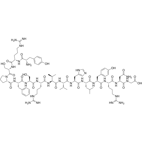
MOG (92–106), mouse, rat is a biological active peptide. (This is amino acids 92 to 106 fragment of the myelin oligodendrocyte glycoprotein (MOG) from mouse/rat. Mice with MOG (92–106)-induced experimental autoimmune encephalomyelitis develop extensive B cell reactivity against secondary myelin antigens. Despite the fact that this MOG peptide induces only weak T cell responses, MOG-induced autoimmunity is very severe. This peptide is encephalitogenic in SJL mice, DA rats, and rhesus monkeys.)
Anti-Monkey/Human CD20 Antibody (2H7) is a mouse-derived IgG2b κ type antibody inhibitor, targeting to monkey/human CD20. Anti-Monkey/Human CD20 Antibody (2H7) specifically depletes B cells. Anti-Monkey/Human CD20 Antibody (2H7) can be used for the researches of inflammation and metabolic disease, such as diabetes and experimental autoimmune encephalomyelitis .
7-Dehydro desmosterol is a sterol and an intermediate in cholesterol biosynthesis, which is found in the marine diatom Pseudo-nitzschia multistriata and the nervous system of rodents .
Geissoschizine methyl ether is an orally active, blood-brain barrier permeable alkaloid, and a partial agonist of the 5-HT1A receptor. It can be isolated from Uncaria hook. Geissoschizine methyl ether potently inhibits the binding of [ 3H]8-OH-DPAT to the 5-HT1A receptor in a concentration-dependent manner, with an IC50 of 0.904 μM. It ameliorates isolation-induced increased aggression and reduced sociability in mice. Geissoschizine methyl ether promotes oligodendrocyte differentiation and remyelination in the medial prefrontal cortex of adult mice .
4-Ethylphenyl sulfate sodium is an orally active and brain-penetrant gut microbial metabolite. 4-Ethylphenyl sulfate sodium downregulates Bcl2 expression, upregulates Bax expression, and induces cancer cell apoptosis via the endogenous apoptotic pathway. 4-Ethylphenyl sulfate sodium induces G2/M cell cycle arrest and reactive oxygen species (ROS) production. 4-Ethylphenyl sulfate sodium impairs oligodendrocyte maturation, reduces oligodendrocyte-neuron interactions, decreases axonal myelination levels, and shifts the oligodendrocyte population toward immature precursor cells. 4-Ethylphenyl sulfate sodium alters brain region-specific neural activity and functional connectivity in mice, and correlates with anxiety-like behaviors in mice .
Oxyphenbutazone (monohydrate) (Standard) is the analytical standard of Oxyphenbutazone (monohydrate). This product is intended for research and analytical applications. Oxyphenbutazone monohydrate is a Phenylbutazone (HY-B0230) metabolite, with anti-inflammatory effect. Oxyphenbutazone monohydrate is an orally active non-selective COX inhibitor. Oxyphenbutazone monohydrate selectively kills non-replicating Mycobaterium tuberculosis .
MOG Protein potentially finalizes and maintains the myelin sheath, contributing to cell-cell communication.It acts as a homophilic cell adhesion molecule, mediating cellular interactions through homodimerization, indicating its involvement in establishing connections within myelin-related processes.MOG Protein, Mouse (His) is the recombinant mouse-derived MOG protein, expressed by E.coli , with C-His labeled tag.
OMGP protein is a cell adhesion molecule that plays a crucial role in the interactive process necessary for myelination in the central nervous system.It accomplishes this by binding to RTN4R, facilitating the formation and maintenance of myelin sheaths.OMGP Protein, Mouse (HEK293, His) is the recombinant mouse-derived OMGP protein, expressed by HEK293 , with C-His labeled tag.
MOG proteins play a key role in homogeneous cell-to-cell adhesion, promoting important junctions. As a minor but integral component of myelin, it contributes to its underlying completion and maintenance. MOG Protein, Human (HEK293,C-His) is the recombinant human-derived MOG protein, expressed by HEK293 , with C-6*His labeled tag.
MOG proteins play a key role in homogeneous cell-to-cell adhesion, promoting important junctions. As a minor but integral component of myelin, it contributes to its underlying completion and maintenance. MOG Protein, Human (HEK293, His, solution) is the recombinant human-derived MOG protein, expressed by HEK293 , with C-6*His labeled tag.
The OMGP protein is a cell adhesion molecule that plays a crucial role in the complex myelination process in the central nervous system. Its function involves binding to RTN4R, which facilitates molecular interactions required for fundamental steps in myelination. OMGP Protein, Human (HEK293, His) is the recombinant human-derived OMGP protein, expressed by HEK293 , with C-His labeled tag.
MOG Protein potentially finalizes and maintains the myelin sheath, contributing to cell-cell communication. It acts as a homophilic cell adhesion molecule, mediating cellular interactions through homodimerization, indicating its involvement in establishing connections within myelin-related processes. MOG Protein, Mouse (N-His) is the recombinant mouse-derived MOG protein, expressed by E. coli , with N-His labeled tag.
MOG Protein potentially finalizes and maintains the myelin sheath, contributing to cell-cell communication. It acts as a homophilic cell adhesion molecule, mediating cellular interactions through homodimerization, indicating its involvement in establishing connections within myelin-related processes. MOG Protein, Mouse (P.pastoris, His) is the recombinant mouse-derived MOG protein, expressed by P. pastoris , with C-His labeled tag.
Basic domain helix loop helix protein class B 1; Basic helix loop helix protein class B 1; BHLHB; bHLHB1; bHLHe19; Class B basic helix loop helix protein 1; Class B basic helix-loop-helix protein 1; class E basic helix loop helix protein 19; Class E basic helix-loop-helix protein 19; Human protein kinase C binding protein RACK17; Olig2; OLIG2_HUMAN; Oligo2; oligodendrocyte lineage transcription factor 2; oligodendrocyte specific bHLH transcription factor 2; oligodendrocyte transcription factor 2; OTTHUMP00000067569; OTTHUMP00000067570; PRKCBP2; Protein kinase C binding protein 2; Protein kinase C binding protein RACK17; Protein kinase C-binding protein 2; Protein kinase C-binding protein RACK17; RACK17.
WB, ICC/IF, ELISA
Human, Mouse
Olig2 Antibody (YA5279) is a Mouse-derived and non-conjugated IgG monoclonal antibody, targeting to Olig2.
"Basic helix loop helix domain containing class B protein 7 antibody; Basic helix-loop-helix domain-containing protein, class B, 7 antibody; bHLHB7 antibody; bHLHe20 antibody; Class B basic helix loop helix protein 7 antibody; Class B basic helix-loop-helix protein 7 antibody; Class E basic helix loop helix protein 20 antibody; Class E basic helix-loop-helix protein 20 antibody; Olig3 antibody; OLIG3_HUMAN antibody; "Basic helix loop helix domain containing class B protein 7 antibody; Basic helix-loop-helix domain-containing protein, class B, 7 antibody; bHLHB7 antibody; bHLHe20 antibody; Class B basic helix loop helix protein 7 antibody; Class B basic helix-loop-helix protein 7 antibody; Class E basic helix loop helix protein 20 antibody; Class E basic helix-loop-helix protein 20 antibody; Olig3 antibody; OLIG3_HUMAN antibody; Oligo3 antibody; oligodendrocyte lineage transcription factor 3 antibody; oligodendrocyte specific bHLH transcription factor 3 antibody; oligodendrocyte transcription factor 3 antibody; "
WB, ICC/IF, IHC-P
Human, Mouse
Olig3 Antibody (YA6692) is a Rabbit-derived and non-conjugated IgG monoclonal antibody, targeting to Olig3.
(5)
Product Comparison
Compare
Clear All
Compare Products
Products
In-stock
-
+
Add to Cart
Cat. No.
Host
Reactivity
Application
Dilution Ratio
Molecular Weight
Conjugation
Clonality
Immunogen
Appearance
Isotype
Gene ID
SwissProt ID
Purity
Formulation
Free Sample
YesNo
Size
* This product has been "discontinued".
Optimized version of product available:
/
In-stock
-
+
Add to Cart
Get quote
Inquiry Online
Your information is safe with us. * Required Fields.
Western blot analysis of extracts from THP-1(lane 2(20μg), Jurkat (lane 3(20μg) and NIH3T3(lane 4(20μg) using FOXO1A (HY-P80132) Rabbit mAb. Proteins were transferred
to a PVDF membrane and blocked with 5% non-fat milk in TBST for 2 hour at room temperature. The primary antibody (1/1000) and Loading control antibody (Beta Actin, HY-P80438, 1/10000) was
used in 5% non-fat milk in TBST at 4°C overnight. Goat Anti-Mouse/Rabbit IgG-HRP Secondary Antibody (1/10000) was used for 1 hour at room temperature.
Western blot analysis of extracts from THP-1(lane 2(20μg), Jurkat (lane 3(20μg) and NIH3T3(lane 4(20μg) using FOXO1A (HY-P80132) Rabbit mAb. Proteins were transferred
to a PVDF membrane and blocked with 5% non-fat milk in TBST for 2 hour at room temperature. The primary antibody (1/1000) and Loading control antibody (Beta Actin, HY-P80438, 1/10000) was
MedchemExpress Validation 03
Western blot analysis of extracts from THP-1(lane 2(20μg), Jurkat (lane 3(20μg) and NIH3T3(lane 4(20μg) using FOXO1A (HY-P80132) Rabbit mAb. Proteins were transferred
MedchemExpress Validation 04
Western blot analysis of extracts from THP-1(lane 2(20μg), Jurkat (lane 3(20μg) and NIH3T3(lane 4(20μg) using FOXO1A (HY-P80132) Rabbit mAb. Proteins were transferred
to a PVDF membrane and blocked with 5% non-fat milk in TBST for 2 hour at room temperature. The primary antibody (1/1000) and Loading control antibody (Beta Actin, HY-P80438, 1/10000) was
used in 5% non-fat milk in TBST at 4°C overnight. Goat Anti-Mouse/Rabbit IgG-HRP Secondary Antibody (1/10000) was used for 1 hour at room temperature.
MedchemExpress Validation
Western blot analysis of extracts from THP-1(lane 2(20μg), Jurkat (lane 3(20μg) and NIH3T3(lane 4(20μg) using FOXO1A (HY-P80132) Rabbit mAb. Proteins were transferred
to a PVDF membrane and blocked with 5% non-fat milk in TBST for 2 hour at room temperature. The primary antibody (1/1000) and Loading control antibody (Beta Actin, HY-P80438, 1/10000) was
used in 5% non-fat milk in TBST at 4°C overnight. Goat Anti-Mouse/Rabbit IgG-HRP Secondary Antibody (1/10000) was used for 1 hour at room temperature.
MedchemExpress Validation
Western blot analysis of extracts from THP-1(lane 2(20μg), Jurkat (lane 3(20μg) and NIH3T3(lane 4(20μg) using FOXO1A (HY-P80132) Rabbit mAb. Proteins were transferred
to a PVDF membrane and blocked with 5% non-fat milk in TBST for 2 hour at room temperature. The primary antibody (1/1000) and Loading control antibody (Beta Actin, HY-P80438, 1/10000) was
used in 5% non-fat milk in TBST at 4°C overnight. Goat Anti-Mouse/Rabbit IgG-HRP Secondary Antibody (1/10000) was used for 1 hour at room temperature.
MedChemExpress values your privacy and your trust is important to us. We use cookies to enhance your website experience. Some cookies are necessary to run the website.
Privacy and Cookie Policy