



MOG (92–106), mouse, rat
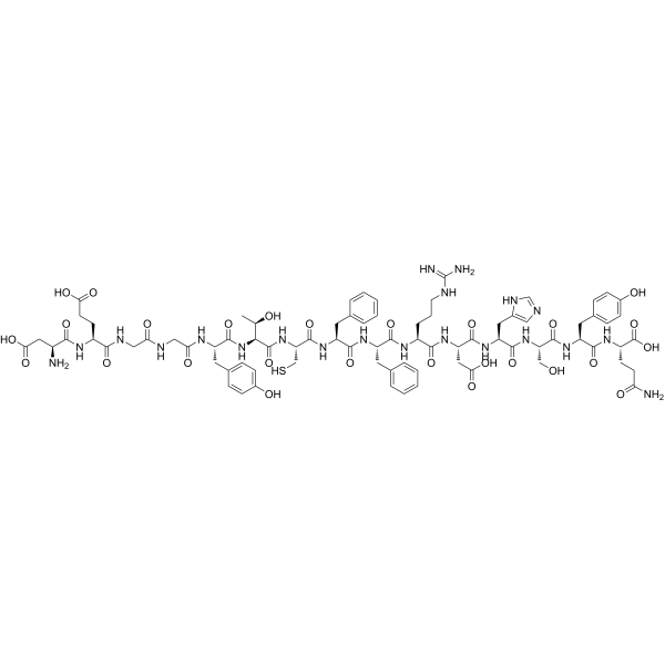
MOG (92-106), mouse, rat is a 15-peptide encephalitogenic epitope composed of amino acid residues 92-106 of myelin oligodendrocyte glycoprotein (MOG). MOG (92-106), mouse, rat serves as an I-As-restricted CD4+ T cell antigen. MOG (92-106), mouse, rat also induces extensive B cell responses against secondary myelin antigens such as MBP and PLP. MOG (92-106), mouse, rat is applicable to studies on multiple sclerosis, EAE, autoimmune demyelination, and antigen-specific T/B cell responses.
For research use only. We do not sell to patients.
- CAS No.: 159507-82-1
- Formula: C80H105N21O27S
- Molecular Weight:1824.88
-
Storage:
Please store the product under the recommended conditions in the Certificate of Analysis.

To place orders, for customer services and technical support, please contact: MedChemExpress USA
Tel: 609-228-6898 E-mail: [email protected] [email protected]


-
 Biological Activity
-
Chemical Information
- Protocols
- Purity & Documentation
- References
-
 Help & FAQs


All Endogenous Metabolite Isoforms
More

Biological Activity
Description
In Vitro
MOG (92-106), mouse, rat induces a weak antigen-specific proliferative response in draining lymph node cells of immunized SJL mice, with a stimulation index of 5.8, but this peptide still induces severe autoimmune disease in vivo[1].
MOG (92-106), mouse, rat (0.01-100 μg/mL) dose-dependently promotes the proliferation of splenocytes from MOG (92-106)/I-As-specific TCR transgenic SJL/J mice[3].
MedChemExpress (MCE) has not independently confirmed the accuracy of these methods. They are for reference only. Further protocols information, click here.
In Vivo
MOG (92-106) (200 μg; s.c.; single dose; administered in CFA with 200 μg Mycobacterium tuberculosis H37RA) has its induced experimental autoimmune encephalomyelitis severity enhanced by adoptive transfer of PLP139-151-specific serum in SJL mice, demonstrating pathogenicity of secondary autoantibodies induced by MOG (92-106) immunization[1].
MOG (92-106) (100 nmol; s.c.; single injection) induces relapsing-remitting EAE with combined classical and ataxic signs, prominent spinal cord demyelination, and T cell-predominant CNS infiltration in female SJL/J mice without Bordetella pertussis supplementation[2].
MOG (92-106) (100 nmol; s.c.; single injection) induces relapsing-remitting EAE with classical signs, prominent spinal cord demyelination, and T cell-predominant CNS infiltration in female SJL/J mice with Bordetella pertussis supplementation[2].
MOG (92-106) (100 nmol; s.c.; single injection) induces fatal primary progressive EAE with severe ataxic signs, large plaque-like demyelination, neutrophil-predominant CNS infiltrates, prominent Ig deposition, and high serum anti-MOG antibody titers in female A.SW mice without Bordetella pertussis supplementation, with serum IgG2a/IgG1 ratio strongly correlating with survival length[2].
MOG (92-106) (100 nmol; s.c.; single injection) induces secondary progressive EAE with combined early classical and later ataxic signs, severe brain demyelination (including chronic lesions), and sparse T cell CNS infiltration in female A.SW mice with Bordetella pertussis supplementation[2].
MedChemExpress (MCE) has not independently confirmed the accuracy of these methods. They are for reference only.
-
Animal Model:SJL/J (7-8 week-old female; experimental allergic encephalomyelitis induced without Bordetella pertussis supplementation)[2]
-
Dosage:100 nmol
-
Administration:s.c.; single injection
-
Result:Induced relapsing-remitting (RR) EAE.
Recorded 1/22 mice with acute disease and a mean maximum clinical score of 0.2.
Recorded 15/22 mice with chronic disease, a mean maximum clinical score of 3.8, and chronic disease onset at 45.1 days post-immunization.
Recorded 1/22 mice dead at day 32.
Observed both classical EAE signs (tail atony, hind limb paralysis) and ataxic signs (head/body turning, waddling gait).
Detected more prominent spinal cord lesions than brain lesions, with large subpial and perivenular demyelinating lesions, meningitis, mild perivascular cuffing, and predominantly mononuclear cell infiltrates.
Found CD3+ T cells predominated in meningeal and perivascular CNS infiltrates and parenchymal demyelinating lesions; B cells comprised ~10% of meningeal infiltrates but were absent in CNS parenchyma.
Detected Ig deposition only in meninges and endothelial cells.
Measured low serum anti-MOG (92-106) antibody titers.
-
Animal Model:SJL/J (7-8 week-old female; experimental allergic encephalomyelitis induced with intravenous Bordetella pertussis supplementation on days 0 and 2)[2]
-
Dosage:100 nmol
-
Administration:s.c.; single injection
-
Result:Induced relapsing-remitting (RR) EAE.
Recorded 5/8 mice with acute disease and a mean maximum clinical score of 3.8.
Recorded 7/8 mice with chronic disease, a mean maximum clinical score of 3.8, and chronic disease onset at 45.1 days post-immunization.
Recorded 1/8 mice dead at day 81.
Observed classical EAE signs (tail atony, hind limb paralysis) with mild or no ataxic signs.
Detected more prominent spinal cord lesions than brain lesions, with large subpial and perivenular demyelinating lesions, meningitis, mild perivascular cuffing, and predominantly mononuclear cell infiltrates.
Found CD3+ T cells predominated in meningeal and perivascular CNS infiltrates and parenchymal demyelinating lesions; B cells comprised ~10% of meningeal infiltrates but were absent in CNS parenchyma.
Detected Ig deposition only in meninges and endothelial cells.
Measured low serum anti-MOG (92-106) antibody titers.
-
Animal Model:A.SW (7-8 week-old female; experimental allergic encephalomyelitis induced without Bordetella pertussis supplementation)[2]
-
Dosage:100 nmol
-
Administration:s.c.; single injection
-
Result:Induced primary progressive (PP) EAE with no acute disease.
Recorded 14/16 mice with chronic disease, a mean maximum clinical score of 4.7, and chronic disease onset at 26.4 days post-immunization.
Recorded 13/16 mice dead at a mean of 42.8 days.
Observed severe ataxic EAE signs (head/body turning, waddling gait, rolling, inability to stand) with moderate spastic limb paralysis, and rarely classical EAE signs.
Detected large plaque-like demyelinating lesions in cerebellar white matter, vestibulocochlear nerve root/nucleus, optic nerve, and spinal cord, with mild meningitis, sparse perivascular cuffing (≤2 layers of inflammatory cells), and infiltrates dominated by neutrophils and macrophages (few lymphocytes).
Found CD3+ T cells were sparse in meningeal/perivascular CNS infiltrates and absent in parenchymal demyelinating lesions; B cells comprised ~10% of meningeal infiltrates but were absent in CNS parenchyma.
Detected intense Ig deposition at the margin of demyelinating plaques, on myelin, and on endothelial cells.
Measured high serum anti-MOG (92-106) IgG1 and IgG2a titers, with a significant positive correlation between serum IgG2a/IgG1 ratio and survival duration.
Detected no TUNEL-positive cells in CNS lesions.
-
Animal Model:A.SW (7-8 week-old female; experimental allergic encephalomyelitis induced with intravenous Bordetella pertussis supplementation on days 0 and 2)[2]
-
Dosage:100 nmol
-
Administration:s.c.; single injection
-
Result:Induced secondary progressive (SP) EAE.
Recorded 3/8 mice with acute disease and a mean maximum clinical score of 0.4.
Recorded 8/8 mice with chronic disease, a mean maximum clinical score of 4.3, and chronic disease onset at 46.3 days post-immunization.
Recorded 6/8 mice dead at a mean of 84.8 days.
Observed ataxic signs (disequilibrium, waddling gait, body rotation) in the latter stage, with half showing classical tail atony in the early acute stage.
Detected severe brain lesions (similar to PP-EAE mice) including active demyelination and chronic lesions with foam cells and cholesterol crystals; spinal cord lesions were less marked.
Found CD3+ T cells were sparse in meningeal/perivascular CNS infiltrates and absent in parenchymal demyelinating lesions; B cells comprised ~10% of meningeal infiltrates but were absent in CNS parenchyma.
Detected Ig deposition in demyelinating lesions with foam cells and cholesterol crystals.
Measured low serum anti-MOG (92-106) antibody titers.
Detected a small number of TUNEL-positive cells in CNS lesions.


Chemical Information
-
CAS No. 159507-82-1
-
Molecular Weight 1824.88
-
Formula C80H105N21O27S
-
Sequence
Asp-Glu-Gly-Gly-Tyr-Thr-Cys-Phe-Phe-Arg-Asp- His-Ser-Tyr-Gln
-
Sequence Shortening
DEGGYTCFFRDHSYQ
-
Shipping
Room temperature in continental US; may vary elsewhere.
-
Storage
Please store the product under the recommended conditions in the Certificate of Analysis.

Protocols
-
Research Protocol for Cancer Immunology
Cancer immunology studies how the immune system recognizes, suppresses, edits, or fails to eliminate malignant cells through tumor antigen release, antigen presentation, T-cell priming, immune trafficking, tumor-cell killing, and feedback inhibition in the tumor microenvironment. The cancer-immunity cycle links tumor antigenicity, dendritic-cell priming, CD8+ T-cell infiltration, cytotoxic function, and immune-checkpoint regulation to tumor rejection or immune escape. Immune-checkpoint pathways such as PD-1/PD-L1 and CTLA-4 suppress antitumor T-cell activity and can be therapeutically blocked, but many tumors remain resistant because of poor antigen presentation, weak T-cell infiltration, suppressive myeloid cells, regulatory T cells, and tumor-intrinsic immune-exclusion programs. Unresolved questions include which immune-cell states predict response, how tumor-intrinsic pathways exclude immune cells, how myeloid suppression limits checkpoint blockade, and which combination strategies


Purity & Documentation
References
[1]. Bischof F, et al. A structurally available encephalitogenic epitope of myelin oligodendrocyte glycoprotein specifically induces a diversified pathogenic autoimmune response. Journal of immunology (Baltimore, Md. : 1950). 2004 Jul 01;173(1):600-6. [Content Brief]
[2]. Tsunoda I, et al. Antibody association with a novel model for primary progressive multiple sclerosis: induction of relapsing-remitting and progressive forms of EAE in H2s mouse strains. Brain pathology (Zurich, Switzerland). 2000 Jul;10(3):402-18. [Content Brief]
[3]. Pöllinger B, et al. Spontaneous relapsing-remitting EAE in the SJL/J mouse: MOG-reactive transgenic T cells recruit endogenous MOG-specific B cells. The Journal of experimental medicine. 2009 Jun 08;206(6):1303-16. [Content Brief]

Calculators
Concentration (start) × Volume (start) = Concentration (final) × Volume (final)


MOG (92–106), mouse, rat Related Classifications
HY-P5448 Related Classifications
 Powered by Bioz