



MOG (35-55) acetate
Based on 9 publication(s) in Google Scholar
MOG (35-55) (Myelin Oligodendrocyte Glycoprotein Peptide (35-55), mouse, rat) acetate is a minor component of CNS myelin. MOG (35-55) (acetate) has encephalitogenic activity and induces T cell proliferative. MOG (35-55) (acetate) induces Th1 cytokine response as well as relatively high levels of IgG antibodies. MOG (35-55) (acetate) produces a relapsing-remitting neurological disease with extensive plaque-like demyelination. MOG (35-55) (Myelin Oligodendrocyte Glycoprotein Peptide (35-55), mouse, rat) acetate can be used for experimental autoimmune encephalomyelitis (EAE) modeling.
For research use only. We do not sell to patients.
- Purity : 99.86%
- Formula: C120H181N35O31S
- Molecular Weight:2642.00
-
Storage:
Sealed storage, away from moisture and light, under nitrogen.
Powder -80°C, 2 years , -20°C, 1 year* In solvent : -80°C, 6 months; -20°C, 1 month (sealed storage, away from moisture and light, under nitrogen)


To place orders, for customer services and technical support, please contact: MedChemExpress USA
Tel: 609-228-6898 E-mail: [email protected] [email protected]


-
 Biological Activity
-
Chemical Information
-
Solvent & Solubility
- Protocols
- Purity & Documentation
- References
-
 Help & FAQs



Publications Citing Use of MedChemExpress (MCE) MOG (35-55) acetate
More- Adv Sci (Weinh). 2025 Apr;12(14):e2409086. [Abstract]
- Cell Death Dis. 2022 Sep 2;13(9):759. [Abstract]
- EMBO Mol Med. 2025 Sep 29. [Abstract]
- Acta Physiol. 2023 Jun;238(2):e13974. [Abstract]
- Int Immunopharmacol. 2022 Apr:105:108566. [Abstract]
- Front Biosci (Landmark Ed). 2025 May 23;30(5):37838. [Abstract]
- Mol Biol Rep. 2025 Nov 15;53(1):92. [Abstract]
- Cornell University. 2025.
- Research Square Print. September 28th, 2022.
 Customer Validation & Images

-
Others
-
Histological Imaging/Staining
-
Others
-
Cell Proliferation/Viability Assay
-
ELISA


Biological Activity
Description
In Vitro
MOG (35-55) (acetate) (Myelin Oligodendrocyte Glycoprotein Peptide (35-55), mouse, rat (acetate)); 0-50 μg/mL; 72 h; lymph nodes cells) acetate induces T cell proliferative and secretes Th1 cytokines including IFN-γ, TNF-α, IL-10, IL-4 and IL-5. MOG (35-55) (acetate) increases the level of IgG[1].
MedChemExpress (MCE) has not independently confirmed the accuracy of these methods. They are for reference only. Further protocols information, click here.
In Vivo
-
Animal Model:HLA-DR2 (DRB1*1501) mice[1]
-
Dosage:200 μg (0.2 mL)
-
Administration:Intraperitoneal injection; once, for 38 days
-
Result:Resulted in paralysis of both hind and forelimbs.
Induced Disease Models
Please do not refer to only one article to determine the experimental conditions. It is recommended to determine the optimal experimental conditions (animal strain, age, dosage, frequency and cycle, detection time and indicators, etc.) through preliminary experiments before the formal experiment.
Myelin oligodendrocyte glycoprotein (MOG) peptide 35-55 is one of the targets of self-reactive T cell responses, leading to acute and later chronic autoimmune processes. MOG 35-55 is capable of inducing experimental autoimmune encephalomyelitis (EAE) in B6 mice[1].
Administration: On day 0: 3 mg/mL peptide plus CFA (4 mg/mL) • Mix thoroughly 1: 1 peptide and CFA solutions • s.c. • 100 μL for each hind flank; • On day 0 and day 2: 2.5 μg/mL of Bordetella pertussis toxin • i.p. • 200 μL
2. Symptoms appear approximately 10-15 days after immunization.
3. When mice have clinical symptoms of EAE, hydrated food is placed on the cage floor to facilitate access to these mice.
4. Record parameters every other day at the beginning, and daily from 7th postimmunization day. Body weight decreases usually precedes EAE clinical signs and is a very objective and valuable measure.
prototypical histopathological changes: Luxol fast blue staining of spinal cord for myelin evaluation shows infiltrates and demyelination in EAE animals.
MedChemExpress (MCE) has not independently confirmed the accuracy of these methods. They are for reference only.


Chemical Information
-
Appearance Solid
-
Molecular Weight 2642.00
-
Formula C120H181N35O31S
-
Color White to off-white
-
Synonyms
Myelin Oligodendrocyte Glycoprotein Peptide (35-55), mouse, rat acetate
-
Sequence
Met-Glu-Val-Gly-Trp-Tyr-Arg-Ser-Pro-Phe-Ser-Arg-Val-Val-His-Leu-Tyr-Arg-Asn-Gly-Lys
-
Sequence Shortening
MEVGWYRSPFSRVVHLYRNGK
-
Shipping
Room temperature in continental US; may vary elsewhere.
-
Storage
Sealed storage, away from moisture and light, under nitrogen
Powder -80°C 2 years -20°C 1 year * In solvent : -80°C, 6 months; -20°C, 1 month (sealed storage, away from moisture and light, under nitrogen)


Publications (9)
-
Journal Impact Factor
-
Most Recent
-
Adv Sci (Weinh)
TIA1-Mediated Stress Granules Promote the Neuroinflammation and Demyelination in Experimental Autoimmune Encephalomyelitis through Upregulating IL-31RA Signaling. [Abstract]2025 Apr;12(14):e2409086. PMID: 39804990 -
Cell Death Dis
Astrocytic SARM1 promotes neuroinflammation and axonal demyelination in experimental autoimmune encephalomyelitis through inhibiting GDNF signaling. [Abstract]2022 Sep 2;13(9):759. PMID: 36055989 -
EMBO Mol Med
Selective inhibition of mitochondrial Kv1.3 prevents and alleviates multiple sclerosis in vivo. [Abstract]2025 Sep 29. PMID: 41023199
MOG (35-55) acetate purchased from MedChemExpress. Usage Cited in: EMBO Mol Med. 2025 Sep 29. [Abstract]
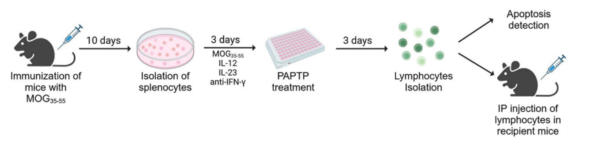
Scheme of the EAE Adoptive Transfer Model: Mice underwent immunization via subcutaneous injection of MOG35–55 in complete Freund’s adjuvant. Ten days post-immunization, spleens were harvested, and single-cell suspensions were prepared. These splenocytes were treated for 3 days with IL-12, IL-23, and anti-IFN-γ. Subsequently, the splenocytes were subjected to a 3-day treatment with 1 μM PAPTP, or left untreated. Antigen-specificity of the cells was confirmed by additional proliferation assays of isolated donor lymphocytes (see Methods). Lymphocytes were isolated, and the induction of apoptosis was assessed using flow cytometry. Additionally, these lymphocytes were transferred into wild-type recipient animals, and the animals’ clinical scores were monitored daily for 10 days. Following the observation period, the animals were euthanized for further analysis.
MOG (35-55) acetate purchased from MedChemExpress. Usage Cited in: EMBO Mol Med. 2025 Sep 29. [Abstract]
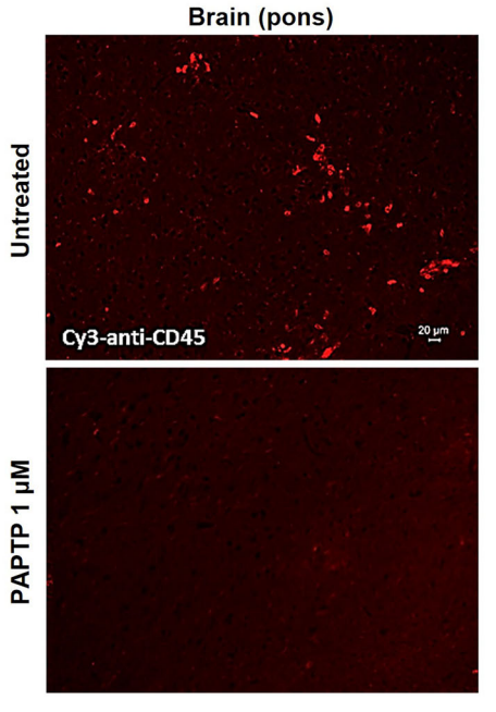
Representative image (of 5 sections) of brains (upper part of the pons region) stained with PE-anti-CD45, from wild-type mice intraperitoneally injected with either untreated or PAPTP 1 μM treated, MOG35–55 activated lymphocytes.3J
MOG (35-55) acetate purchased from MedChemExpress. Usage Cited in: EMBO Mol Med. 2025 Sep 29. [Abstract]
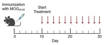
Treatment scheme for wild-type mice immunized via subcutaneous injection of MOG35–55: upon the manifestation of initial symptoms, typically around day 10 post-immunization, treatment commenced. The treatment regimen was administered every 48 h, totaling 11 injections. Mice were euthanized the day following the final injection for subsequent analysis of peripheral blood, brains, and spinal cords.
-
Acta Physiol
SARM1 promotes the neuroinflammation and demyelination through IGFBP2/NF-κB pathway in experimental autoimmune encephalomyelitis mice. [Abstract]2023 Jun;238(2):e13974. PMID: 37186158 -
Int Immunopharmacol
High-density lipoprotein reduces microglia activation and protects against experimental autoimmune encephalomyelitis in mice. [Abstract]2022 Apr:105:108566. PMID: 35101849 -
Front Biosci (Landmark Ed)
HMGB1 Mediates Microglia-Astrocyte/Neuron Crosstalk and Pyroptosis by the TLR4/NF-κB Pathway in Multiple Sclerosis. [Abstract]2025 May 23;30(5):37838. PMID: 40464519
MOG (35-55) acetate purchased from MedChemExpress. Usage Cited in: Front Biosci (Landmark Ed). 2025 May 23;30(5):37838. [Abstract]
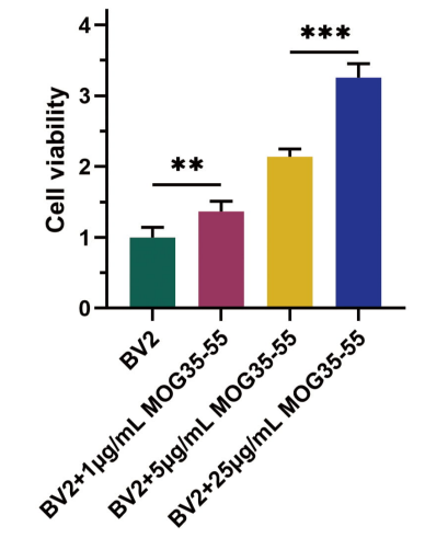
The BV-2 cells were cultured in designated concentrations of MOG35-55 (0-25μg/mL, 24 h). MTT analysis of BV-2 cell viability.
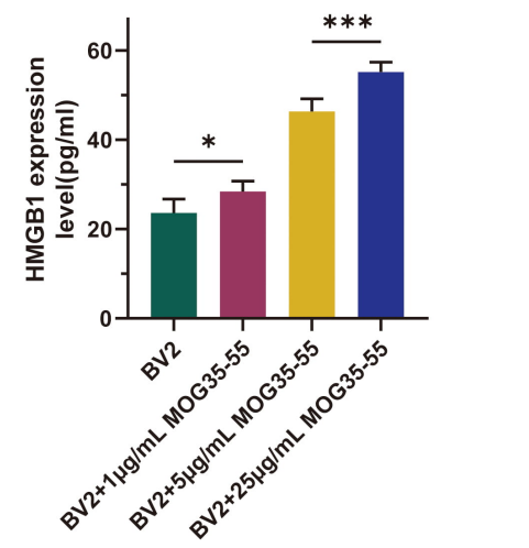
MOG (35-55) acetate purchased from MedChemExpress. Usage Cited in: Front Biosci (Landmark Ed). 2025 May 23;30(5):37838. [Abstract]
The BV-2 cells were cultured in designated concentrations of MOG35-55 (0-25μg/mL, 24 h). ELISA analysis of the release of HMGB1.
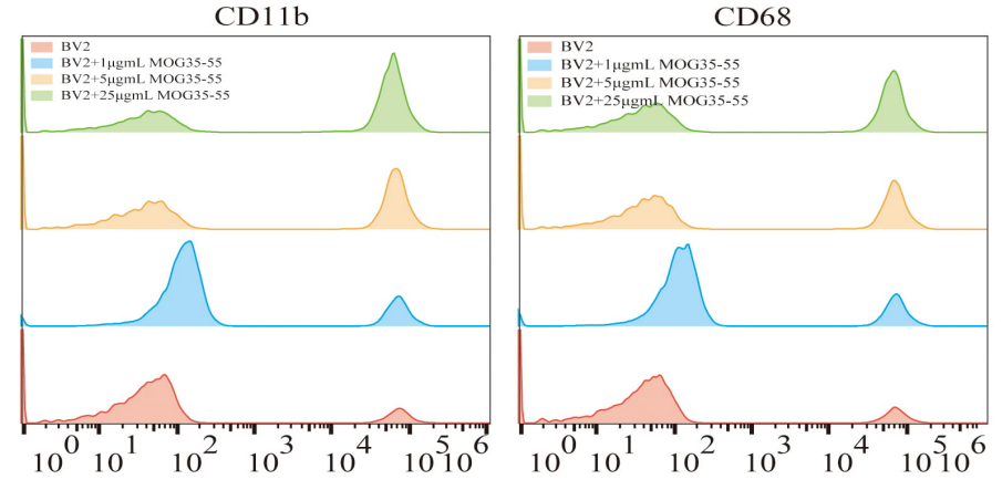
MOG (35-55) acetate purchased from MedChemExpress. Usage Cited in: Front Biosci (Landmark Ed). 2025 May 23;30(5):37838. [Abstract]
The BV-2 cells were cultured in designated concentrations of MOG35-55 (0-25μg/mL). Cytometry analysis for CD11b+ and CD68+ cells.
-
Mol Biol Rep
Th2 cell-derived exosomes alleviate central nervous system inflammation and demyelination in experimental autoimmune encephalomyelitis by regulating inflammasomes to inhibit microglia activation. [Abstract]2025 Nov 15;53(1):92. PMID: 41240231 -
-


Solvent & Solubility
In Vitro:
H2O : 100 mg/mL (37.85 mM; Need ultrasonic)
Please refer to the solubility information to select the appropriate solvent. Once prepared, please aliquot and store the solution to prevent product inactivation from repeated freeze-thaw cycles.
Storage method and period of stock solution: -80°C, 6 months; -20°C, 1 month (sealed storage, away from moisture and light, under nitrogen). When stored at -80°C, please use it within 6 months. When stored at -20°C, please use it within 1 month.
* Note: If you choose water as the stock solution, please dilute it to the working solution, then filter and sterilize it with a 0.22 μm filter before use.
Please refer to the solubility information to select the appropriate solvent. Once prepared, please aliquot and store the solution to prevent product inactivation from repeated freeze-thaw cycles.
Storage method and period of stock solution: -80°C, 6 months; -20°C, 1 month (sealed storage, away from moisture and light, under nitrogen). When stored at -80°C, please use it within 6 months. When stored at -20°C, please use it within 1 month.
* Note: If you choose water as the stock solution, please dilute it to the working solution, then filter and sterilize it with a 0.22 μm filter before use.
Concentration (start) × Volume (start) = Concentration (final) × Volume (final)

Protocols
-
Naïve CD4+ T-cell subset differentiation/polarization
Naïve CD4+ T-cell subset differentiation/polarization is an in vitro assay in which purified naïve CD4+ T cells are activated through TCR and CD28 costimulation and cultured with defined cytokines and neutralizing antibodies to generate Th0, Th1, Th2, Th17, or induced Treg-like populations. Differentiation is detected by subset-associated cytokines and transcription factors: IFN-γ/T-bet for Th1, IL-4/GATA3 for Th2, IL-17A/RORγt for Th17, and Foxp3 for induced Treg cells. The assay readout is usually generated by intracellular cytokine staining after restimulation, transcription-factor staining by flow cytometry, ELISA of secreted cytokines, or gene-expression analysis. The result reflects cytokine-directed lineage commitment or polarization rather than antigen-specific immune protection by itself.
-
Research Protocol for Inflammation-related Diseases
The NLRP3 inflammasome is a cytosolic innate immune signaling platform that integrates priming signals and danger-signal activation to promote caspase-1 activation, maturation of IL-1β and IL-18, and gasdermin D-mediated pyroptotic cell death. The core experimental logic is to determine whether inflammatory disease phenotypes are driven by increased NLRP3 expression, ASC-containing inflammasome assembly, caspase-1 cleavage, GSDMD cleavage, and extracellular release of IL-1β/IL-18 rather than by nonspecific cell injury alone. The pathway is strongly linked to inflammation-related disease phenotypes because monosodium urate crystals activate NALP3/NLRP3 inflammasome signaling in gout-like crystal inflammation, cholesterol crystals activate NLRP3 inflammasomes in atherogenesis models, and DSS-induced intestinal inflammation has been reported to involve NLRP3 inflammasome activity. However, experimental colitis studies also show context-dependent protective effects of NLRP3 inflammasome co
-
Research Protocol for Neurological Diseases
PINK1/Parkin-mediated mitophagy pathway is a mitochondrial quality-control signaling axis in which mitochondrial depolarization stabilizes PINK1 on damaged mitochondria, activates Parkin recruitment and E3 ubiquitin ligase activity, promotes ubiquitination of outer mitochondrial membrane proteins, recruits selective autophagy adaptors, and drives lysosomal degradation of damaged mitochondria. In neurological disease research, this pathway is experimentally important because neurons, especially dopaminergic neurons, are highly dependent on mitochondrial integrity, and defective mitochondrial turnover can lead to mitochondrial dysfunction, oxidative stress, impaired neuronal survival, α-synuclein accumulation, and neuroinflammatory damage-associated signals. The genetic disease link is strongest in Parkinson’s disease because mutations in PRKN/parkin cause autosomal recessive juvenile parkinsonism, mutations in PINK1 cause hereditary early-onset Parkinson’s disease, and Drosophila studie
-
Research Protocol for Cancer Immunology
Cancer immunology studies how the immune system recognizes, suppresses, edits, or fails to eliminate malignant cells through tumor antigen release, antigen presentation, T-cell priming, immune trafficking, tumor-cell killing, and feedback inhibition in the tumor microenvironment. The cancer-immunity cycle links tumor antigenicity, dendritic-cell priming, CD8+ T-cell infiltration, cytotoxic function, and immune-checkpoint regulation to tumor rejection or immune escape. Immune-checkpoint pathways such as PD-1/PD-L1 and CTLA-4 suppress antitumor T-cell activity and can be therapeutically blocked, but many tumors remain resistant because of poor antigen presentation, weak T-cell infiltration, suppressive myeloid cells, regulatory T cells, and tumor-intrinsic immune-exclusion programs. Unresolved questions include which immune-cell states predict response, how tumor-intrinsic pathways exclude immune cells, how myeloid suppression limits checkpoint blockade, and which combination strategies


Purity & Documentation
-
Data Sheet (286 KB)
-
SDS (252 KB)
- English - EN (252 KB)
- Français - FR (252 KB)
- Deutsch - DE (252 KB)
- Norwegian - NO (252 KB)
- Español - ES (252 KB)
- Swedish - SV (252 KB)
- Italian - IT (252 KB)
- Korean - KR (252 KB)
- Portuguese - PT (252 KB)
-
Handling Instructions (2659 KB)
References
[1]. Rich C, et, al. Myelin oligodendrocyte glycoprotein-35-55 peptide induces severe chronic experimental autoimmune encephalomyelitis in HLA-DR2-transgenic mice. Eur J Immunol. 2004 May;34(5):1251-61. [Content Brief]
[2]. Slavin A, et, al. Induction of a multiple sclerosis-like disease in mice with an immunodominant epitope of myelin oligodendrocyte glycoprotein. Autoimmunity. 1998;28(2):109-20. [Content Brief]
[3]. Giralt M, et, al. Active Induction of Experimental Autoimmune Encephalomyelitis (EAE) with MOG35-55 in the Mouse. Methods Mol Biol. 2018;1791:227-232. [Content Brief]


Complete Stock Solution Preparation Table
Please refer to the solubility information to select the appropriate solvent. Once prepared, please aliquot and store the solution to prevent product inactivation from repeated freeze-thaw cycles.
Storage method and period of stock solution: -80°C, 6 months; -20°C, 1 month (sealed storage, away from moisture and light, under nitrogen). When stored at -80°C, please use it within 6 months. When stored at -20°C, please use it within 1 month.
| Optional Solvent | Concentration Solvent Mass | 1 mg | 5 mg | 10 mg | 25 mg |
|---|---|---|---|---|---|
| H2O | 1 mM | 0.3785 mL | 1.8925 mL | 3.7850 mL | 9.4625 mL |
| 5 mM | 0.0757 mL | 0.3785 mL | 0.7570 mL | 1.8925 mL | |
| 10 mM | 0.0379 mL | 0.1893 mL | 0.3785 mL | 0.9463 mL | |
| 15 mM | 0.0252 mL | 0.1262 mL | 0.2523 mL | 0.6308 mL | |
| 20 mM | 0.0189 mL | 0.0946 mL | 0.1893 mL | 0.4731 mL | |
| 25 mM | 0.0151 mL | 0.0757 mL | 0.1514 mL | 0.3785 mL | |
| 30 mM | 0.0126 mL | 0.0631 mL | 0.1262 mL | 0.3154 mL |
* Note: If you choose water as the stock solution, please dilute it to the working solution, then filter and sterilize it with a 0.22 μm filter before use.


MOG (35-55) acetate Related Classifications
HY-P1240B Related Classifications
 Powered by Bioz