



BMS-561392
Based on 2 publication(s) in Google Scholar
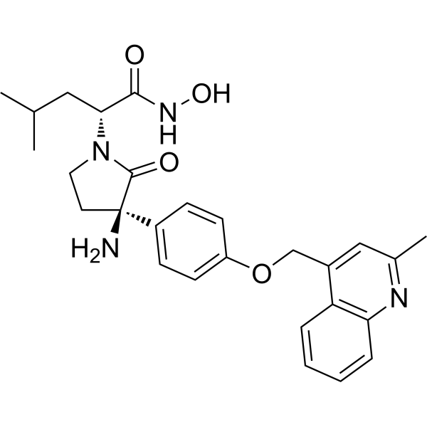
BMS-561392 (BMS-561392) is a selective ADAM17(TACE) inhibitor. BMS-561392 inhibits TNF-α secretion by regulating signaling pathways such as p44 MAPK and NF-κB. BMS-561392 also affects the survival of central nervous system-related cells including oligodendrocytes and microglia. BMS-561392 promotes microglial apoptosis, enlarges the injury area and exacerbates astrogliosis in a mouse spinal cord injury model. BMS-561392 can be used in research related to spinal cord injury and inflammatory diseases.
For research use only. We do not sell to patients.
- CAS No.: 611227-74-8
- Formula: C27H32N4O4
- Molecular Weight:476.57
-
Storage:
Please store the product under the recommended conditions in the Certificate of Analysis.

To place orders, for customer services and technical support, please contact: MedChemExpress USA
Tel: 609-228-6898 E-mail: [email protected] [email protected]


-
 Biological Activity
-
Chemical Information
- Protocols
- Purity & Documentation
- References
-
 Help & FAQs


Publications Citing Use of MedChemExpress (MCE) BMS-561392
More

Biological Activity
Description
In Vitro
BMS-561392 (10 nM; 1-2.5 h) potently inhibits recombinant human TACE (ADAM17) enzymatic activity by ~80%[1].
BMS-561392 (10 μM; 1 h pre-incubation followed by 4 h LPS stimulation) effectively suppresses LPS-induced TNF-α secretion in RAW264.7 macrophage cells[1].
BMS-561392 (1-100 μM; 1 h pre-incubation followed by 4 h LPS stimulation) dose-dependently inhibits LPS-induced p-IκB expression, and thus NFκB pathway activation, in RAW264.7 macrophage cells[1].
BMS-561392 (1-100 μM; 1 h pre-incubation followed by 1 h LPS stimulation) inhibits LPS-induced p-NFκB expression in RAW264.7 macrophage cells[1].
BMS-561392 (0.3-2.7 mM; 48 h) reduces viability of undifferentiated HOG cells in a concentration-dependent manner, with the greatest reduction (89.5-93.5%) observed at 2.7 mM after 48 h incubation[2].
BMS-561392 (0.3-2.7 mM; 48 h) modulates viability of BV-2 microglial cells in a concentration-dependent manner after 48 h incubation, with a slight increase at 0.3 mM, 50% reduction at 1.3 mM, and 94% reduction at 2.7 mM[2].
BMS-561392 (1.3-2.7 mM) induces apoptosis in BV-2 microglial cells, as shown by increased activated caspase-3-positive cells[2].
BMS-561392 (2.7 mM; 3 h) increases membrane TNFR-1 expression in BV-2 microglial cells by >50% after 3 h incubation[2].
BMS-561392 (0.3-2.7 mM; 3 h) modulates phosphorylated p44 MAPK levels in BV-2 microglial cells in a concentration-dependent manner after 3 h incubation, increasing levels at 0.3 mM and eliminating detectable levels at 2.7 mM, with no effect on phosphorylated p42 MAPK[2].
MedChemExpress (MCE) has not independently confirmed the accuracy of these methods. They are for reference only.
-
Cell Line:RAW264.7 macrophage cells
-
Concentration:10 μM (pre-incubation); 1 μg/mL (LPS stimulation)
-
Incubation Time:1 h (pre-incubation); 4 h (LPS stimulation)
-
Result:Reduced LPS-induced TNF-α levels to near-control levels.
-
Cell Line:RAW264.7 macrophage cells
-
Concentration:1, 10 and 100 μM (pre-incubation); 1 μg/mL (LPS stimulation)
-
Incubation Time:1 h (pre-incubation); 4 h (LPS stimulation)
-
Result:Induced a dose-dependent reduction in LPS-stimulated p-IκB levels.
In Vivo
MedChemExpress (MCE) has not independently confirmed the accuracy of these methods. They are for reference only.
-
Animal Model:C57BL/6 (10-week-old)[2]
-
Dosage:2.2 mM
-
Administration:s.c.; daily; 15 days
-
Result:Significantly reduced locomotor recovery as measured by BMS score.
Significantly increased lesion size.
Significantly increased astrogliosis in tissue caudal to the lesion site.
Significantly reduced Iba1 expression in tissue rostro-caudal to the lesion epicenter.
Significantly increased the number of apoptotic microglial/macrophage cells in tissue caudal to the lesion site.
Significantly increased pro-apoptotic Bax expression in spinal cord tissue.
Showed a non-significant decrease in anti-apoptotic Bcl-2 expression in spinal cord tissue.


Chemical Information
-
CAS No. 611227-74-8
-
Molecular Weight 476.57
-
Formula C27H32N4O4
-
SMILES
N[C@]1(C2=CC=C(C=C2)OCC3=CC(C)=NC4=C3C=CC=C4)C(N(CC1)[C@H](CC(C)C)C(NO)=O)=O
-
Synonyms
DPC 333
-
Shipping
Room temperature in continental US; may vary elsewhere.
-
Storage
Please store the product under the recommended conditions in the Certificate of Analysis.


Publications (2)
-
Journal Impact Factor
-
Most Recent
-
Curr Issues Mol Biol
Mechanistic Insights into Vorinostat as a Repositioned Modulator of TACE-Mediated TNF-α Signaling via MAPK and NFκB Pathways. [Abstract]2025 Sep 4;47(9):720. PMID: 41020841 -
PLoS One
Discovery of novel TACE inhibitors using graph convolutional network, molecular docking, molecular dynamics simulation, and Biological evaluation. [Abstract]2024 Dec 27;19(12):e0315245. PMID: 39729480


Purity & Documentation
References
[1]. Park J, et al. Mechanistic Insights into Vorinostat as a Repositioned Modulator of TACE-Mediated TNF-α Signaling via MAPK and NFκB Pathways. Curr Issues Mol Biol. 2025 Sep 4;47(9):720. [Content Brief]
[2]. Vidal PM, et al. ADAM17 is a survival factor for microglial cells in vitro and in vivo after spinal cord injury in mice. Cell Death Dis. 2013;4(12):e954. Published 2013 Dec 12. [Content Brief]

Calculators
Concentration (start) × Volume (start) = Concentration (final) × Volume (final)


BMS-561392 Related Classifications
HY-19667 Related Classifications
 Powered by Bioz