Search Result
Results for "
red fluorescence
" in MedChemExpress (MCE) Product Catalog:
8
Biochemical Assay Reagents
| Cat. No. |
Product Name |
Target |
Research Areas |
Chemical Structure |
-
- HY-D1055
-
MitoSOX Red
Maximum Cited Publications
263 Publications Verification
|
Fluorescent Dye
Reactive Oxygen Species (ROS)
Mitochondrial Metabolism
|
Cancer
|
MitoSOX Red is a live cell fluorescent probe that specifically targets mitochondria and is cell membrane permeable. MitoSOX Red enters mitochondria and is oxidized by superoxide but not by other ROS or RNS generating systems. The oxidized MitoSOX Red then binds to nucleic acids in mitochondria/nucleus, producing strong red fluorescence. MitoSOX Red can be used as a fluorescent indicator to specifically detect superoxide. In addition, superoxide dismutase (SOD) can prevent the oxidation of MitoSOX Red.
Excitation/emission wavelength: 510/580 nm.
|
-
-
- HY-D0815
-
|
|
Fluorescent Dye
DNA/RNA Synthesis
|
Others
|
|
Propidium Iodide (PI) is a nuclear staining agent that stains DNA. Propidium Iodide is an analogue of ethidine bromide that emits red fluorescence upon embedding in double-stranded DNA. Propidium Iodide cannot pass through living cell membranes, but it can pass through damaged cell membranes to stain the nucleus. Propidium Iodide has a fluorescence wavelength of 493/617 nm and a wavelength of 536/635 nm after Mosaic with DNA. Propidium Iodide is commonly used in the detection of apoptosis (apoptosis) or necrosis (necrosis), and is often used in flow cytometry analysis.
|
-
-
- HY-15534
-
|
CBIC2
|
Fluorescent Dye
|
Others
|
|
JC-1 (CBIC2) is an ideal fluorescent probe widely used to detect mitochondrial membrane potential. JC-1 accumulates in mitochondria in a potential dependent manner and can be used to detect the membrane potential of cells, tissues or purified mitochondria. In normal mitochondria, JC-1 aggregates in the mitochondrial matrix to form a polymer, which emits strong red fluorescence (Ex=585 nm, Em=590 nm); When the mitochondrial membrane potential is low, JC-1 cannot aggregate in the matrix of mitochondria and produce green fluorescence (ex=510 nm, em= 527 nm) .
|
-
-
- HY-D0718
-
|
Nile Blue A oxazone; Phenoxazone 9
|
Fluorescent Dye
|
Others
|
|
Nile red (Nile blue oxazone) is a lipophilic stain. Nile red has environment-sensitive fluorescence. Nile red is intensely fluorescent in a lipid-rich environment while it has minimal fluorescence in aqueous media. Nile red is an excellent vital stain for the detection of intracellular lipid droplets by fluorescence microscopy and flow cytof uorometry. Nile red stains intracellular lipid droplets red. The fluorescence wavelength is 559/635 nm .
|
-
-
- HY-D0079
-
|
Hydroethidine; PD-MY 003
|
Fluorescent Dye
|
Others
|
|
Dihydroethidium, also known as DHE, is a peroxide indicator. Dihydroethidium penetrates cell membranes to form a fluorescent protein complex with blue fluoresces. After entering the cells, Dihydroethidium is mainly localized in the cell membrane, cytoplasm and nucleus, and the staining effect is the strongest in the nucleus. Dihydroethidium produces inherent blue fluorescence with a maximum excitation wavelength of 370 nm and a maximum emission wavelength of 420 nm; after dehydrogenation, Dihydroethidium combines with RNA or DNA to produce red fluorescence with a maximum excitation wavelength of 300 nm and a maximum emission wavelength of 610 nm. 535 nm can also be used as the excitation wavelength for actual observation .
|
-
-
- HY-D1421
-
|
|
Fluorescent Dye
|
Others
|
|
PKH67 is a fluorescent cell binding dye with green fluorescence. PKH67 can stain the cell membrane and the Ex/Em is 490/502 nm. PKH67 is often used in combination with the non-specific red fluorescent dye PKH26 (Ex/Em=551/567 nm) to label cells, detect cell proliferation in vitro, and trace cells in vitro and in vivo .
|
-

-
- HY-D1451
-
|
|
Fluorescent Dye
|
Others
|
|
PKH 26 is a red fluorescent dye, PKH 26 can stably bind to the lipid region of cell membrane and emit red fluorescence (Ex/Em=551/567 nm), which is mainly used for in vitro cell labeling, in vitro cell proliferation studies and in vivo and in vitro cell tracing studies .
|
-
-
- HY-D2449
-
|
|
Fluorescent Dye
|
Others
|
|
DQ-BSA-Red is a bovine serum albumin labeled with a red fluorescent dye that can be used to detect lysosomal activity. The excitation wavelength and emission wavelength of DQ-BSA-Red are 590 nm and 620 nm, respectively. The BSA molecule in DQ-BSA-Red is labeled with high concentration of red fluorescent dye in multiple sites, which shows high fluorescence self-inhibition. Once DQ-BSA-RED enters the lysosome, DQ-BSA is cleaved by lysosomal proteases, resulting in unquenched and released fluorescent fragments, emitting bright fluorescence. Inactivated lysosomes are unable to degrade the BSA protein and thus have a lower or even no fluorescent signal .
|
-

-
- HY-D1119
-
|
|
Fluorescent Dye
|
Others
|
|
AF647-NHS ester is an analog of Alexa Fluor 647 (AF647). NHS ester can covalently bind to molecules with amino groups (such as proteins, antibodies, etc.). AF647 is a far-red fluorescent dye with an excitation wavelength (λex) of 635 nm (conventional fluorescence detection)/620 nm (instantaneous detection). Storage: Protect from light .
|
-
-
- HY-120601
-
|
ARS sodium
|
Fluorescent Dye
|
Others
|
|
Alizarin Red S sodium is an anthraquinone derivative dye. When combined with cations such as calcium ions, the functional group of Alizarin Red S sodium can form a coordination bond with the cation through the oxygen atom to show orange-red fluorescence. Alizarin Red S sodium can be used for screening of calcium compounds in synovial fluid and detecting osteoblast differentiation, and can also be used for bone staining in mice. Excitation/emission wavelength: 500/570 nm .
|
-
-
- HY-D0048
-
|
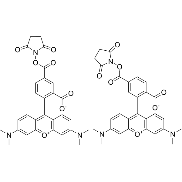
5-TAMRA-NHS ester; 5-Carboxytetramethylrhodamine succinimidyl ester
|
Fluorescent Dye
|
Others
|
|
5-TAMRA-SE is an amine-reactive fluorescent agent, and its conjugate produces bright, pH-insensitive orange-red fluorescence with good photostability (Ex/Em = 565/580 nm).
|
-
-
- HY-D1373
-
HBC
3 Publications Verification
HBC 530
|
Fluorescent Dye
|
Others
|
|
HBC (HBC 530) is a GFP fluorophore-like synthetic dye, with a structurally rigid electron acceptor and a strong electron donor. HBC has a low fluorescence background, and when combined with Pepper (RNA aptamer), HBC forms a tight complex and activates and emits bright fluorescence (Kd of ~3.5 nM). HBC emission peaks vary in different complexes and covers the spectrum from cyan to red. HBC can be used in the live cell imaging of RNA (Em/Ex = 530/485 nm) .
|
-
-
- HY-D0988
-
|
R-PE
|
Fluorescent Dye
Apoptosis
|
Cancer
|
|
R-Phycoerythrin is found in Heterosiphonia japonica. R-Phycoerythrin is an orange-red fluorescent probe with α, β, and γ subunits. R-Phycoerythrin can be used in photodynamic therapy (PDT) to induce apoptosis in tumor cells. R-Phycoerythrin can be used in fluorescence microscopy, flow cytometry, and immunofluorescence analysis (Ex: 495 nm).
|
-

-
- HY-15942
-
5-TAMRA
3 Publications Verification
|
Fluorescent Dye
|
Others
|
|
5-TAMRA can produce bright, pH-insensitive orange-red fluorescence (excitation and emission extremes of 546/579) and has good photostability. 5-TAMRA is mainly used as a fluorescent marker for the synthesis and study of specific oligonucleotide probes .
|
-
-
- HY-D1819
-
|
|
Fluorescent Dye
|
Others
|
|
Vari Fluor 633-Phalloidin is a fluorescent derivative of Phalloidin that specifically labels myofilament proteins and exhibits red fluorescence at 630/650 nm when labeled .
|
-

-
- HY-123630
-
|
FD&C red NO. 40; CI 16035
|
Environmental Pollutants
Fluorescent Dye
5-HT Receptor
Interleukin Related
Reactive Oxygen Species (ROS)
IFNAR
|
Inflammation/Immunology
|
|
Allura Red AC is a food colorant, appearing as a deep red water-soluble powder or granules, used in various applications such as beverages, syrups, candies, and cereals. Allura Red AC can statically quench the intrinsic fluorescence of HSA. Additionally, Allura Red AC is a 5-hydroxytryptamine (5-HT) pathway-associated pro-inflammatory agent, capable of exacerbating experimental colitis. Allura Red AC holds potential for research in inflammatory bowel disease (IBD), intestinal barrier function, and food additive safety .
|
-
-
- HY-D0791
-
TRITC
1 Publications Verification
5(6)-Tetramethylrhodamine isothiocyanate
|
Fluorescent Dye
|
Others
|
|
TRITC (5(6)-Tetramethylrhodamine isothiocyanate) is a commonly used fluorescent dye, belonging to the Rhodamine derivative family. It exhibits red fluorescence properties (Ex/Em ≈ 550/580 nm). TRITC can be used for cell labeling and imaging .
|
-
-
- HY-D0093
-
|
EthD-1
|
DNA Stain
|
Others
|
|
Ethidium homodimer (EthD-1) is a high-affinity fluorescent nucleic acid dye commonly used to stain mammals, bacteria, yeast, and fungi. Ethidium homodimer binds to DNA or RNA, enhancing fluorescence more than 30 times. The Ethidium homodimer has a strong positive charge, so it cannot cross cell membranes and stain living cells; But the Ethidium homodimer can cross the disordered region of the dead cell membrane to reach the nucleus and embed the DNA double strand to produce red fluorescence. Therefore, Ethidium homodimer is a relatively sensitive nucleic acid stain that can accurately detect nucleic acids in solution or in decomposing cells. Ethidium homodimer binds DNA, Ex/Em=528/617 nm .
|
-
-
- HY-D1094
-
|
SNARF 1
|
Fluorescent Dye
|
Others
|
|
Carboxy-SNARF 1 (5/6-mixture) (SNARF 1) is a fluorescent probe that is sensitive to pH (Ex: 488 nm). Carboxy-SNARF 1 (5/6-mixture) can be used for measurement pH. Carboxy-SNARF 1 (5/6-mixture) exhibits a significant emission shift from yellow-orange (Em: 580 nm) to deep red fluorescence (Em: 640 nm) under acidic and basic conditions (pH=7-8), respectively .
|
-
-
- HY-125959
-
Ucf-101
1 Publications Verification
|
Apoptosis
|
Cardiovascular Disease
Neurological Disease
|
|
Ucf-101 is a selective and competitive inhibitor of pro-apoptotic protease Omi/HtrA2, with an IC50 of 9.5 μM for His-Omi. Ucf-101 exhibits very little activity against various other serine proteases (IC50>200 μM). Ucf-101 has a natural red fluorescence at 543 nm that is used to monitor its ability to enter mammalian cells. Ucf-101 has a significant cardioprotective effect against MI/R injury and also has certain neuroprotective effect .
|
-
-
- HY-D0723
-
|
5(6)-Carboxytetramethylrhodamine N-succinimidyl ester
|
Fluorescent Dye
|
Cancer
|
|
5(6)-TAMRA SE is a fluorescent dye that emits red fluorescence. 5(6)-TAMRA SE binds to oligonucleotides and is used in DNA sequencing. 5(6)-TAMRA SE can be used in cancer research (Ex/Em = 565/580 nm) .
|
-
-
- HY-DY1008
-
|
|
Fluorescent Dye
|
Others
|
Nile Red (solution) is a lipophilic stain. Nile red has environment-sensitive fluorescence. Nile red is intensely fluorescent in a lipid-rich environment while it has minimal fluorescence in aqueous media. Nile red is an excellent vital stain for the detection of intracellular lipid droplets by fluorescence microscopy and flow cytof uorometry. Nile red stains intracellular lipid droplets red. The fluorescence wavelength is 559/635 nm .
Solvent and concentration: DMSO: 1 mM
|
-

-
- HY-D1816
-
|
|
Fluorescent Dye
|
Others
|
|
Vari Fluor 555-Phalloidin is a fluorescent derivative of Phalloidin that specifically labels myofilament proteins and exhibits red fluorescence when labeled, allowing for fluorescence imaging using the PE channel (Ex/Em=550 nm/561 nm) .
|
-

-
- HY-D2865
-
|
|
Fluorescent Dye
|
Others
|
|
Celltrack Deep Red is a fluorescent dye with a fluorescence signal that can be maintained for at least 72 h and has good stability. Celltrack Deep Red can be used for cell tracing and multi-generation cell movement tracking. Within a cell population, Celltrack Deep Red is only transferred to daughter cells and not to neighboring cells (Ex/Em = 630/650 nm) .
|
-

-
- HY-120601A
-
|
ARS
|
Fluorescent Dye
|
Others
|
|
Alizarin Red S (ARS) is an anthraquinone derivative dye. When combined with cations such as calcium ions, the functional group of Alizarin Red S can form a coordination bond with the cation through the oxygen atom to show orange-red fluorescence. Alizarin Red S can be used for screening of calcium compounds in synovial fluid and detecting osteoblast differentiation, and can also be used for bone staining in mice. Excitation/emission wavelength: 500/570 nm .
|
-

-
- HY-P2270
-
|
|
Fluorescent Dye
Arp2/3 Complex
|
Others
|
|
Phalloidin-TRITC is a fluorescein derivative of Phalloidin, which can specifically label myof lin and display red fluorescence when labeled and can be observed using Tesred channels .
|
-
-
- HY-D1742
-
|
|
Fluorescent Dye
DNA Stain
|
Others
|
|
DeepRed Nucleus Dye is a novel cell permeant and far red-fluorescing DNA probe. DeepRed Nucleus Dye excites at a wavelength of 647 nm, close to the Ex, and produces a fluorescence spectrum extending from 665 nm out to beyond 780 nm wavelengths. DeepRed Nucleus Dye fluorescence reflects cellular DNA content. DeepRed Nucleus Dye can be used in combination with FITC and RPE-labelled antibodies, without the need for fluorescence compensation .
|
-
-
- HY-DY1011
-
|
|
Fluorescent Dye
|
Others
|
PKH 26 (solution) is a red fluorescent dye, PKH 26 can stably bind to the lipid region of cell membrane and emit red fluorescence (Ex/Em=551/567 nm) , which is mainly used for in vitro cell labeling, in vitro cell proliferation studies and in vivo and in vitro cell tracing studies .
Solvent and concentration: DMSO: 5 mM
|
-

-
- HY-DY1003
-
|
|
Fluorescent Dye
|
Others
|
JC-1 (CBIC2) (solution) is an ideal fluorescent probe widely used to detect mitochondrial membrane potential. JC-1 accumulates in mitochondria in a potential dependent manner and can be used to detect the membrane potential of cells, tissues or purified mitochondria. In normal mitochondria, JC-1 aggregates in the mitochondrial matrix to form a polymer, which emits strong red fluorescence (Ex=585 nm, Em=590 nm) ; When the mitochondrial membrane potential is low, JC-1 cannot aggregate in the matrix of mitochondria and produce green fluorescence (ex=510 nm, em= 527 nm) .
Solvent and concentration: DMSO: 1.5 mM
|
-

-
- HY-DY1006
-
|
|
Fluorescent Dye
DNA/RNA Synthesis
|
Others
|
Propidium Iodide (PI) (solution) is a nuclear staining agent that stains DNA. Propidium Iodide is an analogue of ethidine bromide that emits red fluorescence upon embedding in double-stranded DNA. Propidium Iodide cannot pass through living cell membranes, but it can pass through damaged cell membranes to stain the nucleus. Propidium Iodide has a fluorescence wavelength of 493/617 nm and a wavelength of 536/635 nm after Mosaic with DNA. Propidium Iodide is commonly used in the detection of apoptosis (apoptosis) or necrosis (necrosis) , and is often used in flow cytometry analysis.
Solvent and Concentration: Sterile water: 1 mg/mL
The 1 mL volume is defined as the base specification. All larger sizes correspond to incremental volumes of this base.
|
-

-
- HY-D1359
-
|
|
Fluorescent Dye
|
Others
|
|
Mito Red is a vital dye and mitochondrial stain that can be used to detect and evaluate mitochondrial function and status. Mito Red accumulates in mitochondria, and its fluorescence intensity is positively correlated with mitochondrial membrane potential. When the mitochondrial membrane potential increases, the fluorescence signal of Mito Red increases .
|
-
-
- HY-D0917
-
|
|
DNA Stain
|
Cancer
|
|
NIR-Red Dead Cell-1 Dye is a DNA-binding fluorescent dye for non-living cells (Ex/Em=515 nm/531 nm). NIR-Red Dead Cell-1 Dye can intercalate into base pairs of double-stranded DNA and produce stronger fluorescence. NIR-Red Dead Cell-1 Dye is suitable for necrotic cells or late apoptotic cells with damaged cell membranes, showing green fluorescence under fluorescence microscopy or flow cytometry. NIR-Red Dead Cell-1 Dye can be used to distinguish live cells from dead cells and distinguish cell membrane integrity. NIR-Red Dead Cell-1 Dye can be attached to the surface of Feraheme (FH) nanoparticles (NPs) to obtain fluorescent dye-functionalized NPs for drug delivery studies .
|
-
-
- HY-117468
-
|
|
Fluorescent Dye
|
Others
|
|
Lissamine rhodamine B is a red-fluorescent dye, it is a derivative of rhodamine. Lissamine rhodamine B can be used as a fluorescent probe to develop competitive aptamer fluorescence anisotropy/polarization (FA/FP) assays .
|
-
-
- HY-D1251
-
|
|
Fluorescent Dye
|
Others
|
|
TRFS-red, a red fluorescence emission off-on probe, is selective for thioredoxin reductase (TrxR). TRFS-red exhibits high response rate and sensitivity. TRFS-red can be used for imaging live cells .
|
-
-
- HY-153783
-
|
|
Fluorescent Dye
|
Others
Cancer
|
|
OBI is a Red Broccoli probe with red fluorescence. OBI enables Red Broccoli to be easily detected in living mammalian cells. OBI can be used to monitor intracellular metabolites .
|
-
-
- HY-D1820
-
|
|
Fluorescent Dye
|
Others
|
|
Vari Fluor 594-Phalloidin is a fluorescent derivative of Phalloidin that specifically labels myofilament proteins and exhibits red fluorescence at 585/609 nm when labeled .
|
-

-
- HY-D0016
-
|
CTC
|
Fluorescent Dye
|
Cancer
|
|
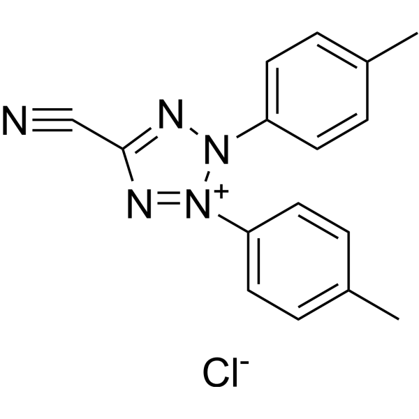
5-Cyano-2,3-di-(p-tolyl)tetrazolium chloride (CTC) is a redox-sensitive red fluorescent dye. 5-Cyano-2,3-di-(p-tolyl)tetrazolium chloride can be used for detecting metabolic activity in microorganisms. The emission maximum of 5-Cyano-2,3-di-(p-tolyl)tetrazolium chloride is 602 nm .
|
-
-
- HY-152073
-
|
|
Fluorescent Dye
|
Others
|
|
BETA-1 is the first twisted intramolecular charge transfer (TICT)-aggregation-induced emission (AIE) integration molecule. BETA-1 emits cyan fluorescence in lipid droplets (LDs) and red fluorescence in mitochondria. BETA-1 can be used for the simultaneous and dual-color imaging of LDs and mitochondria in vivo and in vitro .
|
-
-
- HY-104056
-
|
|
Fluorescent Dye
|
Others
|
|
Fura Red is a Ca 2+-sensitive fluorescent dye, which decreases in fluorescence with rising [Ca 2+] .
|
-
-
- HY-D2346
-
|
|
Fluorescent Dye
|
Others
|
|
HBmito Crimson is a deep red fluorescent probe (λex: 658 nm, λem: 678 nm) for the inner mitochondrial membrane. HBmito Crimson is a cell membrane-permeable probe with high selectivity for the mitochondrial inner membrane, suitable for specific fluorescence staining of the inner mitochondrial membrane in living cells. HBmito Crimson has high photostability and brightness, suitable for long-term dynamic fluorescence imaging.
|
-

-
- HY-D2166
-
|
|
Fluorescent Dye
|
Others
|
|
AF 594 NHS ester is a derivative of the red fluorescent dye AF 594 with high fluorescence quantum yield and high photostability (Ex=594 nm, Em=615 nm). AF 594 NHS ester can form an ester bond by reacting the NHS group with ammonia, with maximum excitation wavelength of 594 nm .
|
-
-
- HY-42984
-
|
|
Biochemical Assay Reagents
|
Others
|
|
BHQ-2 NHS is a dark quencher with no native emission due to the polyaromatic-azo backbone and a terminal NHS ester. UBHQ-2 NHS has a wide and intense quenching range from 560-670 nm, which makes it useful as an acceptor in fluorescence resonance energy transfer (FRET) applications in conjunction with orange to far-red emitting dyes. The NHS ester can be applied to label the primary amines (-NH2) of proteins, amine-modified oligonucleotides, and other amine-containing molecules.
|
-

-
- HY-153843
-
|
|
Biochemical Assay Reagents
|
Others
|
|
RNA Aptamer Corn (sodium) is a 28-nt-long aptamer that is substantially shorter than Spinach and Spinach2 and exhibits bright red fluorescence upon binding DFHO (a soluble analog of the intrinsic fluorophore of red fluorescent protein), RNA Aptamer Corn (sodium) can be used to visualize RNA expression or localization in live cells which have been soaked with chromophores. The Corn-DFHO does not become appreciably cytotoxic when illuminated. And most importantly, Corn-DFHO exhibits markedly increased photostability compared to other aptamer-chromophore complexes both in vitro and in vivo. (36 nt Corn construct: 5'-GGCGCGAGGAAGGAGGUCUGAGGAGGUCACUGCGCC-3'; A 36-nt RNA construct, comprised of the 28-nt minimal Corn sequence extended proximally with a 4 base-pair stem.)
|
-

-
- HY-D1818
-
|
|
Fluorescent Dye
|
Others
|
|
Vari Fluor 680-Phalloidin is a fluorescent derivative of Phalloidin that specifically labels myofilament proteins and exhibits red fluorescence when labeled at 680/700 nm .
|
-

-
- HY-D1723
-
|
|
DNA Stain
|
Others
|
|
EthD-III is a nucleic acid probe. EthD-III is a red fluorescent stain that can be used to detect dead cells. EthD-III enters cells with damaged membranes and binds to nucleic acids, resulting in bright red fluorescence in dead cells (Ex/Em=530/645 nm) .
|
-
-
- HY-P2270Y
-
|
|
Arp2/3 Complex
Fluorescent Dye
|
Others
|
|
Phalloidin-TRITC (solution) is a fluorescein derivative of Phalloidin, which can specifically label myof lin and display red fluorescence when labeled and can be observed using Tesred channels .
|
-

-
- HY-158218
-
|
Gelatin Methacryloyl, 30% methacrylation, red Fluorescent
|
Biochemical Assay Reagents
|
Others
|
Red Fluorescent Gelatin Methacryloyl (Red Fluorescent GelMA) is methacryloyl gelatin (GelMA) with red fluorescence, which is obtained by "grafting" fluorescent molecules on GelMA. Red Fluorescent Gelatin Methacryloyl acts as a scaffold and can be used to engineer tissue analogs from the vasculature to cartilage and bone, allowing cells to proliferate and spread . GelMA, 30% methacrylation, Red Fluorescent needs to self-assemble into fibrous hydrogel under the action of photoinitiator LAP (HY-44076), and target bioactive adhesion sites, play an inherent supporting role for tissue cells and biodegradable activity.
Application: cell culture, biological 3D printing, tissue engineering, etc.
|
-

-
- HY-D2175
-
|
|
Fluorescent Dye
|
Others
|
|
AF 555 NHS ester is a bright, orange-red fluorescence probe. AF 555 NHS ester can be used to label primary amines (R-NH2) of proteins, amine-modified oligonucleotides, and other amine-containing molecules (Ex/Em = 552/566 nm) .
|
-
-
- HY-W854659
-
|
Ce6 trisodium
|
Photosensitizer
|
Cancer
|
|
Chlorin e6 Ce6 (trisodium) is a water-soluble derivative of chlorophyll, belonging to the chlorin class of photosensitizers with an absorption wavelength range of 600-670 nm. Chlorin e6 trisodium emits characteristic red fluorescence upon light excitation, enabling real-time identification of tumor boundaries and progression. Chlorin e6 trisodium can be used for the study of photodynamic therapy (PDT) of cancers (bladder cancer) and fluorescence diagnosis of neoplastic lesions .
|
-

-
- HY-D1190
-
|
|
RAR/RXR
|
Others
|
|
DC271 is a RAR agonist and synthetic retinoid that binds to the retinoid-binding site of cellular retinoic acid-binding protein II (CRBP-II). DC271 exhibits solvatochromic fluorescence properties: it produces intense blue-shifted emission in nonpolar environments and weak red-shifted emission in polar environments, and its severe fluorescence quenching in aqueous solutions can be reversed by embedding in the hydrophobic retinoid-binding protein pocket. DC271 enables direct detection of the binding between unlabeled compounds and related retinoid-binding proteins via fluorescence competition assays (Ex/Em = 355 nm/460 nm) .
|
-
- HY-107864
-
|
Tetraiodofluorescein
|
Fluorescent Dye
|
Infection
|
|
Erythrosine B free acid is a visibly red dye with colorimetric and fluorescent properties that serves as an important dye for many Gram-positive and -negative bacteria. Erythrosine B free acid can be used for live/dead determination in both colorimetric and fluorescence-based assays for low, medium and high-throughput experimentation .
|
-

- HY-D2161A
-
|
|
Fluorescent Dye
|
Others
|
|
AF 594 azide (triethylamine) is an azide derivative of the red fluorescent dye AF 594, which has high fluorescence quantum yield and high photostability (maximum absorption wavelength of 586 nm, maximum emission wavelength of 613 nm). AF 594 azide (triethylamine) forms stable adducts by reaction of the azide group with alkynyl derivatives (terminal alkynes and cyclooctyne) .
|
-
- HY-DY1015
-
|
|
Fluorescent Dye
|
Others
|
Dihydroethidium (solution) , also known as DHE, is a peroxide indicator. Dihydroethidium penetrates cell membranes to form a fluorescent protein complex with blue fluoresces. After entering the cells, Dihydroethidium is mainly localized in the cell membrane, cytoplasm and nucleus, and the staining effect is the strongest in the nucleus. Dihydroethidium produces inherent blue fluorescence with a maximum excitation wavelength of 370 nm and a maximum emission wavelength of 420 nm; after dehydrogenation, Dihydroethidium combines with RNA or DNA to produce red fluorescence with a maximum excitation wavelength of 300 nm and a maximum emission wavelength of 610 nm. 535 nm can also be used as the excitation wavelength for actual observation .
Solvent and concentration: DMSO: 5 mM
|
-

- HY-P2496
-
|
|
Endothelin Receptor
|
Cardiovascular Disease
|
|
Endothelin 1 (swine, human), Alexa Fluor 488-labeled is a synthetic Endothelin 1 peptide labled with Alexa Fluor 488. Endothelin 1 (swine, human) is a synthetic peptide with the sequence of human and swine Endothelin 1, which is a potent endogenous vasoconstrictor. Endothelin 1 acts through two types of receptors ETA and ETB .
|
-

- HY-173308
-
|
|
Fluorescent Dye
|
Others
|
|
QSY-21 is a fluorescence quencher. QSY-21 possesses broad absorption in far red and NIR range, and can quench fluorescence of dyes that emit in this region. This is a carboxylic acid derivative. QSY-21 has intense absorption maximum at 661 nm, making it useful as an acceptor in fluorescence resonance energy transfer (FRET) applications. It is a common quencher for Cyanine5, Cyanine5.5, AF 647, or other spectrally similar fluorescent dyes.
|
-

- HY-158218A
-
|
Gelatin Methacryloyl, 60% methacrylation, red fluorescent
|
Biochemical Assay Reagents
|
Others
|
Red Fluorescent Gelatin Methacryloyl (Red Fluorescent GelMA) is methacryloyl gelatin (GelMA) with red fluorescence, which is obtained by "grafting" fluorescent molecules on GelMA. Red Fluorescent Gelatin Methacryloyl acts as a scaffold and can be used to engineer tissue analogs from the vasculature to cartilage and bone, allowing cells to proliferate and spread . GelMA, 60% methacrylation, Red Fluorescent needs to self-assemble into fibrous hydrogel under the action of photoinitiator LAP (HY-44076), and target bioactive adhesion sites, play an inherent supporting role for tissue cells and biodegradable activity.
Application: cell culture, biological 3D printing, tissue engineering, etc.
|
-

- HY-158218B
-
|
Gelatin Methacryloyl, 90% methacrylation, red fluorescent
|
Biochemical Assay Reagents
|
Others
|
Red Fluorescent Gelatin Methacryloyl (Red Fluorescent GelMA) is methacryloyl gelatin (GelMA) with red fluorescence, which is obtained by "grafting" fluorescent molecules on GelMA. Red Fluorescent Gelatin Methacryloyl acts as a scaffold and can be used to engineer tissue analogs from the vasculature to cartilage and bone, allowing cells to proliferate and spread . GelMA, 90% methacrylation, Red Fluorescent needs to self-assemble into fibrous hydrogel under the action of photoinitiator LAP (HY-44076), and target bioactive adhesion sites, play an inherent supporting role for tissue cells and biodegradable activity.
Application: cell culture, biological 3D printing, tissue engineering, etc.
|
-

- HY-D2444
-
|
|
Fluorescent Dye
|
Cancer
|
|
AF555 NHS is a red fluorescent dye with excellent fluorescence properties and light stability. The excitation wavelength is 556 nm and the emission wavelength is 571 nm, which can be used for protein labeling, antibody labeling, and cell imaging .
|
-

- HY-D2396
-
|
Sulfo SMCC R-PE
|
Fluorescent Dye
|
Cancer
|
|
Sulfo SMCC R-phycoerythrin is a conjugate composed of the protein crosslinker SMCC (HY-42360) and R-PE (R-Phycoerythrin) (HY-D0988) that can be used to label proteins to make them carry red fluorescence. Among them, SMCC is able to engage antigen-coupled spleen cells to induce antigen-specific immune responses .
|
-

- HY-W127781
-
|
|
Fluorescent Dye
|
Others
|
|
Rhod-2 triammonium is a cell impermeant, red fluorescent calcium indicator. Rhod-2 triammonium exhibits a significant shift in fluorescence intensity upon calcium binding (ex max=549 nm; calcium-free v. ex/em max=552/581 nm; calcium-bound). Unlike the UV-excitable indicators Fura-2 and Indo-1 (HY-D0121), there is no accompanying spectral shift .
|
-
- HY-D1671
-
|
|
Fluorescent Dye
|
Others
|
|
TRITC-DHPE is a rhodamine-labeled glycerophosphate ethanolamine lipid, with head groups marked with bright red fluorescent TRITC dye (λEx/λEm=514/580 nm). TRITC-DHPE can be used for membrane fusion assay to trace lipid processing in intracellular phagocytosis. TRITC-DHPE can serves as an energy transfer receptor for NBD, BODIPY and fluorescein lipid probes .
|
-
- HY-D1678
-
|
|
Fluorescent Dye
|
Others
|
|
5(6)-Carboxynaphthofluorescein diacetate is a fluorescent substrate for esterase assays that can be cleaved by intracellular esterases, producing red fluorescence to measure enzyme activity (Ex = 590 nm; Em = 645 nm) .
|
-
- HY-D3000
-
|
|
Fluorescent Dye
|
Metabolic Disease
|
|
NIR-RED ROS-H2O2 Probe is a near-infrared fluorescent probe used for detecting hydrogen peroxide (H₂O₂) and featuring dual-modal fluorescence/photoacoustic imaging capabilities. NIR-RED ROS-H2O2 Probe successfully detects the upregulated fluorescence signal of H₂O₂ in HepG2 cells and a mouse liver injury model. NIR-RED ROS-H2O2 Probe can be used as a biomarker detection tool for drug-induced liver injury (DILI) .
|
-

- HY-123630R
-
|
FD&C red NO. 40 (Standard); CI 16035 (Standard)
|
Fluorescent Dye
Interleukin Related
5-HT Receptor
IFNAR
Reactive Oxygen Species (ROS)
Reference Standards
|
Inflammation/Immunology
|
|
Allura Red AC (Standard) is an analytical standard of Allura Red AC. This product is intended for research and analytical applications. Allura Red AC is a food colorant, appearing as a deep red water-soluble powder or granules, used in various applications such as beverages, syrups, candies, and cereals. Allura Red AC can statically quench the intrinsic fluorescence of HSA. Additionally, Allura Red AC is a 5-hydroxytryptamine (5-HT) pathway-associated pro-inflammatory agent, capable of exacerbating experimental colitis. Allura Red AC holds potential for research in inflammatory bowel disease (IBD), intestinal barrier function, and food additive safety .
|
-

- HY-D1366
-
|
|
Fluorescent Dye
|
|
|
Sulfo-Cyanine5.5 carboxylic acidCI Pigment violet 32 is a water-soluble, far-red emitting fluorophore. Due to its four sulfo groups, this dye has a negative charge at neutral pH and is very hydrophilic. As a cyanine dye, sulfo-Cyanine5.5 shows a very low dependence of fluorescence on pH and a very high extinction coefficient.
|
-
- HY-D2338
-
|
|
Fluorescent Dye
|
Others
|
|
PMBD is a lysosome (Lyso)-targeting fluorescent probe. PMBD selectively and sensitively detects endogenous N-acylethanolamine amidase (NAAA), allowing real-time visual monitoring of endogenous NAAA in living cells. PMBD has a maximum absorption peak at 350 nm. After the metabolism of NAAA, the maximum absorption peak of the product AMBD shifts red to 450 nm, and a significant fluorescence emission signal appears at 550 nm .
|
-

- HY-D1119A
-
|
|
Fluorescent Dye
|
Others
|
|
AF647-NHS ester tripotassium is an analogue of Alexa Fluor 647 (AF647) (HY-D1119). NHS ester can covalently bind to molecules with amino groups (such as proteins, antibodies, etc.). AF647 is a bright, far-red fluorescent dye with an excitation wavelength (λex) of 635 nm (conventional fluorescence detection)/620 nm (instantaneous detection). Storage: protect from light .
|
-
- HY-125959R
-
|
|
Apoptosis
|
Cardiovascular Disease
Neurological Disease
|
|
Ucf-101 (Standard) is the analytical standard of Ucf-101. This product is intended for research and analytical applications. Ucf-101 is a selective and competitive inhibitor of pro-apoptotic protease Omi/HtrA2, with an IC50 of 9.5 μM for His-Omi. Ucf-101 exhibits very little activity against various other serine proteases (IC50>200 μM). Ucf-101 has a natural red fluorescence at 543 nm that is used to monitor its ability to enter mammalian cells. Ucf-101 has a significant cardioprotective effect against MI/R injury and also has certain neuroprotective effect .
|
-

- HY-D2333
-
|
|
Fluorescent Dye
|
Inflammation/Immunology
|
|
RhFNMB is a dualchannel/localization single-molecule fluorescence probe for ATP and HOCl, with independent fluorescence responses in the light red channel with ATP (λex = 520 nm, λem = 586 nm) and deep red channel with HOCl (λex = 620 nm, λem = 688 nm) .
|
-
- HY-D1349
-
|
|
Fluorescent Dye
|
|
|
Bodipy TR alkyneis one of a boron dipyrromethene fluorophore for the ROX (Texas Red) channel. This is a versatile fluorophore that can be used in microscopy, fluorescence polarization measurements, and other applications. This derivative is a terminal alkyne of copper-catalyzed click chemistry.
|
-
- HY-W800808
-
|
|
Fluorescent Dye
|
Others
|
|
ROX azide, 5-isomer is a red-emitting rhodamine dye possessing high brightness and fluorescence quantum yield. The azide group can react with alkyne, BCN, DBCO via Click Chemistry to yield a stable triazole linkage.
|
-

- HY-D2094
-
|
|
Fluorescent Dye
|
Inflammation/Immunology
Cancer
|
|
PerCP Maleimide is a fluorescent dye that reacts with free sulfhydryl groups on proteins. PerCP is a red fluorescence albuminous dye for immunostaining and Maleimide can be leveraged for the preparation of fluorogenic probe, which is mainly used for the specific detection of thiol analytes .
|
-

- HY-D2336
-
|
|
PROTACs
|
Cancer
|
|
PROTAC Aster-A degrader-1 (compound NGF3) is a degrader of the sterol transport protein Aster-A. PROTAC Aster-A degrader-1 can be used as a fluorescence probe. (Red: Aster-A inhibitor, black: linker, Blue: E3 ligase ligand) .
|
-

- HY-W800832
-
|
|
Fluorescent Dye
|
Others
|
|
MB 660R DBCO is a bright and photostable far-red dye that emits fluorescence at about 685 nm in the borderline spectral region between far-red and near-IR. Although the absorption maximum is at around 665 nm, this dye can be sufficiently excited by the 633 or 635 nm laser. MB 660R DBCO is water soluble and pH-insensitive from pH 4 to pH 10. MB 660R DBCO is a rhodamine-based dye, and like rhodamine dyes in general, it is very bright and exceptionally photostable.
|
-

- HY-D2579
-
|
|
Biochemical Assay Reagents
|
Others
|
|
Sulfo DBCO-UBQ-2 is a click chemistry reagent combining a dark quencher, UBQ-2, with a polyaromatic-azo backbone, offering no native emission. UBQ-2 effectively quenches fluorescence in the 560-670 nm range, ideal for qPCR probes and FRET applications with orange to far-red dyes .
|
-

- HY-D2040
-
|
|
Fluorescent Dye
|
Others
|
|
ROX tetrazine is a derivative of ROX (Rhodamine X, Rhodamine 101) dye, a red-emitting fluorophore possessing high brightness and fluorescence quantum yield. This compound contains tetrazine moiety that reacts with trans-cycloalkenes and other strained olefins in inverse electron demand Diels-Alder reaction (IEDDA). The reaction is very quick and specific.
|
-

- HY-D2162
-
|
|
Fluorescent Dye
|
Others
|
|
AF 594 carboxylic acid is a carboxyl derivative of the red fluorescent dye AF 594, which has high fluorescence quantum yield and high photostability (Ex=594 nm, Em=615 nm). AF 594 carboxylic acid can form stable covalent bonds through the reaction of carboxylic acid groups with molecules with amino groups .
|
-
- HY-W440936
-
|
|
Liposome
|
Others
|
|
Stearic acid-PEG5000-Rhodamine is a fatty acid containing PEG polymer which can self assemble in an aqueous solution to form micelles. The polymer can be used to prepare nanoparticles for drug encapsulation. The red dye rhodamine can be easily traced by fluorescence microscopy. Rhodamine has maximum absorption at 570 nm and emission around 595 nm.
|
-

- HY-W440935
-
|
|
Liposome
|
Others
|
|
Stearic acid-PEG3400-Rhodamine is a fatty acid containing PEG polymer which can self assemble in an aqueous solution to form micelles. The polymer can be used to prepare nanoparticles for drug encapsulation. The red dye rhodamine can be easily traced by fluorescence microscopy. Rhodamine has maximum absorption at 570 nm and emission around 595 nm.
|
-
- HY-D2740
-
|
|
Fluorescent Dye
|
Others
|
|
ROX azide, 6-isomer is an alkyne-reactive derivative of ROX (Rhodamine X, Rhodamine 101) dye. ROX is a red-emitting fluorophore possessing high brightness and fluorescence quantum yield. This reagent is a pure 6-isomer. It is used for labeling alkyne and cycloalkyne-containing biomolecules via copper-catalyzed and copper-free click chemistry reactions.
|
-

- HY-155070
-
|
|
DNA/RNA Synthesis
Apoptosis
|
Cancer
|
|
SRE-II, an amide derivative, is an activatable photosensitizer for photodynamic cancer research with decreased fluorescence and photosensitizing capabilities. SRE-II can be further converted into the active photosensitizer SDU Red via carboxylesterase-catalyzed amide bond cleavage. SRE-II induces DNA damage and cell apoptosis in the presence of light. SRE-II can act as a promising theranostic agent for triple-negative breast cancer .
|
-
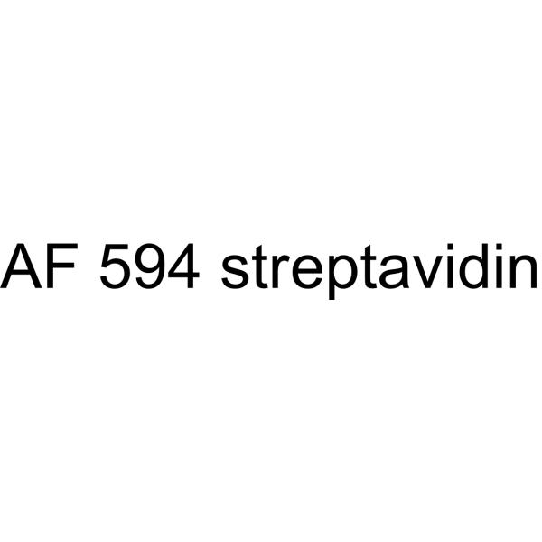
- HY-D2165
-
|
|
Fluorescent Dye
|
Others
|
|
AF 594 streptavidin is a bioconjugating agent. It consists of AF 594 and streptomycin, a streptomycin derivative of the red fluorescent dye AF 594. AF 594 has high fluorescence quantum yield and high photostability (Ex=594 nm, Em=615 nm). AF 594 streptavidin can be selectively conjugated to streptavidin-modified molecules via a streptomycin-modifying group for fluorescent labeling and spectroscopic analysis .
|
-
- HY-D1119B
-
|
|
Fluorescent Dye
|
Others
|
|
AF647-NHS ester (trisodium) is an analogue of Alexa Fluor 647 (AF647) (HY-D1119). NHS ester can covalently bind to molecules with amino groups (such as proteins, antibodies, etc.). AF647 is a bright, far-red fluorescent dye with an excitation wavelength (λex) of 635 nm (conventional fluorescence detection)/620 nm (instantaneous detection). Storage: protect from light .
|
-
- HY-136784A
-
|
|
Fluorescent Dye
|
Others
|
|
Rhod-2 sodium is a cell impermeant, red fluorescent calcium indicator. Rhod-2 sodium exhibits a significant shift in fluorescence intensity upon calcium binding (ex max=549 nm; calcium-free v. ex/em max=552/581 nm; calcium-bound). Unlike the UV-excitable indicators Fura-2 and Indo-1 (HY-D0121), there is no accompanying spectral shift .
|
-

- HY-136784
-
|
|
Fluorescent Dye
|
Others
|
|
Rhod-2 potassium is a cell impermeant, red fluorescent calcium indicator. Rhod-2 potassium exhibits a significant shift in fluorescence intensity upon calcium binding (ex max=549 nm; calcium-free v. ex/em max=552/581 nm; calcium-bound). Unlike the UV-excitable indicators Fura-2 and Indo-1 (HY-D0121), there is no accompanying spectral shift .
|
-

- HY-172309
-
|
|
Fluorescent Dye
|
Others
|
|
UBQ-3 NHS Ester is a fluroescent agent with a terminal NHS ester group. UBQ-3 NHS Ester has a wide quenching range from 620-730 nm, which makes the compound useful as an acceptor in fluorescence resonance energy transfer (FRET) applications in conjunction with far-red to near-IR emitting dyes such as Cy5, Cy5.5, Alexa Fluor 633, 647, 700. The NHS ester can be applied to label the primary amines (-NH2) of proteins, amine-modified oligonucleotides, and other amine-containing molecules.
|
-

- HY-167255
-
|
|
Fluorescent Dye
|
Others
|
|
JC-10 is a lipophilic mitochondrial membrane potential indicator and is a fluorescent dye. JC-10 accumulates and aggregates in healthy mitochondria to emit red fluorescence; exists as a monomer emitting green fluorescence in the cytosol or apoptotic cells with collapsed mitochondrial membrane potential, enabling measurement of mitochondrial depolarization via the green/red fluorescence ratio .
|
-

- HY-D3420
-
|
|
Fluorescent Dye
|
Neurological Disease
|
|
Neuro-DiI is a red retrograde Fluorescent tracer. Neuro-DiI is transported retrogradely to the cell bodies in the ventral tegmental area and labels ventral tegmental area neurons with red fluorescence .
|
-

- HY-D3012
-
|
|
Fluorescent Dye
|
Others
|
|
12-AS is a 9-anthracenoxy fatty acid probe. 12-AS’s fluorescence decay is not a single index, and its lifetime increases with the red shift of the emission wavelength .
|
-

- HY-203233
-
|
|
Fluorescent Dye
|
Others
|
|
Rhodamine-DHPE is a fluorescently labeled phosphatidylethanolamine lipid that labels phospholipid bilayers. Rhodamine-DHPE serves as a fluorescence quenching substrate and membrane stain. The fluorescence lifetime of Rhodamine-DHPE decreases significantly in the presence of Cu 2+-PS complexes. Rhodamine-DHPE effectively stains the membranes of human red blood cells and mouse fibroblasts, and supports lifetime-resolved imaging via pump-probe fluorescence microscopy .
|
-

- HY-D3226
-
|
|
Fluorescent Dye
|
Neurological Disease
|
|
Zinc (II) probe-1 (Compound DNP) is a dual-color Fluorescent probe that can simultaneously monitor Zn 2+ and H +. Upon interaction with Zn 2+, Zinc (II) probe-1 produces bright blue fluorescence (excitation wavelength: 405 nm; blue channel wavelength: 420-500 nm). Upon interaction with H +, Zinc (II) probe-1 exhibits red fluorescence (excitation wavelength: 561 nm; red channel emission wavelength: 630-730 nm). Zinc (II) probe-1 can be used in studies related to depression .
|
-

- HY-D3251
-
|
|
Fluorescent Dye
|
Cancer
|
|
LCP is a fluorescent probe applicable for subcellular localization. LCP responds to polarity changes in the cellular microenvironment via fluorescence resonance energy transfer, emitting blue fluorescence in low-polarity environments and red fluorescence in high-polarity environments. LCP enables dual-color visualization of dynamic changes in lysosomes and cytoplasmic membranes during drug-induced cell apoptosis, and monitors cell viability through localization and emission color changes. LCP can be used in cancer research .
|
-

- HY-D3275
-
|
|
Fluorescent Dye
|
Others
|
|
PE-Cy5.5 is a far-red emitting tandem dye designed based on the principle of fluorescence resonance energy transfer (FRET), and is widely used in multicolor flow cytometry. PE-Cy5.5 consists of phycoerythrin (PE) as the energy donor and the cyanine dye Cy5.5 as the energy acceptor. Upon excitation by blue or green laser light, PE absorbs energy and transfers it to Cy5.5 via FRET, ultimately resulting in Cy5.5 emitting characteristic far-red fluorescence (Ex/Em = 450-500 nm/698 nm) .
|
-

- HY-107864A
-
|
Tetraiodofluorescein aluminum
|
Fluorescent Dye
|
Infection
|
|
Erythrosine B aluminum is a visibly red dye with colorimetric and fluorescent properties that serves as an important dye for many Gram-positive and -negative bacteria. Erythrosine B aluminum can be used for live/dead determination in both colorimetric and fluorescence-based assays for low, medium and high-throughput experimentation .
|
-

- HY-D2755
-
|
|
Fluorescent Dye
|
Others
|
|
BP Light 650 carboxylic acid is an vibrant far-red fluorochrome with comparable or improved performance over other dyes, including BP Fluor 647 and Cy5 dye, for fluorescent applications. It is used to label antibodies and other proteins as molecular probes for cellular imaging and other fluorescence detection methods application.
|
-

- HY-D3230
-
|
|
Fluorescent Dye
|
Others
|
|
FLCS1 is a BODIPY-based Fluorescent probe and selective copper (I) ion binder (λex = 630 nm, λemis = 660 nm). Binding of FLCS1 to copper (I) ions induces fluorescence turn-on. Addition of copper (I) to a methanolic solution of FLCS1 causes a slight red shift (4 nm) in the maximum absorption wavelength (λmax) in the UV-Vis spectrum .
|
-

- HY-D0988A
-
|
R-PE (concentrated solution)
|
Fluorescent Dye
Apoptosis
|
Others
|
|
R-Phycoerythrin (R-PE) (concentrated solution) is found in Heterosiphonia japonica. R-Phycoerythrin (concentrated solution) is an orange-red fluorescent probe with α, β, and γ subunits. R-Phycoerythrin (concentrated solution) can be used in photodynamic therapy (PDT) to induce apoptosis in tumor cells. R-Phycoerythrin (concentrated solution) can be used in fluorescence microscopy, flow cytometry, and immunofluorescence analysis (Ex/Em = 496/578 nm) .
|
-

- HY-107864R
-
|
Tetraiodofluorescein (Standard)
|
Reference Standards
Fluorescent Dye
|
Infection
|
|
Erythrosine B free acid (Standard) is the analytical standard of Erythrosine B (free acid) (HY-107864). This product is intended for research and analytical applications. Erythrosine B free acid is a visibly red dye with colorimetric and fluorescent properties that serves as an important dye for many Gram-positive and -negative bacteria. Erythrosine B free acid can be used for live/dead determination in both colorimetric and fluorescence-based assays for low, medium and high-throughput experimentation .
|
-

- HY-D0988B
-
|
R-PE ammonium sulfate precipitate
|
Fluorescent Dye
Apoptosis
|
Others
|
|
R-Phycoerythrin (R-PE) (ammonium sulfate precipitate) is found in Heterosiphonia japonica. R-Phycoerythrin (ammonium sulfate precipitate) is an orange-red fluorescent probe with α, β, and γ subunits. R-Phycoerythrin (concentrated solution) can be used in photodynamic therapy (PDT) to induce apoptosis in tumor cells. R-Phycoerythrin (ammonium sulfate precipitate) can be used in fluorescence microscopy, flow cytometry, and immunofluorescence analysis (Ex/Em = 496/578 nm) .
|
-

- HY-D3404
-
|
|
DNA Stain
|
Others
|
|
BODi-1 is a fluorescent modulator targeting dsDNA, which binds to dsDNA via a bis-intercalation mechanism (Ex=465 nm, Em=490 nm). BODi-1 exhibits a fluorescence enhancement effect upon binding to nucleic acids, but its fluorescence intensity, anisotropy and average lifetime decrease at higher dye/DNA ratios. When BODi-1 binds to DNA in liposome complexes, it also shows red-shifted emission spectra, along with reduced quantum yield and average lifetime. BODi-1 does not induce significant DNA conformational changes when the dye/DNA ratio is below 0.01. BODi-1 can be used as a fluorescent probe for the characterization of liposome complexes and FRET studies at this ratio .
|
-

- HY-D2161
-
|
|
Fluorescent Dye
|
Others
|
|
AF 594 azide is an azide derivative of the red fluorescent dye AF 594, which has high fluorescence quantum yield and high photostability (maximum absorption wavelength of 586 nm, maximum emission wavelength of 613 nm). AF 594 azide forms stable adducts by reaction of the azide group with alkynyl derivatives (terminal alkynes and cyclooctyne). It contains an azide group and can undergo copper-catalyzed azide-alkyne cycloaddition (CuAAc) with molecules containing alkyne groups. It can also undergo ring strain-promoted alkyne-azide cycloaddition (SPAAC) with molecules containing DBCO or BCN groups .
|
-
- HY-D2365
-
|
|
Fluorescent Dye
|
Others
|
|
QSY 21 NHS is a dark quencher and an efficient energy transfer acceptor for far-red and near-infrared fluorescent probes. QSY 21 NHS operates at a wavelength range of 540-750 nm and is commonly used in FRET applications. QSY 21 NHS does not fluoresce under normal conditions. The NHS ester can be used to label primary amines (R-NH2) of proteins, amine-modified oligonucleotides and other amine-containing molecules. QSY 21 NHS can be conjugated with dendritic poly-L-lysine to achieve intramolecular quenching of Cy5 fluorescence .
|
-

- HY-D3182
-
|
|
Fluorescent Dye
Aldehyde Dehydrogenase (ALDH)
|
Cancer
|
|
AldeRed 588-A is a fluorescent labeling reagent and a substrate for aldehyde dehydrogenase (ALDH). AldeRed 588-A is metabolized by functionally active ALDH enzymes, thereby specifically labeling viable ALDH bright cell populations with red-shifted fluorescence. AldeRed 588-A supports one-step isolation and sorting of ALDH-expressing cells (including normal stem cells and cancer stem cells), and can be used in combination with green fluorophores for multicolor experimental applications. AldeRed 588-A is widely applicable to research related to various cancers such as bladder cancer, breast cancer, and head and neck cancer .
|
-

- HY-D0952
-
|
|
Parasite
|
Others
|
|
Acridine Orange base is a cell-permeable fluorescent dye that stains organisms (bacteria, parasites, viruses, etc.) bright orange and, when used under appropriate conditions (pH=3.5, Ex=460 nm), distinguishes human cells in green for detection by fluorescence microscopy. Acridine Orange base fluoresces green when bound to dsDNA (Ex=488, Em=520-524) and red when bound to ssDNA (Ex=457, Em=630-644) or ssRNA (Ex=457, Em=630-644), also can be used in cell cycle assays .
|
-
- HY-B2235B
-
|
L-α-Phosphatidylcholine (egg yolk, Type XVI-E), 99%, lyophilized powder; 1,2-Diacyl-sn-glycero-3-phosphocholine (egg yolk, Type XVI-E), 99%; egg yolk Lecithins, Type XVI-E, 99%
|
Environmental Pollutants
Biochemical Assay Reagents
Liposome
|
Others
|
|
L-α-Lecithin (egg yolk, Type XVI-E), 99% (L-α-Phosphatidylcholine (egg yolk, Type XVI-E), 99%) is an active biomaterial. L-α-Lecithin (egg yolk, Type XVI-E), 99% forms liposomes with compounds (PF or BA). L-α-Lecithin (egg yolk, Type XVI-E), 99% increases membrane fluidity and affects microemulsion stability and fluorescence intensity stained with Nile red (HY-D0718). L-α-Lecithin (egg yolk, Type XVI-E), 99% It can be used for cell membrane structure research, biological membrane potential research, and liposome research .
|
-

- HY-B1247
-
|
PPIX
|
Endogenous Metabolite
|
Others
|
|
Protoporphyrin IX is a final intermediate in the heme biosynthetic pathway, which acts as a radiation sensitizer enhancing ROS generation even in a hypoxic state and inducing DNA damage. Protoporphyrin IX also acts as a photo sensitizer undergoing photobleaching that occurs through direct degradation by light irradiation. Protoporphyrin IX is formed and accumulated following 5-aminolevulinic acid (5-ALA) (HY-W000450) administration in the tumor cells of rats. Protoporphyrin IX causes selective improvement of basal cell carcinoma when activated red fluorescence of a peak wavelength at 405 nm. Protoporphyrin IX is promising for research of sonodynamic and photodynamic agents for a wide range of cancers, such as bladder cancer and nodular basal cell carcinoma .
|
-
- HY-B1247A
-
|
PPIX disodium
|
Endogenous Metabolite
|
Cancer
|
|
Protoporphyrin IX disodium is a final intermediate in the heme biosynthetic pathway, which acts as a radiation sensitizer enhancing ROS generation even in a hypoxic state and inducing DNA damage. Protoporphyrin IX disodium also acts as a photo sensitizer undergoing photobleaching that occurs through direct degradation by light irradiation. Protoporphyrin IX disodium is formed and accumulated following 5-aminolevulinic acid (5-ALA) (HY-W000450) administration in the tumor cells of rats. Protoporphyrin IX disodium causes selective improvement of basal cell carcinoma when activated red fluorescence of a peak wavelength at 405 nm. Protoporphyrin IX disodium is promising for research of sonodynamic and photodynamic agents for a wide range of cancers, such as bladder cancer and nodular basal cell carcinoma .
|
-

- HY-W014394R
-
|
|
TRP Channel
Reference Standards
Parasite
|
Cardiovascular Disease
|
|
Protoporphyrin IX (Standard) is the analytical standard of Protoporphyrin IX. This product is intended for research and analytical applications. Protoporphyrin IX is a final intermediate in the heme biosynthetic pathway, which acts as a radiation sensitizer enhancing ROS generation even in a hypoxic state and inducing DNA damage. Protoporphyrin IX also acts as a photo sensitizer undergoing photobleaching that occurs through direct degradation by light irradiation. Protoporphyrin IX is formed and accumulated following 5-aminolevulinic acid (5-ALA) (HY-W000450) administration in the tumor cells of rats. Protoporphyrin IX causes selective improvement of basal cell carcinoma when activated red fluorescence of a peak wavelength at 405 nm. Protoporphyrin IX is promising for research of sonodynamic and photodynamic agents for a wide range of cancers, such as bladder cancer and nodular basal cell carcinoma .
|
-

- HY-B1247R
-
|
PPIX (Standard)
|
Reference Standards
Endogenous Metabolite
|
Others
|
|
Protoporphyrin IX (Standard) is the analytical standard of Protoporphyrin IX. This product is intended for research and analytical applications. Protoporphyrin IX is a final intermediate in the heme biosynthetic pathway, which acts as a radiation sensitizer enhancing ROS generation even in a hypoxic state and inducing DNA damage. Protoporphyrin IX also acts as a photo sensitizer undergoing photobleaching that occurs through direct degradation by light irradiation. Protoporphyrin IX is formed and accumulated following 5-aminolevulinic acid (5-ALA) (HY-W000450) administration in the tumor cells of rats. Protoporphyrin IX causes selective improvement of basal cell carcinoma when activated red fluorescence of a peak wavelength at 405 nm. Protoporphyrin IX is promising for research of sonodynamic and photodynamic agents for a wide range of cancers, such as bladder cancer and nodular basal cell carcinoma .
|
-

- HY-D0996
-
|
|
DNA Stain
|
Others
|
|
Lds-751 is a nucleic acid stain that mainly detects DNA. Lds-751 is a nucleic acid stain that mainly detects DNA. Lds-751 has a high affinity for DNA and fluorescence is enhanced after binding, but the maximum emission wavelength is 670nm. Lds-751 and Thiazole orange can be used for the differentiation of red blood cells, platelets, reticulocytes, and nucleated cells and can be stimulated at 488nm. Studies have shown that LDS-751 binds almost exclusively to mitochondria when incubated with nucleated living cells. After nucleated Acridine Orange (HY-101879) staining and LDS-751 treatment of cells, confocal microscopy revealed almost no co-location of the cells. Staining with Rhodamine 123 (HY-D0816), a dye known to bind polarized mitochondria, was almost identical to the pattern observed with LDS-751 .
|
-
- HY-D1737
-
|
|
Bacterial
|
Infection
|
|
RADA is a fluorescent D-amino acid (FDAA) with high photostability and thermostability, which emits yellow-to-orange fluorescence. RADA shows low outer membrane permeability in wild-type Gram-negative Escherichia coli, but it targets penicillin-binding proteins and L,D-transpeptidases, mimics the interaction between acyl acceptors and enzyme intermediates, and integrates into peptidoglycan during biosynthesis. As a peptidoglycan labeling reagent, RADA metabolically integrates into the nascent peptidoglycan of live bacterial cells, labels the peptidoglycan at the poles and lateral walls of mycobacteria, and enables visualization of peptidoglycan synthesis and remodeling processes. RADA serves as a non-specific stain for fixed cells, is non-toxic to bacterial cells, and its red-shifted excitation/emission spectra reduce phototoxicity. RADA also supports virtual pulse-chase labeling experiments and stochastic optical reconstruction microscopy for sub-diffraction-limited imaging of bacterial cell walls .
|
-

- HY-D3187
-
|
|
Fluorescent Dye
Glycosidase
|
Infection
Cancer
|
|
HMRef-αMan is a substrate-based green fluorescent probe (Ex/Em=465 nm/515 nm) targeting MAN2C1 (α-mannosidase). HMRef-αMan can be specifically cleaved by MAN2C1 to generate a highly fluorescent product, which thus gets activated to produce green fluorescence in malignant breast tissues, benign lesions and living cancer cells. The signal intensity of HMRef-αMan is directly correlated with MAN2C1 activity, and it can effectively detect tiny breast cancer lesions with a diameter of less than 1 mm. When used in combination with the red-emitting γ-glutamyl transpeptidase (GGT) probe gGlu-2OMe SiR600 (HY-D3188), HMRef-αMan enables precise optical differentiation of breast tissue types via a dual-color imaging strategy. HMRef-αMan has been widely used in the research of breast diseases such as breast cancer, fibroadenoma, phyllodes tumor and various types of papilloma .
|
-

| Cat. No. |
Product Name |
Type |
-
- HY-D1055
-
MitoSOX Red
Maximum Cited Publications
263 Publications Verification
|
Fluorescent Dye
|
MitoSOX Red is a live cell fluorescent probe that specifically targets mitochondria and is cell membrane permeable. MitoSOX Red enters mitochondria and is oxidized by superoxide but not by other ROS or RNS generating systems. The oxidized MitoSOX Red then binds to nucleic acids in mitochondria/nucleus, producing strong red fluorescence. MitoSOX Red can be used as a fluorescent indicator to specifically detect superoxide. In addition, superoxide dismutase (SOD) can prevent the oxidation of MitoSOX Red.
Excitation/emission wavelength: 510/580 nm.
|
-
- HY-D0815
-
|
|
Fluorescent Dye
|
|
Propidium Iodide (PI) is a nuclear staining agent that stains DNA. Propidium Iodide is an analogue of ethidine bromide that emits red fluorescence upon embedding in double-stranded DNA. Propidium Iodide cannot pass through living cell membranes, but it can pass through damaged cell membranes to stain the nucleus. Propidium Iodide has a fluorescence wavelength of 493/617 nm and a wavelength of 536/635 nm after Mosaic with DNA. Propidium Iodide is commonly used in the detection of apoptosis (apoptosis) or necrosis (necrosis), and is often used in flow cytometry analysis.
|
-
- HY-15534
-
|
CBIC2
|
Fluorescent Dye
|
|
JC-1 (CBIC2) is an ideal fluorescent probe widely used to detect mitochondrial membrane potential. JC-1 accumulates in mitochondria in a potential dependent manner and can be used to detect the membrane potential of cells, tissues or purified mitochondria. In normal mitochondria, JC-1 aggregates in the mitochondrial matrix to form a polymer, which emits strong red fluorescence (Ex=585 nm, Em=590 nm); When the mitochondrial membrane potential is low, JC-1 cannot aggregate in the matrix of mitochondria and produce green fluorescence (ex=510 nm, em= 527 nm) .
|
-
- HY-D0718
-
|
Nile Blue A oxazone; Phenoxazone 9
|
Fluorescent Dye
|
|
Nile red (Nile blue oxazone) is a lipophilic stain. Nile red has environment-sensitive fluorescence. Nile red is intensely fluorescent in a lipid-rich environment while it has minimal fluorescence in aqueous media. Nile red is an excellent vital stain for the detection of intracellular lipid droplets by fluorescence microscopy and flow cytof uorometry. Nile red stains intracellular lipid droplets red. The fluorescence wavelength is 559/635 nm .
|
-
- HY-D0079
-
|
Hydroethidine; PD-MY 003
|
Fluorescent Dye
|
|
Dihydroethidium, also known as DHE, is a peroxide indicator. Dihydroethidium penetrates cell membranes to form a fluorescent protein complex with blue fluoresces. After entering the cells, Dihydroethidium is mainly localized in the cell membrane, cytoplasm and nucleus, and the staining effect is the strongest in the nucleus. Dihydroethidium produces inherent blue fluorescence with a maximum excitation wavelength of 370 nm and a maximum emission wavelength of 420 nm; after dehydrogenation, Dihydroethidium combines with RNA or DNA to produce red fluorescence with a maximum excitation wavelength of 300 nm and a maximum emission wavelength of 610 nm. 535 nm can also be used as the excitation wavelength for actual observation .
|
-
- HY-D1421
-
|
|
Fluorescent Dye
|
|
PKH67 is a fluorescent cell binding dye with green fluorescence. PKH67 can stain the cell membrane and the Ex/Em is 490/502 nm. PKH67 is often used in combination with the non-specific red fluorescent dye PKH26 (Ex/Em=551/567 nm) to label cells, detect cell proliferation in vitro, and trace cells in vitro and in vivo .
|
-
- HY-D1451
-
|
|
Fluorescent Dye
|
|
PKH 26 is a red fluorescent dye, PKH 26 can stably bind to the lipid region of cell membrane and emit red fluorescence (Ex/Em=551/567 nm), which is mainly used for in vitro cell labeling, in vitro cell proliferation studies and in vivo and in vitro cell tracing studies .
|
-
- HY-D2449
-
|
|
Fluorescent Dye
|
|
DQ-BSA-Red is a bovine serum albumin labeled with a red fluorescent dye that can be used to detect lysosomal activity. The excitation wavelength and emission wavelength of DQ-BSA-Red are 590 nm and 620 nm, respectively. The BSA molecule in DQ-BSA-Red is labeled with high concentration of red fluorescent dye in multiple sites, which shows high fluorescence self-inhibition. Once DQ-BSA-RED enters the lysosome, DQ-BSA is cleaved by lysosomal proteases, resulting in unquenched and released fluorescent fragments, emitting bright fluorescence. Inactivated lysosomes are unable to degrade the BSA protein and thus have a lower or even no fluorescent signal .
|
-
- HY-D1119
-
|
|
Fluorescent Dye
|
|
AF647-NHS ester is an analog of Alexa Fluor 647 (AF647). NHS ester can covalently bind to molecules with amino groups (such as proteins, antibodies, etc.). AF647 is a far-red fluorescent dye with an excitation wavelength (λex) of 635 nm (conventional fluorescence detection)/620 nm (instantaneous detection). Storage: Protect from light .
|
-
- HY-D0048
-
|
5-TAMRA-NHS ester; 5-Carboxytetramethylrhodamine succinimidyl ester
|
Fluorescent Dye
|
|
5-TAMRA-SE is an amine-reactive fluorescent agent, and its conjugate produces bright, pH-insensitive orange-red fluorescence with good photostability (Ex/Em = 565/580 nm).
|
-
- HY-D1373
-
HBC
3 Publications Verification
HBC 530
|
Fluorescent Dye
|
|
HBC (HBC 530) is a GFP fluorophore-like synthetic dye, with a structurally rigid electron acceptor and a strong electron donor. HBC has a low fluorescence background, and when combined with Pepper (RNA aptamer), HBC forms a tight complex and activates and emits bright fluorescence (Kd of ~3.5 nM). HBC emission peaks vary in different complexes and covers the spectrum from cyan to red. HBC can be used in the live cell imaging of RNA (Em/Ex = 530/485 nm) .
|
-
- HY-D0988
-
|
R-PE
|
Fluorescent Dye
|
|
R-Phycoerythrin is found in Heterosiphonia japonica. R-Phycoerythrin is an orange-red fluorescent probe with α, β, and γ subunits. R-Phycoerythrin can be used in photodynamic therapy (PDT) to induce apoptosis in tumor cells. R-Phycoerythrin can be used in fluorescence microscopy, flow cytometry, and immunofluorescence analysis (Ex: 495 nm).
|
-
- HY-15942
-
5-TAMRA
3 Publications Verification
|
Fluorescent Dye
|
|
5-TAMRA can produce bright, pH-insensitive orange-red fluorescence (excitation and emission extremes of 546/579) and has good photostability. 5-TAMRA is mainly used as a fluorescent marker for the synthesis and study of specific oligonucleotide probes .
|
-
- HY-D0952
-
|
|
Fluorescent Dye
|
|
Acridine Orange base is a cell-permeable fluorescent dye that stains organisms (bacteria, parasites, viruses, etc.) bright orange and, when used under appropriate conditions (pH=3.5, Ex=460 nm), distinguishes human cells in green for detection by fluorescence microscopy. Acridine Orange base fluoresces green when bound to dsDNA (Ex=488, Em=520-524) and red when bound to ssDNA (Ex=457, Em=630-644) or ssRNA (Ex=457, Em=630-644), also can be used in cell cycle assays .
|
-
- HY-D1819
-
|
|
Fluorescent Dye
|
|
Vari Fluor 633-Phalloidin is a fluorescent derivative of Phalloidin that specifically labels myofilament proteins and exhibits red fluorescence at 630/650 nm when labeled .
|
-
- HY-123630
-
|
FD&C red NO. 40; CI 16035
|
Fluorescent Dye
|
|
Allura Red AC is a food colorant, appearing as a deep red water-soluble powder or granules, used in various applications such as beverages, syrups, candies, and cereals. Allura Red AC can statically quench the intrinsic fluorescence of HSA. Additionally, Allura Red AC is a 5-hydroxytryptamine (5-HT) pathway-associated pro-inflammatory agent, capable of exacerbating experimental colitis. Allura Red AC holds potential for research in inflammatory bowel disease (IBD), intestinal barrier function, and food additive safety .
|
-
- HY-D0791
-
TRITC
1 Publications Verification
5(6)-Tetramethylrhodamine isothiocyanate
|
Fluorescent Dye
|
|
TRITC (5(6)-Tetramethylrhodamine isothiocyanate) is a commonly used fluorescent dye, belonging to the Rhodamine derivative family. It exhibits red fluorescence properties (Ex/Em ≈ 550/580 nm). TRITC can be used for cell labeling and imaging .
|
-
- HY-D0093
-
|
EthD-1
|
Fluorescent Dye
|
|
Ethidium homodimer (EthD-1) is a high-affinity fluorescent nucleic acid dye commonly used to stain mammals, bacteria, yeast, and fungi. Ethidium homodimer binds to DNA or RNA, enhancing fluorescence more than 30 times. The Ethidium homodimer has a strong positive charge, so it cannot cross cell membranes and stain living cells; But the Ethidium homodimer can cross the disordered region of the dead cell membrane to reach the nucleus and embed the DNA double strand to produce red fluorescence. Therefore, Ethidium homodimer is a relatively sensitive nucleic acid stain that can accurately detect nucleic acids in solution or in decomposing cells. Ethidium homodimer binds DNA, Ex/Em=528/617 nm .
|
-
- HY-D1094
-
|
SNARF 1
|
Fluorescent Dye
|
|
Carboxy-SNARF 1 (5/6-mixture) (SNARF 1) is a fluorescent probe that is sensitive to pH (Ex: 488 nm). Carboxy-SNARF 1 (5/6-mixture) can be used for measurement pH. Carboxy-SNARF 1 (5/6-mixture) exhibits a significant emission shift from yellow-orange (Em: 580 nm) to deep red fluorescence (Em: 640 nm) under acidic and basic conditions (pH=7-8), respectively .
|
-
- HY-D0723
-
|
5(6)-Carboxytetramethylrhodamine N-succinimidyl ester
|
Fluorescent Dye
|
|
5(6)-TAMRA SE is a fluorescent dye that emits red fluorescence. 5(6)-TAMRA SE binds to oligonucleotides and is used in DNA sequencing. 5(6)-TAMRA SE can be used in cancer research (Ex/Em = 565/580 nm) .
|
-
- HY-DY1008
-
|
|
Fluorescent Dye
|
Nile Red (solution) is a lipophilic stain. Nile red has environment-sensitive fluorescence. Nile red is intensely fluorescent in a lipid-rich environment while it has minimal fluorescence in aqueous media. Nile red is an excellent vital stain for the detection of intracellular lipid droplets by fluorescence microscopy and flow cytof uorometry. Nile red stains intracellular lipid droplets red. The fluorescence wavelength is 559/635 nm .
Solvent and concentration: DMSO: 1 mM
|
-
- HY-D1816
-
|
|
Fluorescent Dye
|
|
Vari Fluor 555-Phalloidin is a fluorescent derivative of Phalloidin that specifically labels myofilament proteins and exhibits red fluorescence when labeled, allowing for fluorescence imaging using the PE channel (Ex/Em=550 nm/561 nm) .
|
-
- HY-D2865
-
|
|
Fluorescent Dye
|
|
Celltrack Deep Red is a fluorescent dye with a fluorescence signal that can be maintained for at least 72 h and has good stability. Celltrack Deep Red can be used for cell tracing and multi-generation cell movement tracking. Within a cell population, Celltrack Deep Red is only transferred to daughter cells and not to neighboring cells (Ex/Em = 630/650 nm) .
|
-
- HY-P2270
-
|
|
Fluorescent Dye
|
|
Phalloidin-TRITC is a fluorescein derivative of Phalloidin, which can specifically label myof lin and display red fluorescence when labeled and can be observed using Tesred channels .
|
-
- HY-D1742
-
|
|
Fluorescent Dye
|
|
DeepRed Nucleus Dye is a novel cell permeant and far red-fluorescing DNA probe. DeepRed Nucleus Dye excites at a wavelength of 647 nm, close to the Ex, and produces a fluorescence spectrum extending from 665 nm out to beyond 780 nm wavelengths. DeepRed Nucleus Dye fluorescence reflects cellular DNA content. DeepRed Nucleus Dye can be used in combination with FITC and RPE-labelled antibodies, without the need for fluorescence compensation .
|
-
- HY-DY1011
-
|
|
Fluorescent Dye
|
PKH 26 (solution) is a red fluorescent dye, PKH 26 can stably bind to the lipid region of cell membrane and emit red fluorescence (Ex/Em=551/567 nm) , which is mainly used for in vitro cell labeling, in vitro cell proliferation studies and in vivo and in vitro cell tracing studies .
Solvent and concentration: DMSO: 5 mM
|
-
- HY-DY1003
-
|
|
Fluorescent Dye
|
JC-1 (CBIC2) (solution) is an ideal fluorescent probe widely used to detect mitochondrial membrane potential. JC-1 accumulates in mitochondria in a potential dependent manner and can be used to detect the membrane potential of cells, tissues or purified mitochondria. In normal mitochondria, JC-1 aggregates in the mitochondrial matrix to form a polymer, which emits strong red fluorescence (Ex=585 nm, Em=590 nm) ; When the mitochondrial membrane potential is low, JC-1 cannot aggregate in the matrix of mitochondria and produce green fluorescence (ex=510 nm, em= 527 nm) .
Solvent and concentration: DMSO: 1.5 mM
|
-
- HY-DY1006
-
|
|
Fluorescent Dye
|
Propidium Iodide (PI) (solution) is a nuclear staining agent that stains DNA. Propidium Iodide is an analogue of ethidine bromide that emits red fluorescence upon embedding in double-stranded DNA. Propidium Iodide cannot pass through living cell membranes, but it can pass through damaged cell membranes to stain the nucleus. Propidium Iodide has a fluorescence wavelength of 493/617 nm and a wavelength of 536/635 nm after Mosaic with DNA. Propidium Iodide is commonly used in the detection of apoptosis (apoptosis) or necrosis (necrosis) , and is often used in flow cytometry analysis.
Solvent and Concentration: Sterile water: 1 mg/mL
The 1 mL volume is defined as the base specification. All larger sizes correspond to incremental volumes of this base.
|
-
- HY-D0996
-
|
|
Fluorescent Dye
|
|
Lds-751 is a nucleic acid stain that mainly detects DNA. Lds-751 is a nucleic acid stain that mainly detects DNA. Lds-751 has a high affinity for DNA and fluorescence is enhanced after binding, but the maximum emission wavelength is 670nm. Lds-751 and Thiazole orange can be used for the differentiation of red blood cells, platelets, reticulocytes, and nucleated cells and can be stimulated at 488nm. Studies have shown that LDS-751 binds almost exclusively to mitochondria when incubated with nucleated living cells. After nucleated Acridine Orange (HY-101879) staining and LDS-751 treatment of cells, confocal microscopy revealed almost no co-location of the cells. Staining with Rhodamine 123 (HY-D0816), a dye known to bind polarized mitochondria, was almost identical to the pattern observed with LDS-751 .
|
-
- HY-D1359
-
|
|
Fluorescent Dye
|
|
Mito Red is a vital dye and mitochondrial stain that can be used to detect and evaluate mitochondrial function and status. Mito Red accumulates in mitochondria, and its fluorescence intensity is positively correlated with mitochondrial membrane potential. When the mitochondrial membrane potential increases, the fluorescence signal of Mito Red increases .
|
-
- HY-D0917
-
|
|
Fluorescent Dye
|
|
NIR-Red Dead Cell-1 Dye is a DNA-binding fluorescent dye for non-living cells (Ex/Em=515 nm/531 nm). NIR-Red Dead Cell-1 Dye can intercalate into base pairs of double-stranded DNA and produce stronger fluorescence. NIR-Red Dead Cell-1 Dye is suitable for necrotic cells or late apoptotic cells with damaged cell membranes, showing green fluorescence under fluorescence microscopy or flow cytometry. NIR-Red Dead Cell-1 Dye can be used to distinguish live cells from dead cells and distinguish cell membrane integrity. NIR-Red Dead Cell-1 Dye can be attached to the surface of Feraheme (FH) nanoparticles (NPs) to obtain fluorescent dye-functionalized NPs for drug delivery studies .
|
-
- HY-D2365
-
|
|
Fluorescent Dye
|
|
QSY 21 NHS is a dark quencher and an efficient energy transfer acceptor for far-red and near-infrared fluorescent probes. QSY 21 NHS operates at a wavelength range of 540-750 nm and is commonly used in FRET applications. QSY 21 NHS does not fluoresce under normal conditions. The NHS ester can be used to label primary amines (R-NH2) of proteins, amine-modified oligonucleotides and other amine-containing molecules. QSY 21 NHS can be conjugated with dendritic poly-L-lysine to achieve intramolecular quenching of Cy5 fluorescence .
|
-
- HY-D1251
-
|
|
Fluorescent Dye
|
|
TRFS-red, a red fluorescence emission off-on probe, is selective for thioredoxin reductase (TrxR). TRFS-red exhibits high response rate and sensitivity. TRFS-red can be used for imaging live cells .
|
-
- HY-D1820
-
|
|
Fluorescent Dye
|
|
Vari Fluor 594-Phalloidin is a fluorescent derivative of Phalloidin that specifically labels myofilament proteins and exhibits red fluorescence at 585/609 nm when labeled .
|
-
- HY-D0016
-
|
CTC
|
Fluorescent Dye
|
|
5-Cyano-2,3-di-(p-tolyl)tetrazolium chloride (CTC) is a redox-sensitive red fluorescent dye. 5-Cyano-2,3-di-(p-tolyl)tetrazolium chloride can be used for detecting metabolic activity in microorganisms. The emission maximum of 5-Cyano-2,3-di-(p-tolyl)tetrazolium chloride is 602 nm .
|
-
- HY-104056
-
|
|
Fluorescent Dye
|
|
Fura Red is a Ca 2+-sensitive fluorescent dye, which decreases in fluorescence with rising [Ca 2+] .
|
-
- HY-D2346
-
|
|
Fluorescent Dye
|
|
HBmito Crimson is a deep red fluorescent probe (λex: 658 nm, λem: 678 nm) for the inner mitochondrial membrane. HBmito Crimson is a cell membrane-permeable probe with high selectivity for the mitochondrial inner membrane, suitable for specific fluorescence staining of the inner mitochondrial membrane in living cells. HBmito Crimson has high photostability and brightness, suitable for long-term dynamic fluorescence imaging.
|
-
- HY-D2166
-
|
|
Fluorescent Dye
|
|
AF 594 NHS ester is a derivative of the red fluorescent dye AF 594 with high fluorescence quantum yield and high photostability (Ex=594 nm, Em=615 nm). AF 594 NHS ester can form an ester bond by reacting the NHS group with ammonia, with maximum excitation wavelength of 594 nm .
|
-
- HY-42984
-
|
|
Fluorescent Dye
|
|
BHQ-2 NHS is a dark quencher with no native emission due to the polyaromatic-azo backbone and a terminal NHS ester. UBHQ-2 NHS has a wide and intense quenching range from 560-670 nm, which makes it useful as an acceptor in fluorescence resonance energy transfer (FRET) applications in conjunction with orange to far-red emitting dyes. The NHS ester can be applied to label the primary amines (-NH2) of proteins, amine-modified oligonucleotides, and other amine-containing molecules.
|
-
- HY-D1818
-
|
|
Fluorescent Dye
|
|
Vari Fluor 680-Phalloidin is a fluorescent derivative of Phalloidin that specifically labels myofilament proteins and exhibits red fluorescence when labeled at 680/700 nm .
|
-
- HY-P2270Y
-
|
|
Fluorescent Dye
|
|
Phalloidin-TRITC (solution) is a fluorescein derivative of Phalloidin, which can specifically label myof lin and display red fluorescence when labeled and can be observed using Tesred channels .
|
-
- HY-D2175
-
|
|
Fluorescent Dye
|
|
AF 555 NHS ester is a bright, orange-red fluorescence probe. AF 555 NHS ester can be used to label primary amines (R-NH2) of proteins, amine-modified oligonucleotides, and other amine-containing molecules (Ex/Em = 552/566 nm) .
|
-
- HY-D1190
-
|
|
Fluorescent Dye
|
|
DC271 is a RAR agonist and synthetic retinoid that binds to the retinoid-binding site of cellular retinoic acid-binding protein II (CRBP-II). DC271 exhibits solvatochromic fluorescence properties: it produces intense blue-shifted emission in nonpolar environments and weak red-shifted emission in polar environments, and its severe fluorescence quenching in aqueous solutions can be reversed by embedding in the hydrophobic retinoid-binding protein pocket. DC271 enables direct detection of the binding between unlabeled compounds and related retinoid-binding proteins via fluorescence competition assays (Ex/Em = 355 nm/460 nm) .
|
-
- HY-107864
-
|
Tetraiodofluorescein
|
Fluorescent Dye
|
|
Erythrosine B free acid is a visibly red dye with colorimetric and fluorescent properties that serves as an important dye for many Gram-positive and -negative bacteria. Erythrosine B free acid can be used for live/dead determination in both colorimetric and fluorescence-based assays for low, medium and high-throughput experimentation .
|
-
- HY-D2161A
-
|
|
Fluorescent Dye
|
|
AF 594 azide (triethylamine) is an azide derivative of the red fluorescent dye AF 594, which has high fluorescence quantum yield and high photostability (maximum absorption wavelength of 586 nm, maximum emission wavelength of 613 nm). AF 594 azide (triethylamine) forms stable adducts by reaction of the azide group with alkynyl derivatives (terminal alkynes and cyclooctyne) .
|
-
- HY-DY1015
-
|
|
Fluorescent Dye
|
Dihydroethidium (solution) , also known as DHE, is a peroxide indicator. Dihydroethidium penetrates cell membranes to form a fluorescent protein complex with blue fluoresces. After entering the cells, Dihydroethidium is mainly localized in the cell membrane, cytoplasm and nucleus, and the staining effect is the strongest in the nucleus. Dihydroethidium produces inherent blue fluorescence with a maximum excitation wavelength of 370 nm and a maximum emission wavelength of 420 nm; after dehydrogenation, Dihydroethidium combines with RNA or DNA to produce red fluorescence with a maximum excitation wavelength of 300 nm and a maximum emission wavelength of 610 nm. 535 nm can also be used as the excitation wavelength for actual observation .
Solvent and concentration: DMSO: 5 mM
|
-
- HY-173308
-
|
|
Fluorescent Dye
|
|
QSY-21 is a fluorescence quencher. QSY-21 possesses broad absorption in far red and NIR range, and can quench fluorescence of dyes that emit in this region. This is a carboxylic acid derivative. QSY-21 has intense absorption maximum at 661 nm, making it useful as an acceptor in fluorescence resonance energy transfer (FRET) applications. It is a common quencher for Cyanine5, Cyanine5.5, AF 647, or other spectrally similar fluorescent dyes.
|
-
- HY-D2444
-
|
|
Fluorescent Dye
|
|
AF555 NHS is a red fluorescent dye with excellent fluorescence properties and light stability. The excitation wavelength is 556 nm and the emission wavelength is 571 nm, which can be used for protein labeling, antibody labeling, and cell imaging .
|
-
- HY-D2396
-
|
Sulfo SMCC R-PE
|
Fluorescent Dye
|
|
Sulfo SMCC R-phycoerythrin is a conjugate composed of the protein crosslinker SMCC (HY-42360) and R-PE (R-Phycoerythrin) (HY-D0988) that can be used to label proteins to make them carry red fluorescence. Among them, SMCC is able to engage antigen-coupled spleen cells to induce antigen-specific immune responses .
|
-
- HY-D1671
-
|
|
Fluorescent Dye
|
|
TRITC-DHPE is a rhodamine-labeled glycerophosphate ethanolamine lipid, with head groups marked with bright red fluorescent TRITC dye (λEx/λEm=514/580 nm). TRITC-DHPE can be used for membrane fusion assay to trace lipid processing in intracellular phagocytosis. TRITC-DHPE can serves as an energy transfer receptor for NBD, BODIPY and fluorescein lipid probes .
|
- HY-D1678
-
|
|
Fluorescent Dye
|
|
5(6)-Carboxynaphthofluorescein diacetate is a fluorescent substrate for esterase assays that can be cleaved by intracellular esterases, producing red fluorescence to measure enzyme activity (Ex = 590 nm; Em = 645 nm) .
|
- HY-D3000
-
|
|
Fluorescent Dye
|
|
NIR-RED ROS-H2O2 Probe is a near-infrared fluorescent probe used for detecting hydrogen peroxide (H₂O₂) and featuring dual-modal fluorescence/photoacoustic imaging capabilities. NIR-RED ROS-H2O2 Probe successfully detects the upregulated fluorescence signal of H₂O₂ in HepG2 cells and a mouse liver injury model. NIR-RED ROS-H2O2 Probe can be used as a biomarker detection tool for drug-induced liver injury (DILI) .
|
- HY-123630R
-
|
FD&C red NO. 40 (Standard); CI 16035 (Standard)
|
Fluorescent Dye
|
|
Allura Red AC (Standard) is an analytical standard of Allura Red AC. This product is intended for research and analytical applications. Allura Red AC is a food colorant, appearing as a deep red water-soluble powder or granules, used in various applications such as beverages, syrups, candies, and cereals. Allura Red AC can statically quench the intrinsic fluorescence of HSA. Additionally, Allura Red AC is a 5-hydroxytryptamine (5-HT) pathway-associated pro-inflammatory agent, capable of exacerbating experimental colitis. Allura Red AC holds potential for research in inflammatory bowel disease (IBD), intestinal barrier function, and food additive safety .
|
- HY-D1366
-
|
|
Fluorescent Dye
|
|
Sulfo-Cyanine5.5 carboxylic acidCI Pigment violet 32 is a water-soluble, far-red emitting fluorophore. Due to its four sulfo groups, this dye has a negative charge at neutral pH and is very hydrophilic. As a cyanine dye, sulfo-Cyanine5.5 shows a very low dependence of fluorescence on pH and a very high extinction coefficient.
|
- HY-D2338
-
|
|
Fluorescent Dye
|
|
PMBD is a lysosome (Lyso)-targeting fluorescent probe. PMBD selectively and sensitively detects endogenous N-acylethanolamine amidase (NAAA), allowing real-time visual monitoring of endogenous NAAA in living cells. PMBD has a maximum absorption peak at 350 nm. After the metabolism of NAAA, the maximum absorption peak of the product AMBD shifts red to 450 nm, and a significant fluorescence emission signal appears at 550 nm .
|
- HY-D1119A
-
|
|
Fluorescent Dye
|
|
AF647-NHS ester tripotassium is an analogue of Alexa Fluor 647 (AF647) (HY-D1119). NHS ester can covalently bind to molecules with amino groups (such as proteins, antibodies, etc.). AF647 is a bright, far-red fluorescent dye with an excitation wavelength (λex) of 635 nm (conventional fluorescence detection)/620 nm (instantaneous detection). Storage: protect from light .
|
- HY-D2161
-
|
|
Fluorescent Dye
|
|
AF 594 azide is an azide derivative of the red fluorescent dye AF 594, which has high fluorescence quantum yield and high photostability (maximum absorption wavelength of 586 nm, maximum emission wavelength of 613 nm). AF 594 azide forms stable adducts by reaction of the azide group with alkynyl derivatives (terminal alkynes and cyclooctyne). It contains an azide group and can undergo copper-catalyzed azide-alkyne cycloaddition (CuAAc) with molecules containing alkyne groups. It can also undergo ring strain-promoted alkyne-azide cycloaddition (SPAAC) with molecules containing DBCO or BCN groups .
|
- HY-D2333
-
|
|
Fluorescent Dye
|
|
RhFNMB is a dualchannel/localization single-molecule fluorescence probe for ATP and HOCl, with independent fluorescence responses in the light red channel with ATP (λex = 520 nm, λem = 586 nm) and deep red channel with HOCl (λex = 620 nm, λem = 688 nm) .
|
- HY-D1349
-
|
|
Fluorescent Dye
|
|
Bodipy TR alkyneis one of a boron dipyrromethene fluorophore for the ROX (Texas Red) channel. This is a versatile fluorophore that can be used in microscopy, fluorescence polarization measurements, and other applications. This derivative is a terminal alkyne of copper-catalyzed click chemistry.
|
- HY-W800808
-
|
|
Fluorescent Dye
|
|
ROX azide, 5-isomer is a red-emitting rhodamine dye possessing high brightness and fluorescence quantum yield. The azide group can react with alkyne, BCN, DBCO via Click Chemistry to yield a stable triazole linkage.
|
- HY-D2094
-
|
|
Fluorescent Dye
|
|
PerCP Maleimide is a fluorescent dye that reacts with free sulfhydryl groups on proteins. PerCP is a red fluorescence albuminous dye for immunostaining and Maleimide can be leveraged for the preparation of fluorogenic probe, which is mainly used for the specific detection of thiol analytes .
|
- HY-D2336
-
|
|
Fluorescent Dye
|
|
PROTAC Aster-A degrader-1 (compound NGF3) is a degrader of the sterol transport protein Aster-A. PROTAC Aster-A degrader-1 can be used as a fluorescence probe. (Red: Aster-A inhibitor, black: linker, Blue: E3 ligase ligand) .
|
- HY-D2579
-
|
|
Fluorescent Dye
|
|
Sulfo DBCO-UBQ-2 is a click chemistry reagent combining a dark quencher, UBQ-2, with a polyaromatic-azo backbone, offering no native emission. UBQ-2 effectively quenches fluorescence in the 560-670 nm range, ideal for qPCR probes and FRET applications with orange to far-red dyes .
|
- HY-D2040
-
|
|
Fluorescent Dye
|
|
ROX tetrazine is a derivative of ROX (Rhodamine X, Rhodamine 101) dye, a red-emitting fluorophore possessing high brightness and fluorescence quantum yield. This compound contains tetrazine moiety that reacts with trans-cycloalkenes and other strained olefins in inverse electron demand Diels-Alder reaction (IEDDA). The reaction is very quick and specific.
|
- HY-D2162
-
|
|
Fluorescent Dye
|
|
AF 594 carboxylic acid is a carboxyl derivative of the red fluorescent dye AF 594, which has high fluorescence quantum yield and high photostability (Ex=594 nm, Em=615 nm). AF 594 carboxylic acid can form stable covalent bonds through the reaction of carboxylic acid groups with molecules with amino groups .
|
- HY-D2740
-
|
|
Fluorescent Dye
|
|
ROX azide, 6-isomer is an alkyne-reactive derivative of ROX (Rhodamine X, Rhodamine 101) dye. ROX is a red-emitting fluorophore possessing high brightness and fluorescence quantum yield. This reagent is a pure 6-isomer. It is used for labeling alkyne and cycloalkyne-containing biomolecules via copper-catalyzed and copper-free click chemistry reactions.
|
- HY-D2165
-
|
|
Fluorescent Dye
|
|
AF 594 streptavidin is a bioconjugating agent. It consists of AF 594 and streptomycin, a streptomycin derivative of the red fluorescent dye AF 594. AF 594 has high fluorescence quantum yield and high photostability (Ex=594 nm, Em=615 nm). AF 594 streptavidin can be selectively conjugated to streptavidin-modified molecules via a streptomycin-modifying group for fluorescent labeling and spectroscopic analysis .
|
- HY-D1119B
-
|
|
Fluorescent Dye
|
|
AF647-NHS ester (trisodium) is an analogue of Alexa Fluor 647 (AF647) (HY-D1119). NHS ester can covalently bind to molecules with amino groups (such as proteins, antibodies, etc.). AF647 is a bright, far-red fluorescent dye with an excitation wavelength (λex) of 635 nm (conventional fluorescence detection)/620 nm (instantaneous detection). Storage: protect from light .
|
- HY-136784A
-
|
|
Fluorescent Dye
|
|
Rhod-2 sodium is a cell impermeant, red fluorescent calcium indicator. Rhod-2 sodium exhibits a significant shift in fluorescence intensity upon calcium binding (ex max=549 nm; calcium-free v. ex/em max=552/581 nm; calcium-bound). Unlike the UV-excitable indicators Fura-2 and Indo-1 (HY-D0121), there is no accompanying spectral shift .
|
- HY-136784
-
|
|
Fluorescent Dye
|
|
Rhod-2 potassium is a cell impermeant, red fluorescent calcium indicator. Rhod-2 potassium exhibits a significant shift in fluorescence intensity upon calcium binding (ex max=549 nm; calcium-free v. ex/em max=552/581 nm; calcium-bound). Unlike the UV-excitable indicators Fura-2 and Indo-1 (HY-D0121), there is no accompanying spectral shift .
|
- HY-167255
-
|
|
Fluorescent Dye
|
|
JC-10 is a lipophilic mitochondrial membrane potential indicator and is a fluorescent dye. JC-10 accumulates and aggregates in healthy mitochondria to emit red fluorescence; exists as a monomer emitting green fluorescence in the cytosol or apoptotic cells with collapsed mitochondrial membrane potential, enabling measurement of mitochondrial depolarization via the green/red fluorescence ratio .
|
- HY-D3420
-
|
|
Fluorescent Dye
|
|
Neuro-DiI is a red retrograde Fluorescent tracer. Neuro-DiI is transported retrogradely to the cell bodies in the ventral tegmental area and labels ventral tegmental area neurons with red fluorescence .
|
- HY-D3012
-
|
|
Fluorescent Dye
|
|
12-AS is a 9-anthracenoxy fatty acid probe. 12-AS’s fluorescence decay is not a single index, and its lifetime increases with the red shift of the emission wavelength .
|
- HY-203233
-
|
|
Fluorescent Dye
|
|
Rhodamine-DHPE is a fluorescently labeled phosphatidylethanolamine lipid that labels phospholipid bilayers. Rhodamine-DHPE serves as a fluorescence quenching substrate and membrane stain. The fluorescence lifetime of Rhodamine-DHPE decreases significantly in the presence of Cu 2+-PS complexes. Rhodamine-DHPE effectively stains the membranes of human red blood cells and mouse fibroblasts, and supports lifetime-resolved imaging via pump-probe fluorescence microscopy .
|
- HY-D3226
-
|
|
Fluorescent Dye
|
|
Zinc (II) probe-1 (Compound DNP) is a dual-color Fluorescent probe that can simultaneously monitor Zn 2+ and H +. Upon interaction with Zn 2+, Zinc (II) probe-1 produces bright blue fluorescence (excitation wavelength: 405 nm; blue channel wavelength: 420-500 nm). Upon interaction with H +, Zinc (II) probe-1 exhibits red fluorescence (excitation wavelength: 561 nm; red channel emission wavelength: 630-730 nm). Zinc (II) probe-1 can be used in studies related to depression .
|
- HY-D3251
-
|
|
Fluorescent Dye
|
|
LCP is a fluorescent probe applicable for subcellular localization. LCP responds to polarity changes in the cellular microenvironment via fluorescence resonance energy transfer, emitting blue fluorescence in low-polarity environments and red fluorescence in high-polarity environments. LCP enables dual-color visualization of dynamic changes in lysosomes and cytoplasmic membranes during drug-induced cell apoptosis, and monitors cell viability through localization and emission color changes. LCP can be used in cancer research .
|
- HY-D3275
-
|
|
Fluorescent Dye
|
|
PE-Cy5.5 is a far-red emitting tandem dye designed based on the principle of fluorescence resonance energy transfer (FRET), and is widely used in multicolor flow cytometry. PE-Cy5.5 consists of phycoerythrin (PE) as the energy donor and the cyanine dye Cy5.5 as the energy acceptor. Upon excitation by blue or green laser light, PE absorbs energy and transfers it to Cy5.5 via FRET, ultimately resulting in Cy5.5 emitting characteristic far-red fluorescence (Ex/Em = 450-500 nm/698 nm) .
|
- HY-D2755
-
|
|
Fluorescent Dye
|
|
BP Light 650 carboxylic acid is an vibrant far-red fluorochrome with comparable or improved performance over other dyes, including BP Fluor 647 and Cy5 dye, for fluorescent applications. It is used to label antibodies and other proteins as molecular probes for cellular imaging and other fluorescence detection methods application.
|
- HY-D3230
-
|
|
Fluorescent Dye
|
|
FLCS1 is a BODIPY-based Fluorescent probe and selective copper (I) ion binder (λex = 630 nm, λemis = 660 nm). Binding of FLCS1 to copper (I) ions induces fluorescence turn-on. Addition of copper (I) to a methanolic solution of FLCS1 causes a slight red shift (4 nm) in the maximum absorption wavelength (λmax) in the UV-Vis spectrum .
|
- HY-D0988A
-
|
R-PE (concentrated solution)
|
Fluorescent Dye
|
|
R-Phycoerythrin (R-PE) (concentrated solution) is found in Heterosiphonia japonica. R-Phycoerythrin (concentrated solution) is an orange-red fluorescent probe with α, β, and γ subunits. R-Phycoerythrin (concentrated solution) can be used in photodynamic therapy (PDT) to induce apoptosis in tumor cells. R-Phycoerythrin (concentrated solution) can be used in fluorescence microscopy, flow cytometry, and immunofluorescence analysis (Ex/Em = 496/578 nm) .
|
- HY-107864R
-
|
Tetraiodofluorescein (Standard)
|
Fluorescent Dye
|
|
Erythrosine B free acid (Standard) is the analytical standard of Erythrosine B (free acid) (HY-107864). This product is intended for research and analytical applications. Erythrosine B free acid is a visibly red dye with colorimetric and fluorescent properties that serves as an important dye for many Gram-positive and -negative bacteria. Erythrosine B free acid can be used for live/dead determination in both colorimetric and fluorescence-based assays for low, medium and high-throughput experimentation .
|
- HY-D0988B
-
|
R-PE ammonium sulfate precipitate
|
Fluorescent Dye
|
|
R-Phycoerythrin (R-PE) (ammonium sulfate precipitate) is found in Heterosiphonia japonica. R-Phycoerythrin (ammonium sulfate precipitate) is an orange-red fluorescent probe with α, β, and γ subunits. R-Phycoerythrin (concentrated solution) can be used in photodynamic therapy (PDT) to induce apoptosis in tumor cells. R-Phycoerythrin (ammonium sulfate precipitate) can be used in fluorescence microscopy, flow cytometry, and immunofluorescence analysis (Ex/Em = 496/578 nm) .
|
- HY-D3404
-
|
|
Fluorescent Dye
|
|
BODi-1 is a fluorescent modulator targeting dsDNA, which binds to dsDNA via a bis-intercalation mechanism (Ex=465 nm, Em=490 nm). BODi-1 exhibits a fluorescence enhancement effect upon binding to nucleic acids, but its fluorescence intensity, anisotropy and average lifetime decrease at higher dye/DNA ratios. When BODi-1 binds to DNA in liposome complexes, it also shows red-shifted emission spectra, along with reduced quantum yield and average lifetime. BODi-1 does not induce significant DNA conformational changes when the dye/DNA ratio is below 0.01. BODi-1 can be used as a fluorescent probe for the characterization of liposome complexes and FRET studies at this ratio .
|
- HY-D3182
-
|
|
Fluorescent Dye
|
|
AldeRed 588-A is a fluorescent labeling reagent and a substrate for aldehyde dehydrogenase (ALDH). AldeRed 588-A is metabolized by functionally active ALDH enzymes, thereby specifically labeling viable ALDH bright cell populations with red-shifted fluorescence. AldeRed 588-A supports one-step isolation and sorting of ALDH-expressing cells (including normal stem cells and cancer stem cells), and can be used in combination with green fluorophores for multicolor experimental applications. AldeRed 588-A is widely applicable to research related to various cancers such as bladder cancer, breast cancer, and head and neck cancer .
|
- HY-D1737
-
|
|
Fluorescent Dye
|
|
RADA is a fluorescent D-amino acid (FDAA) with high photostability and thermostability, which emits yellow-to-orange fluorescence. RADA shows low outer membrane permeability in wild-type Gram-negative Escherichia coli, but it targets penicillin-binding proteins and L,D-transpeptidases, mimics the interaction between acyl acceptors and enzyme intermediates, and integrates into peptidoglycan during biosynthesis. As a peptidoglycan labeling reagent, RADA metabolically integrates into the nascent peptidoglycan of live bacterial cells, labels the peptidoglycan at the poles and lateral walls of mycobacteria, and enables visualization of peptidoglycan synthesis and remodeling processes. RADA serves as a non-specific stain for fixed cells, is non-toxic to bacterial cells, and its red-shifted excitation/emission spectra reduce phototoxicity. RADA also supports virtual pulse-chase labeling experiments and stochastic optical reconstruction microscopy for sub-diffraction-limited imaging of bacterial cell walls .
|
- HY-D3187
-
|
|
Fluorescent Dye
|
|
HMRef-αMan is a substrate-based green fluorescent probe (Ex/Em=465 nm/515 nm) targeting MAN2C1 (α-mannosidase). HMRef-αMan can be specifically cleaved by MAN2C1 to generate a highly fluorescent product, which thus gets activated to produce green fluorescence in malignant breast tissues, benign lesions and living cancer cells. The signal intensity of HMRef-αMan is directly correlated with MAN2C1 activity, and it can effectively detect tiny breast cancer lesions with a diameter of less than 1 mm. When used in combination with the red-emitting γ-glutamyl transpeptidase (GGT) probe gGlu-2OMe SiR600 (HY-D3188), HMRef-αMan enables precise optical differentiation of breast tissue types via a dual-color imaging strategy. HMRef-αMan has been widely used in the research of breast diseases such as breast cancer, fibroadenoma, phyllodes tumor and various types of papilloma .
|
| Cat. No. |
Product Name |
Type |
-
- HY-B2235B
-
|
L-α-Phosphatidylcholine (egg yolk, Type XVI-E), 99%, lyophilized powder; 1,2-Diacyl-sn-glycero-3-phosphocholine (egg yolk, Type XVI-E), 99%; egg yolk Lecithins, Type XVI-E, 99%
|
Biochemical Assay Reagents
|
|
L-α-Lecithin (egg yolk, Type XVI-E), 99% (L-α-Phosphatidylcholine (egg yolk, Type XVI-E), 99%) is an active biomaterial. L-α-Lecithin (egg yolk, Type XVI-E), 99% forms liposomes with compounds (PF or BA). L-α-Lecithin (egg yolk, Type XVI-E), 99% increases membrane fluidity and affects microemulsion stability and fluorescence intensity stained with Nile red (HY-D0718). L-α-Lecithin (egg yolk, Type XVI-E), 99% It can be used for cell membrane structure research, biological membrane potential research, and liposome research .
|
-
- HY-158218
-
|
Gelatin Methacryloyl, 30% methacrylation, red Fluorescent
|
Biochemical Assay Reagents
|
Red Fluorescent Gelatin Methacryloyl (Red Fluorescent GelMA) is methacryloyl gelatin (GelMA) with red fluorescence, which is obtained by "grafting" fluorescent molecules on GelMA. Red Fluorescent Gelatin Methacryloyl acts as a scaffold and can be used to engineer tissue analogs from the vasculature to cartilage and bone, allowing cells to proliferate and spread . GelMA, 30% methacrylation, Red Fluorescent needs to self-assemble into fibrous hydrogel under the action of photoinitiator LAP (HY-44076), and target bioactive adhesion sites, play an inherent supporting role for tissue cells and biodegradable activity.
Application: cell culture, biological 3D printing, tissue engineering, etc.
|
-
- HY-W854659
-
|
Ce6 trisodium
|
Biochemical Assay Reagents
|
|
Chlorin e6 Ce6 (trisodium) is a water-soluble derivative of chlorophyll, belonging to the chlorin class of photosensitizers with an absorption wavelength range of 600-670 nm. Chlorin e6 trisodium emits characteristic red fluorescence upon light excitation, enabling real-time identification of tumor boundaries and progression. Chlorin e6 trisodium can be used for the study of photodynamic therapy (PDT) of cancers (bladder cancer) and fluorescence diagnosis of neoplastic lesions .
|
-
- HY-158218A
-
|
Gelatin Methacryloyl, 60% methacrylation, red fluorescent
|
Biochemical Assay Reagents
|
Red Fluorescent Gelatin Methacryloyl (Red Fluorescent GelMA) is methacryloyl gelatin (GelMA) with red fluorescence, which is obtained by "grafting" fluorescent molecules on GelMA. Red Fluorescent Gelatin Methacryloyl acts as a scaffold and can be used to engineer tissue analogs from the vasculature to cartilage and bone, allowing cells to proliferate and spread . GelMA, 60% methacrylation, Red Fluorescent needs to self-assemble into fibrous hydrogel under the action of photoinitiator LAP (HY-44076), and target bioactive adhesion sites, play an inherent supporting role for tissue cells and biodegradable activity.
Application: cell culture, biological 3D printing, tissue engineering, etc.
|
-
- HY-158218B
-
|
Gelatin Methacryloyl, 90% methacrylation, red fluorescent
|
Biochemical Assay Reagents
|
Red Fluorescent Gelatin Methacryloyl (Red Fluorescent GelMA) is methacryloyl gelatin (GelMA) with red fluorescence, which is obtained by "grafting" fluorescent molecules on GelMA. Red Fluorescent Gelatin Methacryloyl acts as a scaffold and can be used to engineer tissue analogs from the vasculature to cartilage and bone, allowing cells to proliferate and spread . GelMA, 90% methacrylation, Red Fluorescent needs to self-assemble into fibrous hydrogel under the action of photoinitiator LAP (HY-44076), and target bioactive adhesion sites, play an inherent supporting role for tissue cells and biodegradable activity.
Application: cell culture, biological 3D printing, tissue engineering, etc.
|
-
- HY-W127781
-
|
|
Biochemical Assay Reagents
|
|
Rhod-2 triammonium is a cell impermeant, red fluorescent calcium indicator. Rhod-2 triammonium exhibits a significant shift in fluorescence intensity upon calcium binding (ex max=549 nm; calcium-free v. ex/em max=552/581 nm; calcium-bound). Unlike the UV-excitable indicators Fura-2 and Indo-1 (HY-D0121), there is no accompanying spectral shift .
|
-
- HY-W440936
-
|
|
Biochemical Assay Reagents
|
|
Stearic acid-PEG5000-Rhodamine is a fatty acid containing PEG polymer which can self assemble in an aqueous solution to form micelles. The polymer can be used to prepare nanoparticles for drug encapsulation. The red dye rhodamine can be easily traced by fluorescence microscopy. Rhodamine has maximum absorption at 570 nm and emission around 595 nm.
|
-
- HY-W440935
-
|
|
Biochemical Assay Reagents
|
|
Stearic acid-PEG3400-Rhodamine is a fatty acid containing PEG polymer which can self assemble in an aqueous solution to form micelles. The polymer can be used to prepare nanoparticles for drug encapsulation. The red dye rhodamine can be easily traced by fluorescence microscopy. Rhodamine has maximum absorption at 570 nm and emission around 595 nm.
|
| Cat. No. |
Product Name |
Target |
Research Area |
-
- HY-P2270
-
|
|
Fluorescent Dye
Arp2/3 Complex
|
Others
|
|
Phalloidin-TRITC is a fluorescein derivative of Phalloidin, which can specifically label myof lin and display red fluorescence when labeled and can be observed using Tesred channels .
|
-
- HY-P2270Y
-
|
|
Arp2/3 Complex
Fluorescent Dye
|
Others
|
|
Phalloidin-TRITC (solution) is a fluorescein derivative of Phalloidin, which can specifically label myof lin and display red fluorescence when labeled and can be observed using Tesred channels .
|
-
- HY-P2496
-
|
|
Endothelin Receptor
|
Cardiovascular Disease
|
|
Endothelin 1 (swine, human), Alexa Fluor 488-labeled is a synthetic Endothelin 1 peptide labled with Alexa Fluor 488. Endothelin 1 (swine, human) is a synthetic peptide with the sequence of human and swine Endothelin 1, which is a potent endogenous vasoconstrictor. Endothelin 1 acts through two types of receptors ETA and ETB .
|
-
- HY-K1077
-
2 Publications Verification
|
|
Annexin V-mCherry/SYTOX Green Apoptosis Detection Kit provides a rapid and convenient method to detect cell apoptosis and necrosis. After staining, live cells show little or no fluorescence, apoptosis cells show red fluorescence, necrosis cells show red and green fluorescence.
|
-
- HY-KD1030
-
|
|
|
Tubulin Deep Red is a fluorescent probe with red fluorescence that can specifically label microtubules in living mammalian cells.
|
-
- HY-K1076
-
|
|
|
Annexin V-mCherry Apoptosis Detection Kit provides a rapid and convenient method to detect cell apoptosis and necrosis. After staining, live cells show little or no fluorescence, apoptosis cells and necrosis cells show red fluorescence.
|
-
- HY-K1079
-
|
|
|
MCE One Step TUNEL Apoptosis Detection Kit (Cyanine 3) provides a rapid and convenient method to detect cell apoptosis. After staining cells with this kit, live cells have no fluorescence, apoptosis cells show red fluorescence.
|
-
- HY-K1075
-
1 Publications Verification
|
|
MCE Annexin V-PE Apoptosis Detection Kit provides a rapid and convenient method to detect cell apoptosis and necrosis. After staining, live cells show little or no fluorescence, apoptosis cells and necrosis cells show red fluorescence.
|
-
- HY-K1073
-
Maximum Cited Publications
66 Publications Verification
|
|
MCE Annexin V-FITC/PI Apoptosis Detection Kit provides a rapid and convenient method to detect cell apoptosis and necrosis. After staining, live cells show little or no fluorescence (Annexin V-/PI-), early apoptosis cells show green fluorescence(Annexin V+/PI-), late apoptosis cells and necrosis cells show red and green fluorescence (Annexin V+/PI+).
|
-
- HY-K1080
-
1 Publications Verification
|
|
MCE Annexin V-iFluor 488/PI Apoptosis Detection Kit provides a rapid and convenient method to detect cell apoptosis and necrosis. After staining, live cells show little or no fluorescence (Annexin V-/PI-),, early apoptosis cells show green fluorescence (Annexin V+/PI-), late apoptosis cells and necrosis cells show red and green fluorescence (Annexin V+/PI+).
|
-
- HY-K1092
-
|
|
|
MCE YO-PRO-1/PI Apoptosis and Necrosis Detection Kit is a dual-fluorescence method based on the green fluorescent dye YO-PRO-1 (YP1) and the red fluorescent dye Propidium Iodide (PI) for detecting cell apoptosis and necrosis.
|
-
- HY-KD1112
-
|
|
|
TheBlue Fluorescence Rapid Labelling Kit (AF350) enables red fluorescent labelling of proteins. Maximum excitation/ emission wavelengths: 346/442 nm. Based on NHS ester chemistry, the NHS ester-activated fluorescent dye reacts with primary amines in the antibody/protein to be labelled at pH 7-9, forming stable amide bonds to achieve conjugation. Typically, one IgG molecule can bind 2-8 molecules of AF350. The entire procedure can be completed within two hours.
|
-
- HY-KD1110
-
|
|
|
The Near-Infrared Fluorescence Rapid Labelling Kit (CY7.5) enables red fluorescent labelling of proteins. Maximum excitation/ emission wavelengths: 788/814 nm. Based on NHS ester chemistry, the NHS ester-activated fluorescent dye reacts with primary amines in the antibody/protein to be labelled at pH 7-9, forming stable amide bonds to achieve conjugation. Typically, one IgG molecule can bind 2-8 molecules of CY7.5. The entire procedure can be completed within two hours.
|
| Cat. No. |
Product Name |
Category |
Target |
Chemical Structure |
-
- HY-B1247
-
-
-
- HY-B1247R
-
|
PPIX (Standard)
|
Structural Classification
Human Gut Microbiota Metabolites
Microorganisms
Ketones, Aldehydes, Acids
Endogenous metabolite
Source Classification
|
Reference Standards
Endogenous Metabolite
|
|
Protoporphyrin IX (Standard) is the analytical standard of Protoporphyrin IX. This product is intended for research and analytical applications. Protoporphyrin IX is a final intermediate in the heme biosynthetic pathway, which acts as a radiation sensitizer enhancing ROS generation even in a hypoxic state and inducing DNA damage. Protoporphyrin IX also acts as a photo sensitizer undergoing photobleaching that occurs through direct degradation by light irradiation. Protoporphyrin IX is formed and accumulated following 5-aminolevulinic acid (5-ALA) (HY-W000450) administration in the tumor cells of rats. Protoporphyrin IX causes selective improvement of basal cell carcinoma when activated red fluorescence of a peak wavelength at 405 nm. Protoporphyrin IX is promising for research of sonodynamic and photodynamic agents for a wide range of cancers, such as bladder cancer and nodular basal cell carcinoma .
|
-
-
- HY-W014394R
-
|
|
Structural Classification
Monophenols
other families
Phenols
Plants
Source Classification
|
TRP Channel
Reference Standards
Parasite
|
|
Protoporphyrin IX (Standard) is the analytical standard of Protoporphyrin IX. This product is intended for research and analytical applications. Protoporphyrin IX is a final intermediate in the heme biosynthetic pathway, which acts as a radiation sensitizer enhancing ROS generation even in a hypoxic state and inducing DNA damage. Protoporphyrin IX also acts as a photo sensitizer undergoing photobleaching that occurs through direct degradation by light irradiation. Protoporphyrin IX is formed and accumulated following 5-aminolevulinic acid (5-ALA) (HY-W000450) administration in the tumor cells of rats. Protoporphyrin IX causes selective improvement of basal cell carcinoma when activated red fluorescence of a peak wavelength at 405 nm. Protoporphyrin IX is promising for research of sonodynamic and photodynamic agents for a wide range of cancers, such as bladder cancer and nodular basal cell carcinoma .
|
-
| Cat. No. |
Product Name |
|
Classification |
-
- HY-D2161
-
|
|
|
Azide
|
|
AF 594 azide is an azide derivative of the red fluorescent dye AF 594, which has high fluorescence quantum yield and high photostability (maximum absorption wavelength of 586 nm, maximum emission wavelength of 613 nm). AF 594 azide forms stable adducts by reaction of the azide group with alkynyl derivatives (terminal alkynes and cyclooctyne). It contains an azide group and can undergo copper-catalyzed azide-alkyne cycloaddition (CuAAc) with molecules containing alkyne groups. It can also undergo ring strain-promoted alkyne-azide cycloaddition (SPAAC) with molecules containing DBCO or BCN groups .
|
-
- HY-D1349
-
|
|
|
Alkynes
|
|
Bodipy TR alkyneis one of a boron dipyrromethene fluorophore for the ROX (Texas Red) channel. This is a versatile fluorophore that can be used in microscopy, fluorescence polarization measurements, and other applications. This derivative is a terminal alkyne of copper-catalyzed click chemistry.
|
-
- HY-D2579
-
|
|
|
DBCO
|
|
Sulfo DBCO-UBQ-2 is a click chemistry reagent combining a dark quencher, UBQ-2, with a polyaromatic-azo backbone, offering no native emission. UBQ-2 effectively quenches fluorescence in the 560-670 nm range, ideal for qPCR probes and FRET applications with orange to far-red dyes .
|
-
- HY-D2040
-
|
|
|
Tetrazine
|
|
ROX tetrazine is a derivative of ROX (Rhodamine X, Rhodamine 101) dye, a red-emitting fluorophore possessing high brightness and fluorescence quantum yield. This compound contains tetrazine moiety that reacts with trans-cycloalkenes and other strained olefins in inverse electron demand Diels-Alder reaction (IEDDA). The reaction is very quick and specific.
|
-
- HY-D2740
-
|
|
|
Azide
|
|
ROX azide, 6-isomer is an alkyne-reactive derivative of ROX (Rhodamine X, Rhodamine 101) dye. ROX is a red-emitting fluorophore possessing high brightness and fluorescence quantum yield. This reagent is a pure 6-isomer. It is used for labeling alkyne and cycloalkyne-containing biomolecules via copper-catalyzed and copper-free click chemistry reactions.
|
| Cat. No. |
Product Name |
|
Classification |
-
- HY-153843
-
|
|
|
Aptamers
|
|
RNA Aptamer Corn (sodium) is a 28-nt-long aptamer that is substantially shorter than Spinach and Spinach2 and exhibits bright red fluorescence upon binding DFHO (a soluble analog of the intrinsic fluorophore of red fluorescent protein), RNA Aptamer Corn (sodium) can be used to visualize RNA expression or localization in live cells which have been soaked with chromophores. The Corn-DFHO does not become appreciably cytotoxic when illuminated. And most importantly, Corn-DFHO exhibits markedly increased photostability compared to other aptamer-chromophore complexes both in vitro and in vivo. (36 nt Corn construct: 5'-GGCGCGAGGAAGGAGGUCUGAGGAGGUCACUGCGCC-3'; A 36-nt RNA construct, comprised of the 28-nt minimal Corn sequence extended proximally with a 4 base-pair stem.)
|
-
- HY-W440936
-
|
|
|
Pegylated Lipids
Fluorescent Lipids
|
|
Stearic acid-PEG5000-Rhodamine is a fatty acid containing PEG polymer which can self assemble in an aqueous solution to form micelles. The polymer can be used to prepare nanoparticles for drug encapsulation. The red dye rhodamine can be easily traced by fluorescence microscopy. Rhodamine has maximum absorption at 570 nm and emission around 595 nm.
|
-
- HY-W440935
-
|
|
|
Pegylated Lipids
Fluorescent Lipids
|
|
Stearic acid-PEG3400-Rhodamine is a fatty acid containing PEG polymer which can self assemble in an aqueous solution to form micelles. The polymer can be used to prepare nanoparticles for drug encapsulation. The red dye rhodamine can be easily traced by fluorescence microscopy. Rhodamine has maximum absorption at 570 nm and emission around 595 nm.
|
Your information is safe with us. * Required Fields.
Inquiry Information
- Product Name:
- Cat. No.:
- Quantity:
- MCE Japan Authorized Agent:


