



Acridine Orange base
Based on 44 publication(s) in Google Scholar
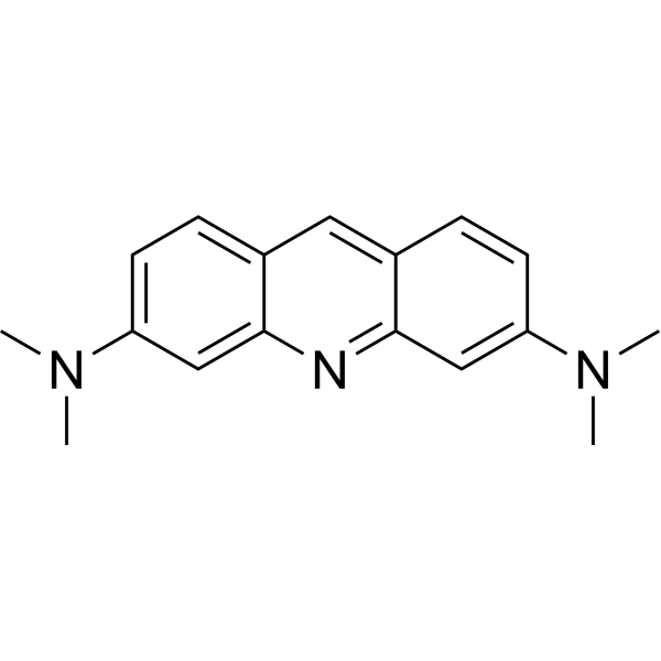
Acridine Orange base is a cell-permeable fluorescent nucleic acid dye and lysosomal probe with a pKa of 9.65. Acridine Orange base enters cells via passive diffusion, is protonated and accumulates in acidic lysosomes, and forms aggregates at high concentrations, producing orange-red fluorescence; when the lysosomal proton gradient or membrane integrity is impaired, Acridine Orange base is released into the cytoplasm, green fluorescence increases and red fluorescence decreases, so it can be used to monitor lysosomal acidification status and changes in membrane permeability. Acridine Orange base binds to nucleic acids through intercalation or electrostatic interactions, exhibiting mainly green fluorescence when bound to double-stranded DNA and mainly red fluorescence when bound to RNA or single-stranded DNA, and can be used for nucleic acid staining and differential DNA/RNA detection. The Ex/Em of Acridine Orange base monomer is approximately 492/525 nm; when bound to double-stranded DNA it is approximately 488/520-524 nm, and when bound to RNA or single-stranded DNA it is approximately 457/630-650 nm.
Nur für Forschungszwecke. Wir verkaufen nicht an Patienten.
- Reinheit : 97.04%
- CAS. Nr.: 494-38-2
- Formel: C17H19N3
- Molecular Weight:265.36
-
Speicherung:
4°C, protect from light
* In solvent : -80°C, 6 months; -20°C, 1 month (protect from light)

To place orders, for customer services and technical support, please contact: MedChemExpress USA
Tel: 609-228-6898 E-mail: [email protected] [email protected]


-
 Biologische Aktivität
-
Chemical Information
-
Lösungsmittel & Löslichkeit
- Protokoll
- Reinheit & Dokumentation
- Verweise
-
 Help & FAQs


-
HY-D0815
Propidium IodideFluorochrome -

HY-DY1006
Propidium Iodide (solution)Fluorochrome -
HY-W016412
Coenzyme Q0Apoptosis Inducer -
HY-118135
4-Methylumbelliferyl-α-D-galactopyranosideGLA Pro-fluorogenic Substrate -
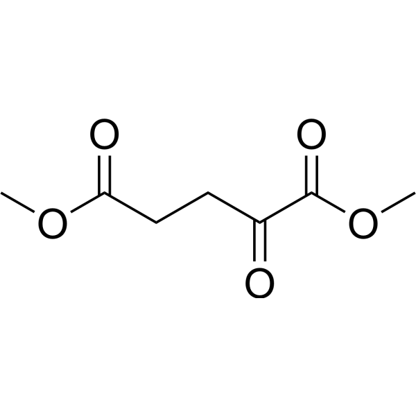
HY-44134
Dimethyl 2-oxoglutarate2-oxoglutarate Derivative -
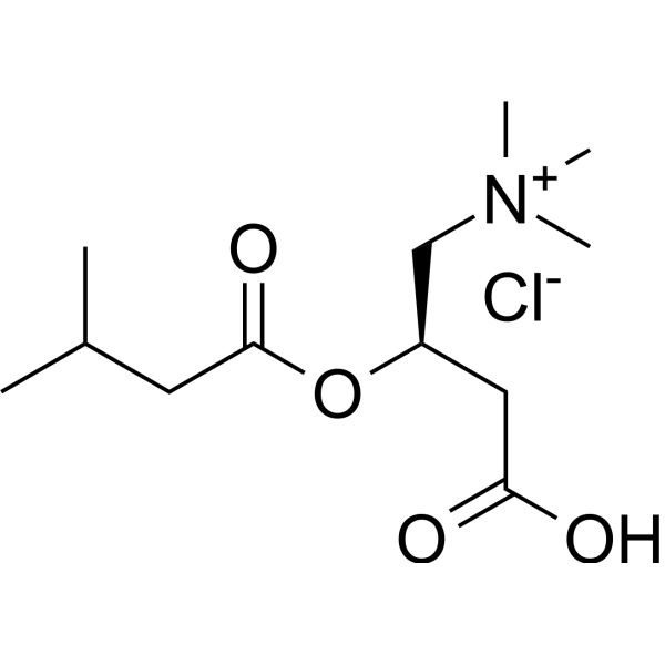
HY-145542
Isovalerylcarnitine chlorideCalpain Activator -
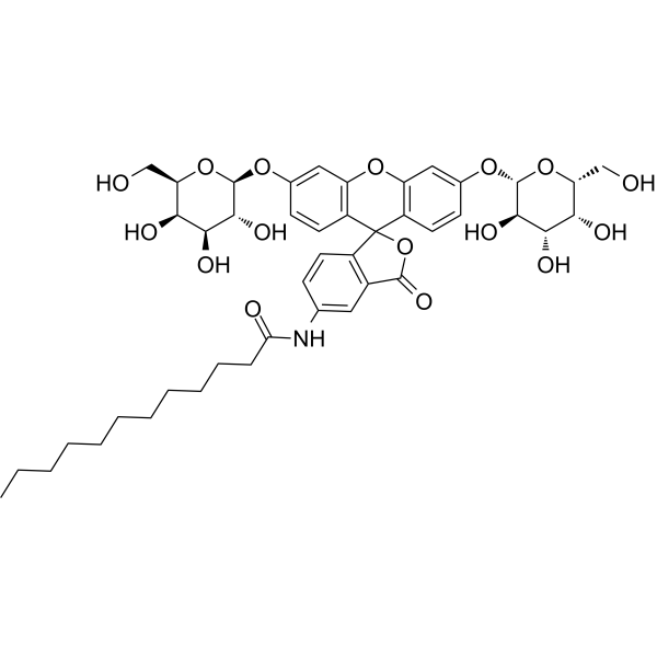
HY-126839
C12FDGβ-Galactosidase Fluorescent Substrate -
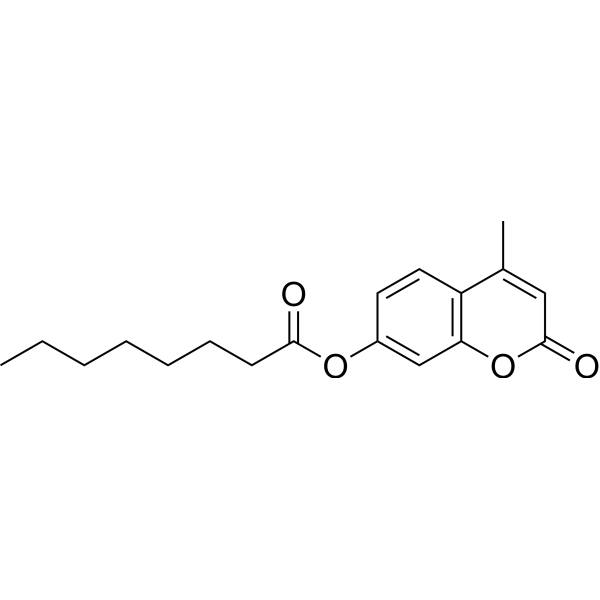
HY-W099648
4-Methylumbelliferyl octanoateOctoate -
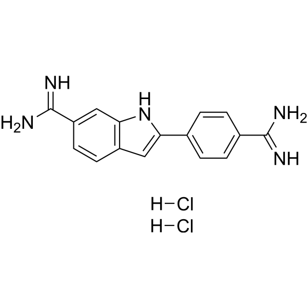
HY-D0814
DAPI dihydrochlorideFluorescent DNA Stain -
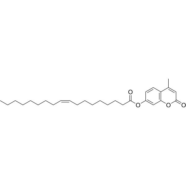
HY-117095
4-Methylumbelliferyl oleateFluorescent Substrate -
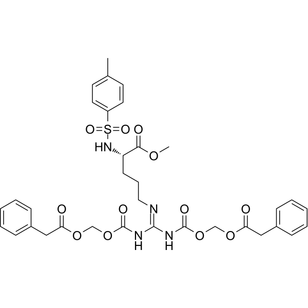
HY-124955
proTAMEAPC Inhibitor -

HY-W778830
Bodipy C12-ceramideFluorescently Tagged Form of C12-Ceramide
Publications Citing Use of MedChemExpress (MCE) Acridine Orange base
More- Signal Transduct Target Ther. 2025 Mar 19;10(1):94. [Abstract]
- Cell Mol Immunol. 2025 Mar;22(3):282-299. [Abstract]
- Adv Funct Mater. 2023 Apr 14.
- Nat Commun. 2023 Jun 30;14(1):3877. [Abstract]
- J Adv Res. 2025 Nov 7:S2090-1232(25)00877-X. [Abstract]
- Acta Pharm Sin B. 2021 Oct;11(10):3178-3192. [Abstract]
- Adv Sci (Weinh). 2024 Oct;11(38):e2404693. [Abstract]
- Biomaterials. 2025 Dec:323:123415. [Abstract]
- J Control Release. 2026 Feb 10:390:114523. [Abstract]
- J Control Release. 2024 Dec:376:167-183. [Abstract]
- Pharmacol Res. 2026 Apr 30:108225. [Abstract]
- Cell Death Dis. 2021 Jan 13;12(1):80. [Abstract]
- Cell Death Dis. 2020 Jul 30;11(7):607. [Abstract]
- Phytomedicine. 2026 Oct:160:158642.
- J Transl Med. 2026 May 22. [Abstract]
- Int J Mol Med. 2023 May;51(5):43. [Abstract]
- Free Radic Biol Med. 2023 Nov 1:208:402-417. [Abstract]
- Clin Transl Med. 2023 Mar;13(3):e1229. [Abstract]
- ACS Appl Mater Interfaces. 2025 Feb 19;17(7):10446-10456. [Abstract]
- Cell Rep. 2026 Feb 24;45(2):116914. [Abstract]
- Cell Prolif. 2022 Jan;55(1):e13172. [Abstract]
- Anal Chem. 2025 Jul 15;97(27):14512-14520. [Abstract]
- Environ Pollut. 2026 Apr 15:395:127832. [Abstract]
- Eur J Med Chem. 2024 Dec 5:279:116866. [Abstract]
- Eur J Med Chem. 2024 Jul 5:273:116507. [Abstract]
- Ecotoxicol Environ Saf. 2025 Sep 15:303:119000. [Abstract]
- Pharmaceuticals (Basel). 2025 Nov 11;18(11):1714. [Abstract]
- Int J Mol Sci. 2023 Sep 7;24(18):13812. [Abstract]
- Invest Ophthalmol Vis Sci. 2025 Feb 3;66(2):5. [Abstract]
- Chem Biol Interact. 2026 Mar 25:427:111932. [Abstract]
- Chem Biol Interact. 2026 Mar 1:426:111911. [Abstract]
- J Nutr Biochem. 2025 Aug 20:110082. [Abstract]
- Antiviral Res. 2026 May:249:106403. [Abstract]
- FASEB J. 2025 Dec 31;39(24):e71372. [Abstract]
- Cell Stress Chaperones. 2025 Sep 17:100112. [Abstract]
- Microbiol Spectr. 2023 Jun 15;11(3):e0313822. [Abstract]
- Neurotoxicology. 2026 Apr 28:103462. [Abstract]
- J Dent Sci. 2025 Jul;20(3):1782-1791. [Abstract]
- STAR Protoc. 2024 Sep 12;5(3):103309. [Abstract]
- SSRN. 2023 Nov 15.
- Environ Toxicol. 2023 Sep;38(9):2100-2110. [Abstract]
- Chemosphere. 2023 Apr:320:138071. [Abstract]
- SSRN. 2023 Mar 9.
- Research Square Print. December 12th, 2022.
 Customer Validation & Images

-
Cell Imaging/Staining
-
Cell Imaging/Staining
-
Cell Imaging/Staining
-
Cell Imaging/Staining
-
Cell Imaging/Staining


Biologische Aktivität
Beschreibung
In Vitro
1. Stock solution preparation
1.1 Solvent: H2O.
1.2 Concentration: Prepare a 1 mg/mL Acridine Orange (AO) stock solution.
2. Working solution preparation
2.1 In complete cell culture medium: Dilute the Acridine Orange stock solution with complete cell culture medium to a final concentration of 2-5 µg/mL. Adjust the working concentration as needed and prepare fresh before use.
2.2 In aqueous phosphate-citric acid buffer: Prepare a buffer containing 37 mM citric acid, 126 mM Na2HPO4, 150 mM NaCl, and 1 mM Na2EDTA. Mix 370 mL 0.1 M citric acid, 630 mL 0.2 M Na2HPO4, 8.77 g NaCl, 340 mg Na2EDTA, 6.0 mL 1 mg/mL Acridine Orange stock solution with H2O to 1 L, so that the AO concentration in the final staining solution is 6 µg/mL. Test 5-20 µg/mL as needed to adjust the working concentration. Store the Acridine Orange staining solution at 4°C protected from light or in a foil-wrapped bottle for up to 6 months.
2.3 For parasite labeling: Use 5 µg/mL, 50 µg/mL, or 100 µg/mL Acridine Orange staining solution. Use a low Acridine Orange concentration to reduce staining of host cell DNA and RNA.
3. Staining procedure
3.1 For adherent primary cells or cell lines[1]:
3.1.1 Culture cells in DMEM containing 10% FBS, 100 IU/mL penicillin, and 100 µg/mL streptomycin at 37°C, 5% CO2.
3.1.2 Trypsinize the cells and seed them in a black 96-well plate for fluorescence reading at a density that reaches 80-90% confluence on the day of the experiment.
3.1.3 Allow the cells to adhere for at least 24 h.
3.1.4 Remove the medium and add 100 µL of 2-5 µg/mL Acridine Orange staining solution to each well.
3.1.5 Incubate at 37°C for 15 min.
3.1.6 Wash the cells twice with 100 µL complete phenol-free medium for 5 min each.
3.1.7 Place the cells in 100 µL complete phenol-free medium.
3.2 For unfixed cells[2]:
3.2.1 Use cells suspended in tissue culture medium containing 10% (v/v) serum or 1% (w/v) BSA.
3.2.2 Transfer the cells to a small glass or plastic tube and cool on ice.
3.2.3 Avoid pipetting, vortexing, or mechanically agitating the cells to prevent cell lysis.
3.2.4 Add 0.4 mL ice-cold cell permeabilization solution (0.1% Triton X-100, 80 mM HCl, 150 mM NaCl), keep the cells on ice, and wait 15 sec.
3.2.5 Gently add 1.2 mL ice-cold 6 µg/mL Acridine Orange staining solution and keep the cells on ice protected from light.
3.2.6 Measure and record cell fluorescence within 2-10 min after adding the Acridine Orange staining solution, keeping the samples on ice before and during measurement.
3.3 For fixed cells[2]:
3.3.1 Rinse the cells once with ice-cold PBS and suspend to approximately 106 cells/mL.
3.3.2 For adherent cells, collect by trypsinization, combine the trypsinized cells with the suspended cells, and rinse once with serum-containing medium, and do not keep on ice for more than 30 min before fixation.
3.3.3 Fix by rapidly injecting 1 mL of cell suspension into 10 mL ice-cold 70% ethanol and fix on ice for ≥2 h.
3.3.4 Before staining, centrifuge the fixed cells at 300 × g for 5 min at 4°C; remove all ethanol; rinse once with ice-cold PBS; transfer the cells to a small tube; and cool on ice.
3.3.5 Avoid pipetting, vortexing, or mechanically agitating the cells to prevent cell lysis.
3.3.6 Add 0.4 mL ice-cold cell permeabilization solution (0.1% Triton X-100, 80 mM HCl, 150 mM NaCl), keep the cells on ice, and wait 15 sec.
3.3.7 Gently add 1.2 mL ice-cold 6 µg/mL Acridine Orange staining solution and keep the cells on ice protected from light.
3.3.8 Measure and record cell fluorescence within 2-10 min after adding the Acridine Orange staining solution, keeping the samples on ice before and during measurement.
3.4 For L. major promastigotes in suspension[3]:
3.4.1 Label 1 × 10⁶ parasites in suspension.
3.4.2 Add the Acridine Orange staining solution to the parasite suspension.
3.4.3 Incubate the parasite-Acridine Orange mixture for 10 min.
3.4.4 Centrifuge the stained parasites and wash seven times with PBS.
3.4.5 After washing, incubate the stained promastigotes with fibroblasts for 6-8 h.
4. Controls
4.1 Include at least one cell-free well for background fluorescence.
4.2 Include unexposed controls for all conditions used in the experiment.
4.3 Optional positive control 1: 5-50 µg/mL glucose oxidase.
4.4 Optional positive control 2: 0.1-5 mM LLOMe.
4.5 Determine the concentration of LMP inducer applicable to the selected cell type.
4.6 To assess RNA stainability, centrifuge ethanol-fixed cells at 300 × g for 5 min at 4°C, remove all ethanol, resuspend the cells in 1 mL RNase solution (100 µg/mL RNase A in PBS containing Mg2+ ), and incubate at 37°C for 30 min. Use DNase-free RNase, or boil the RNase solution for 5 min and cool to 37°C before use.
4.7 To assess DNA stainability, centrifuge the cells as above, resuspend the pellet in 1 mL DNase solution (500 µg/mL DNase I in PBS containing Mg2+ ), and incubate at 37°C for 30 min.
4.8 Stain control cells with Acridine Orange as above and assess the percentage loss of red luminescence and green fluorescence as indicators of RNA and DNA staining specificity, respectively.
4.9 For unfixed Acridine Orange-stained cells, after permeabilization and staining, treat with 100 µg/mL RNase A and incubate at 24°C for 30 min, then perform fluorescence measurement.
4.10 Use Acridine Orange-stained free fibroblasts on day 3 of culture as a control for comparison with infected fibroblasts.
5. Detection and analysis
5.1 Instrument types:
5.1.1 Fluorescence plate reader, flow cytometer, fluorescence microscope.
5.2 Wavelength settings:
5.2.1 Plate reader, green fluorescence: excitation 485 nm, emission 535 nm. Red fluorescence: excitation 465 nm, emission 650-710 nm.
5.2.2 Flow cytometry, DNA detection: excite at 488 nm and collect green fluorescence at 530 nm. The AO-dsDNA absorption peak is 502 nm; recommended excitation 488 nm, emission 520-524 nm, using a 520 nm filter.
5.2.3 Flow cytometry, RNA detection: preferably collect red luminescence above 640 or 650 nm. For dual-laser excitation, use 488 nm for DNA green fluorescence detection and 457 nm for RNA red luminescence detection. AO-ssDNA and AO-ssRNA absorption is 426-458 nm; recommended excitation 457 nm, emission 630-644 nm, using a >640 nm filter.
5.2.4 Fluorescence microscopy: green fluorescence maximum emission wavelength is 525 nm; AO bound to dsDNA emits green fluorescence at 525 nm, and AO bound to RNA or ssDNA emits red fluorescence at 650 nm.
5.3 Measurement and analysis:
5.3.1 Perform a single-point measurement to record the initial AO fluorescence before induction of LMP.
5.3.2 Begin acquiring fluorescence data immediately after adding the LMP inducer at 37°C, using the same settings as the initial measurement.
5.3.3 For LLOMe, acquire fluorescence intensity data once per minute, as maximum leakage typically occurs within 30 min.
5.3.4 For glucose oxidase, acquire data every 2-3 min for up to 2 h.
5.3.5 Control cells show a green monomeric AO emission peak at 535 nm and a red dimeric lysosomal emission peak at 650 nm.
5.3.6 Use the increase in AO green fluorescence as a more sensitive readout of changes in the lysosomal proton gradient, because the decrease in red intensity is less easily detected.
5.3.7 AO intercalated into dsDNA emits green fluorescence, whereas AO bound to ssRNA emits red luminescence with a maximum emission wavelength above 630 nm.
5.3.8 DNA frequency histograms from green fluorescence can be deconvoluted to obtain G1 vs S vs G2/M ratios.
5.3.9 Live promastigotes appear green. After 12 h of culture, free promastigotes appear green before entering fibroblasts.
5.3.10 AO-stained fibroblast nuclei are partially green (approximately 12 μm), nucleoli are green (approximately 0.6-1 μm), and parasites (2.5-5 μm) can be distinguished from nucleoli by size.
MedChemExpress (MCE) has not independently confirmed the accuracy of these methods. They are for reference only.
Emission (Em)
520/570


Chemical Information
-
CAS. Nr. 494-38-2
-
Appearance Solid
-
Molecular Weight 265.36
-
Formel C17H19N3
-
Color Yellow to brown
-
SMILES
CN(C)C1=CC=C2C(N=C3C=C(N(C)C)C=CC3=C2)=C1
-
Versand
Room temperature in continental US; may vary elsewhere.
-
Speicherung
4°C, protect from light
* In solvent : -80°C, 6 months; -20°C, 1 month (protect from light)


Publications (44)
-
Journal Impact Factor
-
Most Recent
-
Signal Transduct Target Ther
2025 Mar 19;10(1):94. PMID: 40102383 -
Cell Mol Immunol
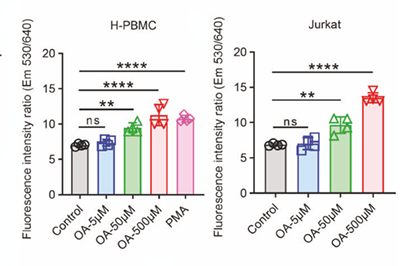
GLP1 alleviates oleic acid-propelled lipocalin-2 generation by tumor-infiltrating CD8+ T cells to reduce polymorphonuclear MDSC recruitment and enhances viral immunotherapy in pancreatic cancer. [Abstract]2025 Mar;22(3):282-299. PMID: 39910336
Acridine Orange base purchased from MedChemExpress. Usage Cited in: Cell Mol Immunol. 2025 Mar;22(3):282-299. [Abstract]
Human PBMCs (H-PBMCs) and Jurkat cells were treated with OA (5, 50, or 500 μM) or 200 ng/mL PMA for 24 hours, after which the cells were stained with acridine orange (AO).
-
-
Nat Commun
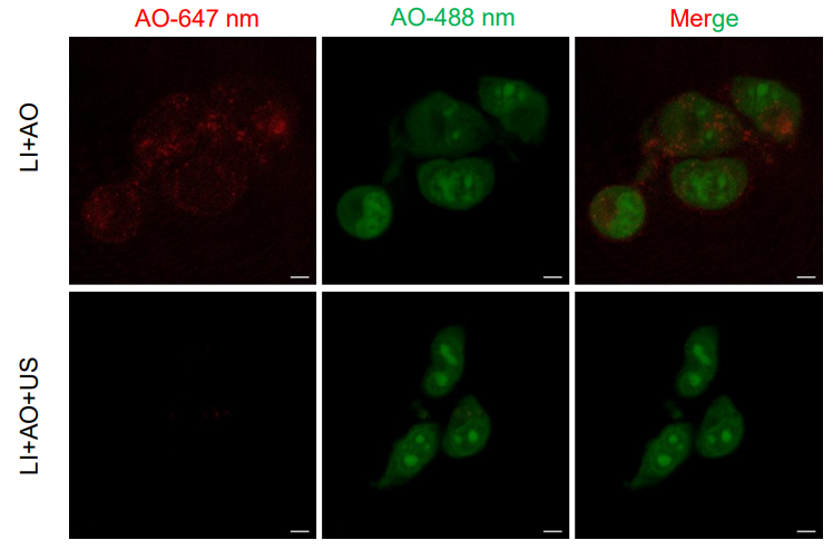
Ultrasound-responsive low-dose doxorubicin liposomes trigger mitochondrial DNA release and activate cGAS-STING-mediated antitumour immunity. [Abstract]2023 Jun 30;14(1):3877. PMID: 37391428
Acridine Orange base purchased from MedChemExpress. Usage Cited in: Nat Commun. 2023 Jun 30;14(1):3877. [Abstract]
Acridine Orange (AO, 5 μM) was added immediately after the ultrasound treatment and was incubated for 30 min at 37°C before confocal microscopy.
-
J Adv Res
Hyperuricemia increases susceptibility to chronic kidney injury exacerbation via autophagic flux blockade-mediated ammonia death pathway. [Abstract]2025 Nov 7:S2090-1232(25)00877-X. PMID: 41205802 -
Acta Pharm Sin B
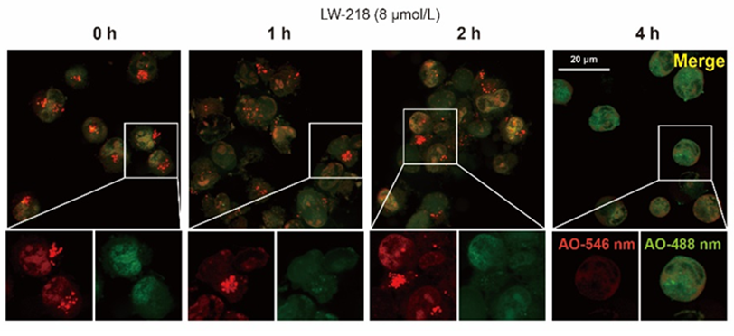
Cholesterol-associated lysosomal disorder triggers cell death of hematological malignancy: Dynamic analysis on cytotoxic effects of LW-218. [Abstract]2021 Oct;11(10):3178-3192. PMID: 34729308
Acridine Orange base purchased from MedChemExpress. Usage Cited in: Acta Pharm Sin B. 2021 Oct;11(10):3178-3192. [Abstract]
The Jurkat cells were treated with LW-218 (8 μmol/L) for 0, 1, 2, and 4 h. Before collecting, the cells were stained with 5 μmol/L Acridine Orange hydrochloride solution for 15 min.
-
Adv Sci (Weinh)
Methuosis Inducer SGI-1027 Cooperates with Everolimus to Promote Apoptosis and Pyroptosis by Triggering Lysosomal Membrane Permeability in Renal Cancer. [Abstract]2024 Oct;11(38):e2404693. PMID: 39119834 -
Biomaterials
Infected microenvironment responsive nanoprodrug for the specific therapeutics of Helicobacter pylori clearance. [Abstract]2025 Dec:323:123415. PMID: 40411983 -
J Control Release
Acid neutralization and metal-phenolic nanomedicine on-demand release microspheres for reversing cartilage degeneration. [Abstract]2026 Feb 10:390:114523. PMID: 41397492 -
J Control Release
An immunostimulatory liponanogel reveals immune activation-enhanced drug delivery and therapeutic efficacy in cancer. [Abstract]2024 Dec:376:167-183. PMID: 39384154 -
Pharmacol Res
Lysosome-triggered nanotherapy packages osteoclast apoptotic bodies with pro-anabolic lipids to couple anti-resorption and bone formation. [Abstract]2026 Apr 30:108225. PMID: 42069319 -
Cell Death Dis
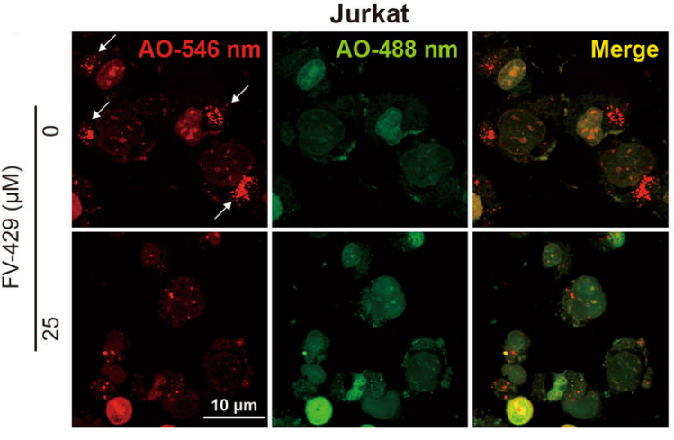
FV-429 induces autophagy blockage and lysosome-dependent cell death of T-cell malignancies via lysosomal dysregulation. [Abstract]2021 Jan 13;12(1):80. PMID: 33441536
Acridine Orange base purchased from MedChemExpress. Usage Cited in: Cell Death Dis. 2021 Jan 13;12(1):80. [Abstract]
The Jurkat cells treated with 25 μM FV-429 for 12 h were stained with Acridine Orange hydrochloride (AO, 5 μM; 15 min) before collection, and the AO fluorescence was determined by a laser confocal microscope. Scale bar: 10 μm.
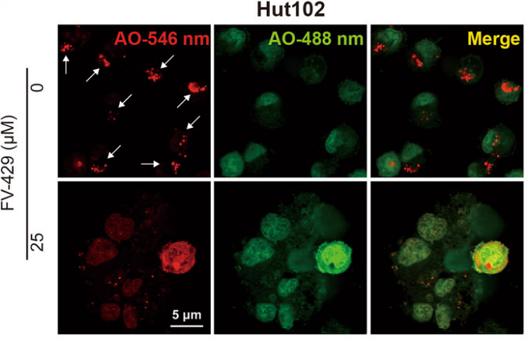
Acridine Orange base purchased from MedChemExpress. Usage Cited in: Cell Death Dis. 2021 Jan 13;12(1):80. [Abstract]
The Jurkat and Hut102 cells treated with 25 μM FV-429 for 12 h were stained with Acridine Orange hydrochloride (AO, 5 μM; 15 min) staining before collecting, and the AO fluorescences were determined by laser confocal microscope. Scale bar: 5 μm.
-
Cell Death Dis
ω-6 Polyunsaturated fatty acids (linoleic acid) activate both autophagy and antioxidation in a synergistic feedback loop via TOR-dependent and TOR-independent signaling pathways. [Abstract]2020 Jul 30;11(7):607. PMID: 32732901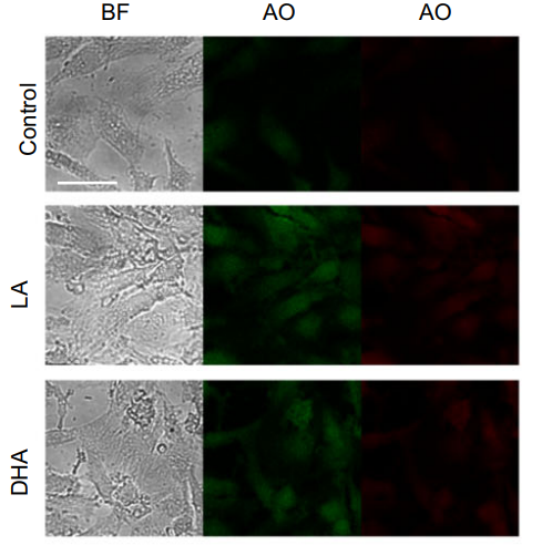
Acridine Orange base purchased from MedChemExpress. Usage Cited in: Cell Death Dis. 2020 Jul 30;11(7):607. [Abstract]
Depending on the acidity, Acridine Orange hydrochloride (AO) causes autophagic lysosomes to appear as orange/red fluorescent vesicles, while it caused nuclei to appear green. AO staining demonstrates that LA dose-dependently increased the numbers of intracellular acidic compartments.
-
-
J Transl Med
Targeting C5AR1 disrupts complement-driven G0-phase maintenance and overcomes metabolic drug resistance in glioma. [Abstract]2026 May 22. PMID: 42174621 -
Int J Mol Med
Casticin suppresses RANKL‑induced osteoclastogenesis and prevents ovariectomy‑induced bone loss by regulating the AKT/ERK and NF‑κB signaling pathways. [Abstract]2023 May;51(5):43. PMID: 37052260 -
Free Radic Biol Med
Phosphatidylethanolamine alleviates OX-LDL-induced macrophage inflammation by upregulating autophagy and inhibiting NLRP1 inflammasome activation. [Abstract]2023 Nov 1:208:402-417. PMID: 37660837 -
Clin Transl Med
Targeting lysosomal HSP70 induces acid sphingomyelinase-mediated disturbance of lipid metabolism and leads to cell death in T cell malignancies. [Abstract]2023 Mar;13(3):e1229. PMID: 36959764 -
ACS Appl Mater Interfaces
DNA Logic Gate-Triggered Membrane Fusion for Accurately Detecting and Killing Cancer Cells. [Abstract]2025 Feb 19;17(7):10446-10456. PMID: 39907089 -
Cell Rep
HRD1 negatively regulates autolysosome formation by inhibiting liquid-liquid phase separation of SNAP29. [Abstract]2026 Feb 24;45(2):116914. PMID: 41615796 -
Cell Prolif
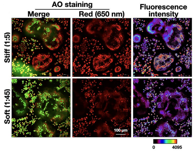
Substrate stiffness regulates the differentiation profile and functions of osteoclasts via cytoskeletal arrangement. [Abstract]2022 Jan;55(1):e13172. PMID: 34953003
Acridine Orange base purchased from MedChemExpress. Usage Cited in: Cell Prolif. 2022 Jan;55(1):e13172. [Abstract]
CLSM images of acridine orange (AO)-stained osteoclasts cultured on PDMS substrates were taken; the cells were stained with 1 μM AO at 37°C for 20 min.
-
Anal Chem
Vesicular Stomatitis Virus Glycoprotein- and Lentiviral RNA-Based Robust Approach for Rapid and Accurate Quantification of Viable Lentiviral Vectors. [Abstract]2025 Jul 15;97(27):14512-14520. PMID: 40600817 -
Environ Pollut
ERα-mediated endoplasmic reticulum stress drives 4-tert-octylphenol-induced cardiac developmental toxicity in zebrafish. [Abstract]2026 Apr 15:395:127832. PMID: 41713771 -
Eur J Med Chem
Rationally designed febuxostat-based hydroxamic acid and its pH-Responsive nanoformulation elicits anti-tumor activity. [Abstract]2024 Dec 5:279:116866. PMID: 39293244 -
Eur J Med Chem
Leveraging a rationally designed veliparib-based anilide eliciting anti-leukemic effects for the design of pH-responsive polymer nanoformulation. [Abstract]2024 Jul 5:273:116507. PMID: 38776806 -
Ecotoxicol Environ Saf
Bisphenol F disrupts lipophagy and lysosomal acidification via ATGL-SIRT1-PPARα signaling in NAFLD-like hepatic changes. [Abstract]2025 Sep 15:303:119000. PMID: 40907412 -
Pharmaceuticals (Basel)
The Effects of Frondanol, a Non-Polar Extract of the Atlantic Sea Cucumber, in Colon Cancer Cells. [Abstract]2025 Nov 11;18(11):1714. PMID: 41304959 -
Int J Mol Sci
The Properties of Linezolid, Rifampicin, and Vancomycin, as Well as the Mechanism of Action of Pentamidine, Determine Their Synergy against Gram-Negative Bacteria. [Abstract]2023 Sep 7;24(18):13812. PMID: 37762115 -
Invest Ophthalmol Vis Sci
HMGB1 Promotes Lysosome-Dependent Cell Death Induced Via Dry Eye by Disrupting Lysosomal Homeostasis. [Abstract]2025 Feb 3;66(2):5. PMID: 39903181 -
Chem Biol Interact
Thalidezine triggers Cathepsin B-mediated cell death in T-cell lymphoma by disrupting lysosomal function. [Abstract]2026 Mar 25:427:111932. PMID: 41570971 -
Chem Biol Interact
Cyflumetofen disrupts the blood-brain barrier and induces neurotoxicity in zebrafish larvae. [Abstract]2026 Mar 1:426:111911. PMID: 41539556 -
J Nutr Biochem
2025 Aug 20:110082. PMID: 40845949 -
Antiviral Res
Salinomycin inhibits orthopoxvirus infection in vitro and in vivo by blocking endosomal acidification. [Abstract]2026 May:249:106403. PMID: 41941992 -
FASEB J
Interaction Between Activating Transcription Factor 4 and Interleukin 6 Dictates Osteoclast Differentiation and Activity. [Abstract]2025 Dec 31;39(24):e71372. PMID: 41420529 -
Cell Stress Chaperones
The role of Atp2a2-mediated calcium imbalance and endoplasmic reticulum stress in hydrocortisone-induced neurotoxicity. [Abstract]2025 Sep 17:100112. PMID: 40972936 -
Microbiol Spectr
When Combined with Pentamidine, Originally Ineffective Linezolid Becomes Active in Carbapenem-Resistant Enterobacteriaceae. [Abstract]2023 Jun 15;11(3):e0313822. PMID: 37125928 -
Neurotoxicology
Lysosomal dysfunction contributes to lead (Pb) and high-fat diet (HFD)-induced neuronal apoptosis: A link with cathepsin B. [Abstract]2026 Apr 28:103462. PMID: 42061594 -
J Dent Sci
3,4-methylenedioxymethamphetamine induces reactive oxygen species-mediated autophagy and thioredoxin-interactive protein/nucleotide-binding domain, leucine-rich containing family, pyrin domain-containing-3 inflammasome activation in dental pulp stem cells. [Abstract]2025 Jul;20(3):1782-1791. PMID: 40654447 -
STAR Protoc
Protocol for detecting lysosome quantity and membrane permeability in acute myeloid leukemia cell lines. [Abstract]2024 Sep 12;5(3):103309. PMID: 39269898 -
-
Environ Toxicol
Autophagy blockage and lysosomal dysfunction are involved in diallyl sulfide-induced inhibition of malignant growth in hepatocellular carcinoma cells. [Abstract]2023 Sep;38(9):2100-2110. PMID: 37209385 -
Chemosphere
Rab7a-mTORC1 signaling-mediated cholesterol trafficking from the lysosome to mitochondria ameliorates hepatic lipotoxicity induced by aflatoxin B1 exposure. [Abstract]2023 Apr:320:138071. PMID: 36754296 -
-


Lösungsmittel & Löslichkeit
In Vitro:
1 M HCl : 200 mg/mL (753.69 mM; Need ultrasonic)
DMSO : 35 mg/mL (131.90 mM; Need ultrasonic; Hygroscopic DMSO has a significant impact on the solubility of product, please use newly opened DMSO)
H2O : 5.83 mg/mL (21.97 mM; ultrasonic and warming and heat to 60°C)
Please refer to the solubility information to select the appropriate solvent. Once prepared, please aliquot and store the solution to prevent product inactivation from repeated freeze-thaw cycles.
Storage method and period of stock solution: -80°C, 6 months; -20°C, 1 month (protect from light). When stored at -80°C, please use it within 6 months. When stored at -20°C, please use it within 1 month.
* Note: If you choose water as the stock solution, please dilute it to the working solution, then filter and sterilize it with a 0.22 μm filter before use.
Please refer to the solubility information to select the appropriate solvent. Once prepared, please aliquot and store the solution to prevent product inactivation from repeated freeze-thaw cycles.
Storage method and period of stock solution: -80°C, 6 months; -20°C, 1 month (protect from light). When stored at -80°C, please use it within 6 months. When stored at -20°C, please use it within 1 month.
* Note: If you choose water as the stock solution, please dilute it to the working solution, then filter and sterilize it with a 0.22 μm filter before use.
Konzentration (Stammlösung) × Volumen (Stammlösung) = Konzentration (Ziellösung) × Volumen (Ziellösung)


Reinheit & Dokumentation
-
Data Sheet (291 KB)
-
SDS (393 KB)
- English - EN (393 KB)
- Français - FR (393 KB)
- Deutsch - DE (393 KB)
- Norwegian - NO (393 KB)
- Español - ES (393 KB)
- Swedish - SV (393 KB)
- Italian - IT (393 KB)
- Korean - KR (393 KB)
- Portuguese - PT (393 KB)
-
Handling Instructions (2659 KB)
Verweise
[1]. Eriksson I, et al. Real-Time Monitoring of Lysosomal Membrane Permeabilization Using Acridine Orange. Methods Protoc. 2023 Aug 9;6(4):72. [Content Brief]
[2]. Darzynkiewicz Z, et al. Differential staining of DNA and RNA. Current protocols in cytometry. 2004 Nov;Chapter 7:Unit 7.3. [Content Brief]
[3]. Yektaeian N, et al. Lipophilic tracer Dil and fluorescence labeling of acridine orange used for Leishmania major tracing in the fibroblast cells. Heliyon. 2019 Dec 18;5(12):e03073. [Content Brief]


Complete Stock Solution Preparation Table
Please refer to the solubility information to select the appropriate solvent. Once prepared, please aliquot and store the solution to prevent product inactivation from repeated freeze-thaw cycles.
Storage method and period of stock solution: -80°C, 6 months; -20°C, 1 month (protect from light). When stored at -80°C, please use it within 6 months. When stored at -20°C, please use it within 1 month.
| Optional Solvent | Concentration Solvent Mass | 1 mg | 5 mg | 10 mg | 25 mg |
|---|---|---|---|---|---|
| H2O / DMSO / 1 M HCl | 1 mM | 3.7685 mL | 18.8423 mL | 37.6847 mL | 94.2116 mL |
| 5 mM | 0.7537 mL | 3.7685 mL | 7.5369 mL | 18.8423 mL | |
| 10 mM | 0.3768 mL | 1.8842 mL | 3.7685 mL | 9.4212 mL | |
| 15 mM | 0.2512 mL | 1.2562 mL | 2.5123 mL | 6.2808 mL | |
| 20 mM | 0.1884 mL | 0.9421 mL | 1.8842 mL | 4.7106 mL | |
| DMSO / 1 M HCl | 25 mM | 0.1507 mL | 0.7537 mL | 1.5074 mL | 3.7685 mL |
| 30 mM | 0.1256 mL | 0.6281 mL | 1.2562 mL | 3.1404 mL | |
| 40 mM | 0.0942 mL | 0.4711 mL | 0.9421 mL | 2.3553 mL | |
| 50 mM | 0.0754 mL | 0.3768 mL | 0.7537 mL | 1.8842 mL | |
| 60 mM | 0.0628 mL | 0.3140 mL | 0.6281 mL | 1.5702 mL | |
| 80 mM | 0.0471 mL | 0.2355 mL | 0.4711 mL | 1.1776 mL | |
| 100 mM | 0.0377 mL | 0.1884 mL | 0.3768 mL | 0.9421 mL |
* Note: If you choose water as the stock solution, please dilute it to the working solution, then filter and sterilize it with a 0.22 μm filter before use.


Acridine Orange base Related Classifications
HY-D0952 Related Classifications
 Powered by Bioz