



Fura Red
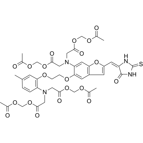
Fura Red is a Ca2+-sensitive fluorescent dye, which decreases in fluorescence with rising [Ca2+].
연구목적의 판매만을 진행합니다. 환자를 대상으로 한 판매는 하지 않습니다.
- CAS No.: 149732-62-7
- 화학식: C41H44N4O20S
- 분자량:944.87
-
보관:
Please store the product under the recommended conditions in the Certificate of Analysis.

To place orders, for customer services and technical support, please contact: MedChemExpress USA
Tel: 609-228-6898 E-mail: [email protected] [email protected]


-
 Biological Activity
-
Chemical Information
- Protocol
- 순도&문서
- References
-
 Help & FAQs




Biological Activity
제품 설명
In Vitro
Guide[2] (Following is our recommended protocol. This protocol only provides a guideline, and should be modified according to your specific needs).
Labeling of Cells:
1. Resuspend the cells in Hanks' Balanced Salt Solution (HBSS), add 1 μM Fura Red AM, supplement with 0.01% Pluronic F127 unless otherwise stated, and incubate in a 37°C water bath for 30 minutes.
2. Wash the cells in HEPES-buffered salt solution (HBSS) containing 1 mmol/L calcium chloride, 0.5 mmol/L magnesium chloride, 0.1% bovine serum albumin, and 10 mmol/L HEPES, then resuspend them in the same HEPES-buffered salt solution supplemented with the vital dye SYTOX Green.
3. Equilibrate the cells in a 37°C water bath for at least 10 minutes.
4. Analyze the samples on a flow cytometer, fluorescence microscope, or fluorescence microplate reader. Ratiometric analysis of Fura Red is performed by excitation with a violet laser (406 nm) and a green laser (502 nm).
MedChemExpress (MCE) has not independently confirmed the accuracy of these methods. They are for reference only.
Emission (Em)
652
Excitation (Ex)
471


Chemical Information
-
CAS No. 149732-62-7
-
분자량 944.87
-
화학식 C41H44N4O20S
-
SMILES
O=C(OCOC(C)=O)CN(C1=CC(OC(/C=C(C(N2)=O)/NC2=S)=C3)=C3C=C1OCCOC(C=C(C)C=C4)=C4N(CC(OCOC(C)=O)=O)CC(OCOC(C)=O)=O)CC(OCOC(C)=O)=O
-
선적
Room temperature in continental US; may vary elsewhere.
-
보관
Please store the product under the recommended conditions in the Certificate of Analysis.


순도&문서
References
[1]. Floto RA, et al. IgG-induced Ca2+ oscillations in differentiated U937 cells; a study using laser scanning confocal microscopy and co-loaded fluo-3 and fura-red fluorescent probes. Cell Calcium. 1995 Nov;18(5):377-89. [Content Brief]
[2]. Wendt ER, et al. Ratiometric analysis of fura red by flow cytometry: a technique for monitoring intracellular calcium flux in primary cell subsets. PLoS One. 2015 Apr 2;10(3):e0119532. [Content Brief]

Calculators
Concentration (start) × Volume (start) = Concentration (final) × Volume (final)


Fura Red Related Classifications
HY-104056 Related Classifications
 Powered by Bioz