From 11:00 pm to 12:00 pm EST ( 8:00 pm to 9:00 pm PST ) on January 6th, the website will be under maintenance. We are sorry for the inconvenience. Please arrange your schedule properly.
HBC (HBC 530) is a GFP fluorophore-like synthetic dye, with a structurally rigid electron acceptor and a strong electron donor. HBC has a low fluorescence background, and when combined with Pepper (RNA aptamer), HBC forms a tight complex and activates and emits bright fluorescence (Kd of ~3.5 nM). HBC emission peaks vary in different complexes and covers the spectrum from cyan to red. HBC can be used in the live cell imaging of RNA (Em/Ex = 530/485 nm) .
BRD-K98645985 is a BAF (mammalian SWI/SNF) transcriptional repression inhibitor with an EC50 of ~2.37 μM. BRD-K98645985 binds ARID1A-specific BAF complexes, prevents nucleosomal positioning, and potently reverses HIV-1 latency, without T cell activation or toxicity .
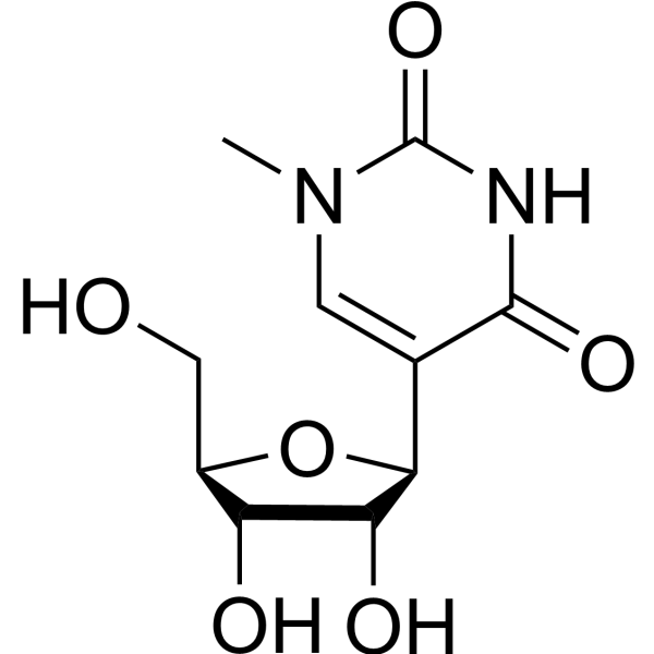
N1-methyl-pseudouridine (1-Methylpseudouridine), a methylpseudouridine, outperforms 5 mC and 5 mC/N1-methyl-pseudouridine in translation. N1-methyl-pseudouridine in mRNA enhances translation through eIF2α-dependent and independent mechanisms by increasing ribosome density .
FP-biotin is a potent organophosphorus toxicant, well-suited for searching for new biomarkers of organophosphorus toxicants exposure. FP-Biotin quantifies FAAH, ABHD6, and MAG-lipase activity. FP-biotin is used for studies with plasma because biotinylated peptides are readily purified by binding to immobilized avidin beads .
DFHBI is a small molecule that resembles the chromophore of green fluorescent protein (GFP). DFHBI can be used for imaging RNA in living cells. . Spinach and DFHBI are essentially nonfluorescent when unbound, whereas the Spinach-DFHBI complex is brightly fluorescent both in vitro and in living cells (Ex/Em = 469/501 nm).
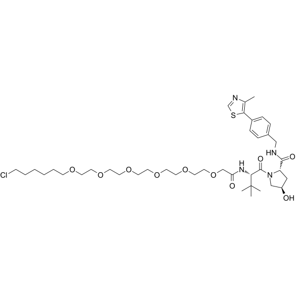
VH285-PEG4-C4-Cl (HaloPROTAC 3) is a conjugate of ligands for E3 and 16-atom-length linker. The connector of linker is Halogen group. VH285-PEG4-C4-Cl incorporates the VH285 based VHL ligand and an alkyl/ether-based linker. VH285-PEG4-C4-Cl is a highly potent and efficacious degrader of GFP-HaloTag7 with a DC50 of 19 nM. VH285-PEG4-C4-Cl is able to induce 90 % degradation of GFP-Halotag at 625 nM. VH285-PEG4-C4-Cl binds to VHL with an IC50 of 0.54 μM .
Senexin A is an inhibitor of CDK8/19 (IC50: 280 nM, CDK8) and an inhibitor downstream of p21 transcription. It only inhibits p21-induced transcription but does not inhibit other biological effects of p21. Senexin A inhibits CMV-GFP induction as well as the p21 stimulatory activity of the consensus NF-κB-dependent promoters .
GFP11 is a protein fusion tag consisting of 16 amino acids. When GFP11 and GFP1-10 are sufficiently close in physical space, they undergo spontaneous, irreversible complementation to reassemble into an intact, functional GFP protein that emits green fluorescence. GFP11 can be used to form a split GFP system for high-throughput biotechnology and flow cytometry applications .
Blue dextran (Dextran blue) (MW 2000000) is a high-molecular-weight long-chain polymer of D-glucose. Blue dextran (MW 2000000) serves as an important model macromolecular drug and molecular weight estimation marker, and can be used as a standard for gel permeation chromatography. The release of Blue dextran (MW 2000000) from alginate microspheres is regulated by preparation conditions; its release rate in a pH 6.8 environment is significantly faster than that in pH 1.2, and it exhibits release characteristics close to zero-order kinetics under this condition .
Ribocil is a selective inhibitor targeting the bacterial FMN riboswitch, regulating the bacterial riboflavin riboswitch. Ribocil competitively binds to the FMN binding site, mimicking the natural ligand FMN to induce conformational changes in the riboswitch, inhibiting ribB gene expression, reducing riboflavin synthesis, and thus inhibiting bacterial growth. Ribocil strongly inhibits GFP expression (EC50=0.3 μM). Ribocil exhibits in vivo antibacterial activity in a mouse model and can be used to study antibacterial drugs related to drug-resistant bacterial infections and bacterial riboflavin metabolic pathways[1][2].
Braco-19 trihydrochloride is a potent telomerase/telomere inhibitor, preventing the capping and catalytic action of telomerase. Braco-19 acts as G-quadruplex (GQ) binding ligand, stabilizing G-quadruplexes formation at the 3V telomeric DNA overhang and produce rapid senescence or selective cell death. Braco-19 is also a HAdV virus replication inhibitor .
LNP Lipid-8 (11-A-M) is an ionizable single-tail multi-head lipid that can be used as a lipid nanoparticle (LNP) to deliver siRNA to T cells without targeting ligands. LNP Lipid-8 is more selective for T cells than other cell types such as hepatocytes. LNP Lipid-8 selectively delivers siRNA/sgRNA to T cells (especially CD8+ T cells) through endogenous lipid transport pathways, and can enter cells and release RNA through endocytosis to achieve gene silencing. LNP Lipid-8 loaded with GFP siRNA (siGFP) significantly led to GFP gene silencing in mouse models. LNP Lipid-8 showed good efficacy and safety in both cells and animals, without obvious liver targeting and toxicity. LNP Lipid-8 can be used for RNA delivery research in the fields of tumor immunotherapy and T cell-mediated autoimmune diseases .
Filastatin is a long-lasting inhibitor of Candida albicans filamentation. Filastatin inhibits adhesion by multiple pathogenic Candida species with an IC50 of ~3 μM in the GFP-based adhesion assay. Filastatin inhibits fungal adhesion to polystyrene and human cells, the yeast-to-hyphal morphological transition, induction of the hyphal-specific HWP1 promoter. Filastatin has potent antifungal effect .
Vps34-IN-2 is a novel, potent and selective inhibitor of Vps34 with IC50s of 2 and 82 nM on the Vps34 enzymatic assay and the GFP-FYVE cellular assay, respectively . Vps34-IN-2 shows antiviral activity against SARS-CoV-2 (IC50 of 3.1 μM), HCoV-229E (IC50 of 0.7 μM) and HCoV-OC43 .
(S,R,S)-AHPC-(C3-PEG)2-C6-Cl is a small molecule HaloPROTAC that incorporates the (S,R,S)-AHPC based VHL ligand and 2-unit PEG linker. (S,R,S)-AHPC-(C3-PEG)2-C6-Cl is capable of inducing the degradation of GFP-HaloTag7 in cell-based assays .
VH032-PEG5-C6-Cl (HaloPROTAC 2) is a conjugate of ligands for E3 and 21-atom-length linker. The connector of linker is Halogen group. VH032-PEG5-C6-Cl incorporates the VH032 based VHL ligand and 5-unit PEG linker. VH032-PEG5-C6-Cl is capable of inducing the degradation of GFP-HaloTag7 in cell-based assays .
ATTO 647 is a carborhodamine fluorophore and imaging tracer with photostable properties. ATTO 647 serves as a fluorescent probe to investigate cell membrane structure and diffusion characteristics. When conjugated with wheat germ agglutinin, ATTO 647 specifically binds to N-acetyl-β-D-glucosamine and sialic acid residues on membrane glycoproteins, enabling single-molecule tracing of glycoprotein diffusion. ATTO 647 exhibits highly stable fluorescence properties with significantly reduced blinking in mounting media such as ROXS (AA/MV) and ROXS (TX/TQ), whereas its brightness properties vary in Ibidi-MM and Vectashield. ATTO 647 can also be used to label histone H2B-GFP in fixed cells for confocal microscopy photobleaching experiments .
Biotin-PEG8-azide is a bioconjugation reagent. Biotin-PEG8-azide consists of biotin, PEG8 linker and azide group and serves as a modifying linker in click reactions. Biotin-PEG8-azide can be used for research related to click chemistry .
(S,R,S)-AHPC-PEG6-C4-Cl is a conjugate of ligands for E3 and 25-atom-length linker. The connector of linker is Halogen group. (S,R,S)-AHPC-PEG6-C4-Cl incorporates the (S,R,S)-AHPC based VHL ligand and 6-unit PEG linker. (S,R,S)-AHPC-PEG6-C4-Cl is capable of inducing the degradation of GFP-HaloTag7 in cell-based assays .
(S,R,S)-AHPC-C6-PEG3-C4-Cl (VH032-C6-PEG3-C4-Cl) is a conjugate of ligands for E3 and 20-atom-length linker. The connector of linker is Halogen group. (S,R,S)-AHPC-C6-PEG3-C4-Cl incorporates the (S,R,S)-AHPC based VHL ligand and an alkyl/ether-based linker. (S,R,S)-AHPC-C6-PEG3-C4-Cl is capable of inducing the degradation of GFP-HaloTag7 in cell-based assays .
iPAF1C is a inhibitor of the polymerase-associated factor 1 complex (PAF1C) with specific targeting to the PAF1 binding groove of CTR9 (a key subunit of PAF1C). iPAF1C disrupts PAF1C assembly by interfering with the PAF1-CTR9 interaction. iPAF1C selectively impairs BRD4-mediated recruitment of PAF1 to chromatin at hypoxia-responsive genes and inhibits RNA polymerase II (RNAPII) pause release. iPAF1C increases the population of HIV-1 NL4.3 Nef-IRES-GFP infected primary human CD4 +T cells in a dose-dependent manner. PAF1C can be used for the study of infection and diseases associated with abnormal hypoxic adaptation (e.g., cancers, neurological disorders) .
PNGase F (Immobilized, Microspin) is a resin in which PNGase F (peptide N-glycosidase F) is covalently coupled to agarose beads, and it is used to remove N-glycans from antibodies, fusion proteins and other N-glycosylated proteins. The enzyme is recombinantly expressed in Escherichia coli, with its sequence derived from Flavobacterium meningsepticum .
BODIPY TR methyl ester is a lipophilic GFP Counterstain. BODIPY TR methyl ester dye readily permeates cell membranes and localizes in endomembranous organelles but not localize strongly in plasma membranes. BODIPY TR methyl ester is an excellent red fluorescent vital dye (Ex=568 nm, Em=625 nm), can be used to reveal the location and shapes of cell nuclei, the shapes of cells within embryonic tissues, as well as the bound aries of organ-forming tissues within the whole embryo .
Ganodermic acid S is an oxygenated triterpenoid, that can be isolated from the Chinese medicinal fungus Ganoderma lucidum (Fr.) Karst (Polyporaceae). Ganodermic acid S exerts a concentration dependent inhibition on the response of human gel-filtered platelets (GFP) to U-46619 (HY-108566), a thromboxane (TX) A2 mimetic .
(S,R,S)-AHPC-PEG2-C4-Cl (VH032-PEG2-C4-Cl) is a conjugate of ligands for E3 and 13-atom-length linker. The connector of linker is Halogen group. (S,R,S)-AHPC-PEG2-C4-Cl incorporates the (S,R,S)-AHPC based VHL ligand and an alkyl/ether-based linker. (S,R,S)-AHPC-PEG2-C4-Cl is capable of inducing the degradation of GFP-HaloTag7 in cell-based assays .
The Cas9-T2A-GFP mRNA encodes a Cas9 nuclease gene with two nuclear localization signals (NLS) and a green fluorescent protein (GFP), which could be used in genome engineering experiments.
The Cas9-T2A-GFP mRNA encodes a Cas9 nuclease gene with two nuclear localization signals (NLS) and a green fluorescent protein (GFP), which could be used in genome engineering experiments. The incorporation of N1-Methylpseudo-UTP can reduce the immunogenicity of the resulting mRNA.
SMU-V18 is a vesicular stomatitis virus (VSV) inhibitor, with an EC50 of 6.2 μM. SMU-V18 inhibits VSV-GFP fluorescence intensity, viral mRNA/protein expression, and progeny virus replication. SMU-V18 interferes with early viral infection stages, also effective against wild-type VSV (VSV-WT). SMU-V18 inhibits VSV-GFP in mouse tissues and prolongs survival. SMU-V18 can be used for the study of vesicular stomatitis virus (VSV) infection .
Stewart-Grubbs catalyst is an effective catalyst for the cross-metathesis of olefins with a large number of allylic substituents. In addition, ChemBeads are chemically coated glass beads that improve flowability and chemical homogeneity, making them ideal for automated solid dispensing and high-throughput experiments. Notably, the manufacture of ChemBeads does not require additional chemicals or surfactants, allowing for precise dispensing of sub-milligram amounts of catalyst.
The Cre-T2A-GFP mRNA is a capped and polyadenylated messenger RNA encoding a Cre recombinase with a nuclear localization sequence (NLS) and a green fluorescent protein (GFP).
Icrocaptide (ITF1697) is a stable Lys-Pro-containing peptide that inhibits the intracellular Ca 2+-dependent fusion of Weibel-Palade bodies with the plasma membrane. Icrocaptide exerts its activity at the early stages of endothelial activation and inhibits P-selectin and von Willebrand factor secretion. Icrocaptide can be used for the study of a variety of microvascular disorders .
Cetzole (Compound 1) is a ferroptosis inducer that induces cell death through ROS accumulation. The CC50 values of Cetzole for NCI-H522, NCI-H522 GFP-SCL7A11 #8, NCI-H522 RV-GFP, HT-1080, NARF2, and MDA-MB-231 are 2.56, 10.31, 2.71, 3.07, 14.9, and 6.28 μM, respectively. Cetzole holds potential for research in the field of cancer .
Antibiofilm agent-6 (Compound 26c) is a quorum sensing inhibitor with strong antibiofilm effects that can inhibit the fluorescence intensity of PAO1-lasB-gfp and PAO1-pqsA-gfp in a concentration-dependent manner. Antibiofilm agent-6 can inhibit the production of pyocyanin and rhamnolipid. Antibiofilm agent-6 aids helps ciprofloxacin (HY-B0356) effectively eliminate the living bacteria in a mouse model infected with P. aeruginosa PAO1 .
PKC-ε translocation inhibitor peptide is a PKC-ε translocation inhibitor. PKC-ε translocation inhibitor peptide can regulate the rate of FcγR-mediated internalization of opsonized beads, has no effect of FcαR trafficking .
Protein kinase affinity probe 1 is a novel protein kinase affinity probe for the functional identification of protein kinases (PKs). Protein kinase affinity probe 1 is a modified Purvalanol B (HY-18299) probe with 50% beads loading (Compound S3) .
IdeS (Immobilized, Microspin) is a resin that covalently couples IdeS protease to agarose beads and cleaves IgG at specific sites to generate F(ab')2 and Fc fragments. After IdeS (Immobilized, Microspin) digestion, F(ab')2 and Fc fragments are obtained in the solution without IdeS enzyme.
Senexin A hydrochloride is an inhibitor of CDK8/19 (IC50: 280 nM, CDK8) and an inhibitor downstream of p21 transcription. It only inhibits p21-induced transcription but does not inhibit other biological effects of p21. Senexin A hydrochloride inhibits CMV-GFP induction as well as the p21 stimulatory activity of the consensus NF-κB-dependent promoters .
AC Antibody purification resin 2 is based on spherical, highly cross-linked agarose beads with a narrow size distribution and high mechanical stability. Used for the separation and purification of complex antibodies such as monoclonal antibody, double antibody, multi-antibody and Fc fusion protein .
Substrate: highly crosslinked agarose microspheres; Particle size: 65μm; Ligand: alkali-resistant recombinant Protein A; ADC purified resin.
ATG16L1 stabilizer-1 (compound A3B) is an FKBP12-independent ATG16L1 stabilizer that promotes cellular Autophagy. ATG16L1 stabilizer-1 inhibits ATG16L1 with an EC50 of 12.1 μM in the presence or absence of FKBP12. ATG16L1 stabilizer-1 alone induces GFP-LC3 puncta formation to a small extent with an EC50 of 12.0 μM .
ε-Biotinamidocaproyl-β-alanyl-β-alanyl-lisinopril is an angiotensin-converting enzyme (ACE) inhibitor. Structurally, ε-Biotinamidocaproyl-β-alanyl-β-alanyl-lisinopril is a biotinylated derivative of lisinopril (HY-18206), with a chemical structure linking the biotin molecule and the lisinopril molecule composed of 19 atoms. ε-Biotinamidocaproyl-β-alanyl-β-alanyl-lisinopril can bind to both ACE and streptavidin (HY-P3152) simultaneously, making it possible to separate and purify ACE using streptavidin-agarose beads .
Si5-N14 is a key component of siloxane-incorporated lipid nanoparticles (SiLNP), possessing pro-vascular repair and anti-tumor activities. In the transgenic GFP mouse model, Si5-N14 can mediate CRISPR-Cas9 editing. In the Lewis lung carcinoma (LLC) tumor-bearing mouse model, Si5-N14 can knock out the expression of Vascular Endothelial Growth Factor Receptor 2 (VEGFR2) to exert an anti-tumor effect. In a mouse model of lung injury induced by viral infection, the delivery of Fibroblast Growth Factor-2 (FGF-2) mRNA via Si5-N14 can promote vascular repair, increase blood oxygen levels, and improve lung function. Si5-N14 shows promise for research in the fields of oncology, pneumonia, and cardiovascular diseases .
The Cre-T2A-GFP mRNA is a capped and polyadenylated messenger RNA encoding a Cre recombinase with a nuclear localization sequence (NLS) and a green fluorescent protein (GFP). The incorporation of N1-Methylpseudo-UTP can reduce the immunogenicity of the resulting mRNA.
51675515 is a YAP inhibitor that inhibits YAP-dependent transcriptional activity. 51675515 reduces YAP1-GFP intensity in cells engineered to express YAP1-GFP. 51675515 can be used for the research of YAP-dependent solid tumors (including hepatocellular carcinoma, malignant mesothelioma, colorectal cancer) .
PE-AF700 is a tandem fluorescent dye used for flow cytometry, consisting of the donor phycoerythrin (PE) and the acceptor Alexa Fluor 700 (AF700) (Ex/Em = 488 nm/715 nm) .
ZK164015 is an estrogen-glucocorticoid receptor chimera that can be used as a compound screening tool to evaluate tissue-selective estrogen activity. ZK164015 was used to evaluate its effects on ER function in osteoblasts in studies based on green fluorescent protein (GFP)-receptor chimeras. In osteoblast-like (ROS and U2OS) and breast cancer (MCF7) cells, ZK164015 showed different effects in response to ER agonists, including modulation of ERE-luc activity and effects on nuclear mobility.
DC-peptoid-1 is a specific binder and crosslinker targeting the phosphorylated Brd4 PDID domain, with a dissociation constant (Kd) of approximately 50-100 μM for human-derived targets. DC-peptoid-1 only binds to phosphorylated PDID and fails to recognize the non-phosphorylated form or other domains (such as Brd4 598-785). DC-peptoid-1 effectively crosslinks with the target protein both in solution and cell lysates. It can successfully capture phosphorylated PDID from complex systems via immobilization, without binding to bacterial-derived non-phosphorylated proteins or other non-specific phosphoproteins. DC-peptoid-1 has the potential to serve as a phosphoprotein-specific antibody substitute for applications such as immunoaffinity purification .
3'-O-(2-Nitrobenzyl)-dGTP is a reversible terminator. 3'-O-(2-Nitrobenzyl)-dGTP can be recognized and incorporated by DNA polymerases, thereby temporarily terminating DNA primer extension; after the 2-nitrobenzyl blocking group is removed via laser irradiation, a free 3'-OH can be regenerated to allow subsequent polymerase-mediated extension. 3'-O-(2-Nitrobenzyl)-dGTP can be used in DNA sequencing studies .
(S,R,S)-AHPC-(C3-PEG)2-C6-Cl (Standard) is the analytical standard of (S,R,S)-AHPC-(C3-PEG)2-C6-Cl (HY-103608). This product is intended for research and analytical applications. (S,R,S)-AHPC-(C3-PEG)2-C6-Cl is a small molecule HaloPROTAC that incorporates the (S,R,S)-AHPC based VHL ligand and 2-unit PEG linker. (S,R,S)-AHPC-(C3-PEG)2-C6-Cl is capable of inducing the degradation of GFP-HaloTag7 in cell-based assays .
(S,R,S)-AHPC-PEG6-C4-Cl (Standard) is the analytical standard of (S,R,S)-AHPC-PEG6-C4-Cl (HY-103606). This product is intended for research and analytical applications. (S,R,S)-AHPC-PEG6-C4-Cl is a conjugate of ligands for E3 and 25-atom-length linker. The connector of linker is Halogen group. (S,R,S)-AHPC-PEG6-C4-Cl incorporates the (S,R,S)-AHPC based VHL ligand and 6-unit PEG linker. (S,R,S)-AHPC-PEG6-C4-Cl is capable of inducing the degradation of GFP-HaloTag7 in cell-based assays .
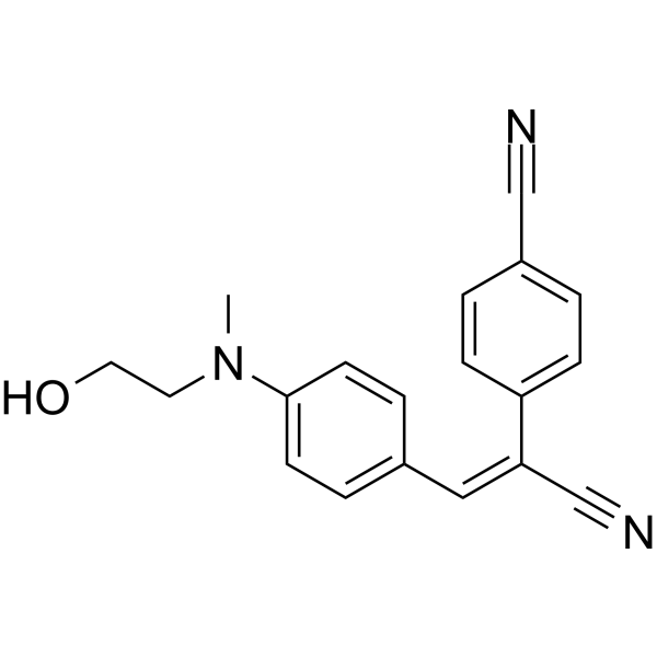
TMR-DN (TAMRA-2,4-dinitroaniline) is a bright orange fluorescent probe that binds to the SRB-2 aptamer, with a Kd value of 35 nM for the SRB-2 aptamer. TMR-DN exhibits low background fluorescence, enabling wash-free live-cell RNA imaging .
(S,R,S)-AHPC-PEG2-C4-Cl (Standard) is the analytical standard of (S,R,S)-AHPC-PEG2-C4-Cl (HY-103607). This product is intended for research and analytical applications. (S,R,S)-AHPC-PEG2-C4-Cl (VH032-PEG2-C4-Cl) is a conjugate of ligands for E3 and 13-atom-length linker. The connector of linker is Halogen group. (S,R,S)-AHPC-PEG2-C4-Cl incorporates the (S,R,S)-AHPC based VHL ligand and an alkyl/ether-based linker. (S,R,S)-AHPC-PEG2-C4-Cl is capable of inducing the degradation of GFP-HaloTag7 in cell-based assays .
(S,R,S)-AHPC-C6-PEG3-C4-Cl (Standard) is the analytical standard of (S,R,S)-AHPC-C6-PEG3-C4-Cl (HY-103605). This product is intended for research and analytical applications. (S,R,S)-AHPC-C6-PEG3-C4-Cl (VH032-C6-PEG3-C4-Cl) is a conjugate of ligands for E3 and 20-atom-length linker. The connector of linker is Halogen group. (S,R,S)-AHPC-C6-PEG3-C4-Cl incorporates the (S,R,S)-AHPC based VHL ligand and an alkyl/ether-based linker. (S,R,S)-AHPC-C6-PEG3-C4-Cl is capable of inducing the degradation of GFP-HaloTag7 in cell-based assays .
HBC (HBC 530) is a GFP fluorophore-like synthetic dye, with a structurally rigid electron acceptor and a strong electron donor. HBC has a low fluorescence background, and when combined with Pepper (RNA aptamer), HBC forms a tight complex and activates and emits bright fluorescence (Kd of ~3.5 nM). HBC emission peaks vary in different complexes and covers the spectrum from cyan to red. HBC can be used in the live cell imaging of RNA (Em/Ex = 530/485 nm) .
DFHBI is a small molecule that resembles the chromophore of green fluorescent protein (GFP). DFHBI can be used for imaging RNA in living cells. . Spinach and DFHBI are essentially nonfluorescent when unbound, whereas the Spinach-DFHBI complex is brightly fluorescent both in vitro and in living cells (Ex/Em = 469/501 nm).
ATTO 647 is a carborhodamine fluorophore and imaging tracer with photostable properties. ATTO 647 serves as a fluorescent probe to investigate cell membrane structure and diffusion characteristics. When conjugated with wheat germ agglutinin, ATTO 647 specifically binds to N-acetyl-β-D-glucosamine and sialic acid residues on membrane glycoproteins, enabling single-molecule tracing of glycoprotein diffusion. ATTO 647 exhibits highly stable fluorescence properties with significantly reduced blinking in mounting media such as ROXS (AA/MV) and ROXS (TX/TQ), whereas its brightness properties vary in Ibidi-MM and Vectashield. ATTO 647 can also be used to label histone H2B-GFP in fixed cells for confocal microscopy photobleaching experiments .
BODIPY TR methyl ester is a lipophilic GFP Counterstain. BODIPY TR methyl ester dye readily permeates cell membranes and localizes in endomembranous organelles but not localize strongly in plasma membranes. BODIPY TR methyl ester is an excellent red fluorescent vital dye (Ex=568 nm, Em=625 nm), can be used to reveal the location and shapes of cell nuclei, the shapes of cells within embryonic tissues, as well as the bound aries of organ-forming tissues within the whole embryo .
PE-AF700 is a tandem fluorescent dye used for flow cytometry, consisting of the donor phycoerythrin (PE) and the acceptor Alexa Fluor 700 (AF700) (Ex/Em = 488 nm/715 nm) .
TMR-DN (TAMRA-2,4-dinitroaniline) is a bright orange fluorescent probe that binds to the SRB-2 aptamer, with a Kd value of 35 nM for the SRB-2 aptamer. TMR-DN exhibits low background fluorescence, enabling wash-free live-cell RNA imaging .
Blue dextran (Dextran blue) (MW 2000000) is a high-molecular-weight long-chain polymer of D-glucose. Blue dextran (MW 2000000) serves as an important model macromolecular drug and molecular weight estimation marker, and can be used as a standard for gel permeation chromatography. The release of Blue dextran (MW 2000000) from alginate microspheres is regulated by preparation conditions; its release rate in a pH 6.8 environment is significantly faster than that in pH 1.2, and it exhibits release characteristics close to zero-order kinetics under this condition .
Stewart-Grubbs catalyst is an effective catalyst for the cross-metathesis of olefins with a large number of allylic substituents. In addition, ChemBeads are chemically coated glass beads that improve flowability and chemical homogeneity, making them ideal for automated solid dispensing and high-throughput experiments. Notably, the manufacture of ChemBeads does not require additional chemicals or surfactants, allowing for precise dispensing of sub-milligram amounts of catalyst.
GFP11 is a protein fusion tag consisting of 16 amino acids. When GFP11 and GFP1-10 are sufficiently close in physical space, they undergo spontaneous, irreversible complementation to reassemble into an intact, functional GFP protein that emits green fluorescence. GFP11 can be used to form a split GFP system for high-throughput biotechnology and flow cytometry applications .
Icrocaptide (ITF1697) is a stable Lys-Pro-containing peptide that inhibits the intracellular Ca 2+-dependent fusion of Weibel-Palade bodies with the plasma membrane. Icrocaptide exerts its activity at the early stages of endothelial activation and inhibits P-selectin and von Willebrand factor secretion. Icrocaptide can be used for the study of a variety of microvascular disorders .
PKC-ε translocation inhibitor peptide is a PKC-ε translocation inhibitor. PKC-ε translocation inhibitor peptide can regulate the rate of FcγR-mediated internalization of opsonized beads, has no effect of FcαR trafficking .
DC-peptoid-1 is a specific binder and crosslinker targeting the phosphorylated Brd4 PDID domain, with a dissociation constant (Kd) of approximately 50-100 μM for human-derived targets. DC-peptoid-1 only binds to phosphorylated PDID and fails to recognize the non-phosphorylated form or other domains (such as Brd4 598-785). DC-peptoid-1 effectively crosslinks with the target protein both in solution and cell lysates. It can successfully capture phosphorylated PDID from complex systems via immobilization, without binding to bacterial-derived non-phosphorylated proteins or other non-specific phosphoproteins. DC-peptoid-1 has the potential to serve as a phosphoprotein-specific antibody substitute for applications such as immunoaffinity purification .
MCE Protein A Plus Magnetic Agarose Beads can be used for the detection and purification of IgG from serum, ascites fluid, cell culture supernatant and other antibody samples.
MCE Protein G Plus Magnetic Agarose Beads can be used for the detection and purification of IgG from serum, ascites fluid, cell culture supernatant and other antibody samples.
Protein L MagneticBeads provide a fast and convenient method for Immunuoprecipitaion, Co-Immunoprecipitation and Chromatin Immunoprecipitation. The 1 mL is defined as the base specification. All larger sizes correspond to incremental volumes of this base.
MCE Hydroxyl Magneticbeads (200 nm, 10 mg/mL) can rapidly isolate nucleic acids from biological samples, which is conducive to the automation and high throughput extraction of nucleic acids.
Protein G MagneticBeads provide a fast and convenient method for Immunoprecipitation and Co-Immunoprecipitation and Chromatin Immunoprecipitation. The 1 mL is defined as the base specification. All larger sizes correspond to incremental volumes of this base.
Protein A MagneticBeads provide a fast and convenient method for Immunoprecipitation and Co-Immunoprecipitation and Chromatin Immunoprecipitation.The 1 mL volume is defined as the base specification. All larger sizes correspond to incremental volumes of this base.
MCE Protein A/G MagneticBeads provide a fast and convenient method for Immunoprecipitation, Co-Immunoprecipitation and Chromatin Immunoprecipitation. The 1 mL volume is defined as the base specification. All larger sizes correspond to incremental volumes of this base.
MCE Glutathione Magnetic Agarose Beads have high protein-binding capacity and stability, making it ideal for high performance purification of GST-tagged fusion proteins expressed in E. coli, yeast, insect and mammalian expression systems.
MCE Anti-GST MagneticBeads are used for immunoprecipitation (IP) of specific GST-tagged proteins expressed in bacterial and mammalian cells and in vitro expression systems. The 1 mL is defined as the base specification. All larger sizes correspond to incremental volumes of this base.
MCE Anti-His MagneticBeads are used for immunoprecipitation (IP) of specific His-tagged proteins expressed in bacterial and mammalian cells and in vitro expression systems. The 1 mL is defined as the base specification. All larger sizes correspond to incremental volumes of this base.
Anti-MBP MagneticBeads are suitable for the detection and purification of MBP-tagged fusion proteins, as well as for immunoprecipitation (IP) and co-immunoprecipitation (Co-IP) applications. The 1 mL is defined as the base specification. All larger sizes correspond to incremental volumes of this base.
MCE Anti-HA MagneticBeads are used for immunoprecipitation (IP) of specific HA-tagged proteins expressed in bacterial and mammalian cells and in vitro expression systems.The 1 mL volume is defined as the base specification. All larger sizes correspond to incremental volumes of this base.
MCE Anti-c-Myc MagneticBeads are used for immunoprecipitation (IP) of specific c-Myc-tagged proteins expressed in bacterial and mammalian cells and in vitro expression systems. The 1 mL is defined as the base specification. All larger sizes correspond to incremental volumes of this base.
MCE CHO MagneticBeads (200 nm, 10 mg/mL) contain CHO functional groups, which react with primary amines on proteins or other molecules to form stable amide linkages,can covalently immobilize proteins for the affinity purification of antibodies, antigens and other biomolecules.
MCE Anti-HA MagneticBeads (1 μm) are used for immunoprecipitation (IP) of specific HA-tagged proteins expressed in bacterial and mammalian cells andin vitro expression systems. The 1 mL is defined as the base specification. All larger sizes correspond to incremental volumes of this base.
MCE NHS MagneticBeads (200 nm, 10 mg/mL) contain N-hydroxysuccinimide (NHS) functional groups, which react with primary amines on proteins or
other molecules to form stable amide linkages,can covalently immobilize proteins for the affinity purification of antibodies, antigens and other
biomolecules.
MCE Streptavidin MagneticBeads provide a fast and convenient method for numerous applications, including purification of proteins and nucleic acids, protein interaction studies, immunoprecipitation, immunoassays, pull-down and cell isolation. The 1 mL is defined as the base specification. All larger sizes correspond to incremental volumes of this base.
MCE ConA MagneticBeads can be used to isolate cells or purify glycoproteins from serum and cell extracts. It is also employed in experiments such as collecting and immobilizing cell nuclei, CUT & Run, and CUT & Tag. The 1 mL is defined as the base specification. All larger sizes correspond to incremental volumes of this base.
MCE Anti-V5 MagneticBeads are well suited for the detection and purification of V5-tagged fusion proteins, as well as for applications such as immunoprecipitation (IP) and co-immunoprecipitation (Co-IP). The 1 mL is defined as the base specification. All larger sizes correspond to incremental volumes of this base.
MCE Amino magneticbeads (200 nm,10 mg/mL) can easily and efficiently combine with a variety of biological ligand in high loads, such as proteins, peptides, oligonucleotides, drug molecules, etc. It can be used as a good basic material for subsequent processing, adsorption, chemical modification and other follow-up processing.
MCE Oligo (dT)30MagneticBeads are designed for the rapid isolation of highly purified, intact mRNA from eukaryotic total RNA or directly from crude extracts of cells, plant and animal tissues. The 1 mL volume is defined as the base specification. All larger sizes correspond to incremental volumes of this base.
MCE Anti-Flag MagneticBeads are used for immunoprecipitation (IP) of specific Flag-tagged (DYKDDDDK) proteins expressed in bacterial and mammalian cells and in vitro expression systems, and also suitable for Co-immunoprecipitation and purification of Flag-tagged protein. The 1 mL is defined as the base specification. All larger sizes correspond to incremental volumes of this base.
MCE Carboxyl Magneticbeads (200 nm, 10 mg/mL) are characterized by superparamagnetism, fast magnetic response, abundant carboxyl functional groups, monodispersity, and submicron scale particle size. Biological ligands (proteins, peptides, oligonucleotides, drug molecules, etc.) can be covalently coupled to the surface of microspheres under the action of special chemical reagents (such as EDC).
MCE Human CD3/CD28 T Cell Activation MagneticBeads are based on the two important co-stimulatory signals, without relying on feeder cells (antigen-presenting cells) or antigens, CD3 and CD28, without relying on feeder cells (antigen-presenting cells) or antigens, enabling simple and rapid T cell activation.
MCE Mouse CD3/CD28 T Cell Activation MagneticBeads are based on the two important co-stimulatory signals, without relying on feeder cells (antigen-presenting cells) or antigens, CD3 and CD28, without relying on feeder cells (antigen-presenting cells) or antigens, enabling simple and rapid T cell activation.
Ganodermic acid S is an oxygenated triterpenoid, that can be isolated from the Chinese medicinal fungus Ganoderma lucidum (Fr.) Karst (Polyporaceae). Ganodermic acid S exerts a concentration dependent inhibition on the response of human gel-filtered platelets (GFP) to U-46619 (HY-108566), a thromboxane (TX) A2 mimetic .
The green fluorescent protein (GFP), Aequorea victoria is a protein that exhibits green fluorescence when exposed to light in the blue to ultraviolet range, found in organisms including Aequorea victoria, corals, sea anemones, zoanithids, copepods and lancelets. GFP Protein has a major excitation peak at a wavelength of 395 nm and a minor one at 475 nm, while its emission peak is at 509 nm, which is in the lower green portion of the visible spectrum. GFP Protein acts as an energy transfer receptor and actively transduces the blue chemiluminescence of the protein aequorin into green fluorescent light by energy transfer, which is used as expression of reporter genes. GFP Protein, Aequorea victoria (His) is a recombinant jellyfish Aequorea victoria GFP protein with N-6*His labeled tag that consists of 238 amino acids, which is expressed in E. coli .
GPR155 Protein, Human (sf9, GFP, Strep, His) is the recombinant human-derived GPR155, expressed by sf9 insect cells , with Strep, His, GFP labeled tag.
Fluorescent VLP with a GFP tag, self-assembles from viral envelope/capsid proteins, serving as a control for VLPs displaying membrane proteins. Formed in HEK293 cultures, these VLPs concentrate membrane proteins on the cell surface, producing high-concentration proteins ideal for immunization and antibody screening.
Prostatic acid phosphatase (ACPP) functions as a multifunctional tyrosine phosphatase under acidic conditions (pH 4-6), dephosphorylating a variety of substrates, including orthophosphate monoesters. ACPP has lipid phosphatase activity and inactivates lysophosphatidic acid in seminal plasma. Prostatic acid Phosphatase/ACPP Protein, Human (HEK293, GFP-His) is the recombinant human-derived Prostatic acid Phosphatase/ACPP protein, expressed by HEK293 , with C-His, GFP labeled tag.
HRH3-VLPs Protein, Human (HEK293, GFP) is recommended for animal immunization, ELISA. It is not recommended for receptor-ligand interaction detection and SPR/BLI assay since there are other irrelevant membrane proteins of the host on the VLP envelope, and the receptor-ligand interaction will have strong background interference. High requirements for chips and experimental protocols are needed for SPR/BLI assays. If VLP control is required, it is recommended HY-P701236. Tags can only be detected under denaturing conditions.
Claudin-6/CLDN6 Protein-VLP is pivotal in targeted elimination of intercellular spaces within tight junctions, ensuring cell barrier integrity and polarity. Moreover, it serves as a receptor for hepatitis C virus (HCV), facilitating viral entry into hepatic cells. Claudin-6 Protein-Nanodisc, Human (HEK293, GFP, His) is the recombinant human-derived Claudin-6 protein, expressed by HEK293, with C-His and C-GFP labeled tag.
Claudin-6/CLDN6 Protein-VLP, Human (HEK293, GFP) is recommended for animal immunization, ELISA. It is not recommended for receptor-ligand interaction detection and SPR/BLI assay since there are other irrelevant membrane proteins of the host on the VLP envelope, and the receptor-ligand interaction will have strong background interference. High requirements for chips and experimental protocols are needed for SPR/BLI assays. If VLP control is required, it is recommended HY-P701236. Tags can only be detected under denaturing conditions.
The GPR158 protein is a key orphan receptor in vision that regulates signaling in retinal depolarizing bipolar cells. As an atypical G protein-coupled receptor, it recruits and regulates the R7 group RGS-GNB5 complex, which is different from traditional G protein activation. GPR158 Protein, Human (HEK293, GFP, FLAG, His) is the recombinant human-derived GPR158 protein, expressed by HEK293 , with C-10*His, C-GFP, C-Flag labeled tag.
Claudin-6/CLDN6 Protein-VLP, Human (HEK293, GFP, solution) is recommended for animal immunization, ELISA. It is not recommended for receptor-ligand interaction detection and SPR/BLI assay since there are other irrelevant membrane proteins of the host on the VLP envelope, and the receptor-ligand interaction will have strong background interference. High requirements for chips and experimental protocols are needed for SPR/BLI assays. If VLP control is required, it is recommended HY-P705433.
EDNRB/Endothelin R Type B Protein-VLP, Human (HEK293, GFP) is recommended for animal immunization, ELISA. It is not recommended for receptor-ligand interaction detection and SPR/BLI assay since there are other irrelevant membrane proteins of the host on the VLP envelope, and the receptor-ligand interaction will have strong background interference. High requirements for chips and experimental protocols are needed for SPR/BLI assays. If VLP control is required, it is recommended HY-P705433.
Claudin-6/CLDN6 Protein-VLP is pivotal in targeted elimination of intercellular spaces within tight junctions, ensuring cell barrier integrity and polarity. Moreover, it serves as a receptor for hepatitis C virus (HCV), facilitating viral entry into hepatic cells. Claudin-6/CLDN6 Protein-Nanodisc, Human (HEK293, His, Flag, GFP) is the recombinant human-derived Claudin-6/CLDN6 protein, expressed by HEK293, with N-His and C-GFP and N-Flag labeled tag.
CD9 protein is an integral membrane protein that plays a critical regulatory role in processes such as sperm-egg fusion, platelet activation, and cell adhesion. CD9 is critical for sperm-egg fusion, coordinating multi-protein complexes on the oocyte surface. CD9 Protein-Nanodisc, Human (HEK293, His, GFP) is the recombinant human-derived CD9 protein, expressed by HEK293, with N-His and N-GFP labeled tag.
Claudin-18/CLDN18.2 Protein-VLP, Human (HEK293, GFP) is recommended for animal immunization, ELISA. It is not recommended for receptor-ligand interaction detection and SPR/BLI assay since there are other irrelevant membrane proteins of the host on the VLP envelope, and the receptor-ligand interaction will have strong background interference. High requirements for chips and experimental protocols are needed for SPR/BLI assays. If VLP control is required, it is recommended HY-P705433.
SSTR2 Protein-VLP, Human (HEK293, GFP) is recommended for animal immunization, ELISA. It is not recommended for receptor-ligand interaction detection and SPR/BLI assay since there are other irrelevant membrane proteins of the host on the VLP envelope, and the receptor-ligand interaction will have strong background interference. High requirements for chips and experimental protocols are needed for SPR/BLI assays. If VLP control is required, it is recommended HY-P705433.
As the catalytic subunit of the VKOR complex, the VKOR1 protein is crucial in vitamin K metabolism and is essential for the reduction of inactive vitamin K 2,3-epoxide to the active form. This process is critical for γ-carboxylation of proteins and is particularly important for blood coagulation and the synthesis of coagulation factors. VKOR1 Protein, Human (HEK293, GFP, His) is the recombinant human-derived VKOR1 protein, expressed by HEK293 , with C-10*His, GFP labeled tag.
The SLC15A2 protein is a proton-coupled amino acid transporter that prefers dipeptides and transports oligopeptides containing 2 to 4 amino acids. The stoichiometric ratio of protons to peptides is 2:1 or 3:1, which helps the kidneys absorb dipeptides and tripeptides. SLC1542 Protein, Human (HEK293, GFP-Strep-His) is the recombinant human-derived SLC15A2 protein, expressed by HEK293 , with C-GFP-Strep-His tag.
SLC12A2 Protein mediates electroneutral transport of chloride, potassium, and/or sodium ions across the membrane, crucial for regulating ionic balance and cell volume. SLC12A2 Protein, Human (HEK293, His, Strep, GFP) is the recombinant human-derived SLC12A2 protein, expressed by HEK293 , with N-Strep, N-EGF, N-His labeled tag.
The ABCC1 protein plays a key role in cellular physiology, mediating the ATP-dependent export of a variety of substrates, including drugs and organic anions. Notably, it confers resistance to anticancer drugs, actively reducing their intracellular accumulation. ABCC1 Protein, Bovine (HEK293, GFP, Strep, His) is the recombinant bovine-derived ABCC1 protein, expressed by HEK293 , with C-10*His, C-GFP, C-StrepII labeled tag.
The hERG protein is the α subunit of voltage-gated potassium channels and is critical for mediating cardiac delayed rectifier potassium current (IKr). It lacks individual channel activity but dynamically affects properties by forming heterotetramers with other isoforms. hERG Protein, Human (HEK293, GFP, His) is the recombinant human-derived hERG protein, expressed by HEK293 , with C-10*His, C-GFP labeled tag.
CCR5 Protein-VLP, Human (HEK293, GFP) is recommended for animal immunization, ELISA. It is not recommended for receptor-ligand interaction detection and SPR/BLI assay since there are other irrelevant membrane proteins of the host on the VLP envelope, and the receptor-ligand interaction will have strong background interference. High requirements for chips and experimental protocols are needed for SPR/BLI assays. If VLP control is required, it is recommended HY-P705433.
Cetzole (Compound 1) is a ferroptosis inducer that induces cell death through ROS accumulation. The CC50 values of Cetzole for NCI-H522, NCI-H522 GFP-SCL7A11 #8, NCI-H522 RV-GFP, HT-1080, NARF2, and MDA-MB-231 are 2.56, 10.31, 2.71, 3.07, 14.9, and 6.28 μM, respectively. Cetzole holds potential for research in the field of cancer .
N1-methyl-pseudouridine (1-Methylpseudouridine), a methylpseudouridine, outperforms 5 mC and 5 mC/N1-methyl-pseudouridine in translation. N1-methyl-pseudouridine in mRNA enhances translation through eIF2α-dependent and independent mechanisms by increasing ribosome density .
LNP Lipid-8 (11-A-M) is an ionizable single-tail multi-head lipid that can be used as a lipid nanoparticle (LNP) to deliver siRNA to T cells without targeting ligands. LNP Lipid-8 is more selective for T cells than other cell types such as hepatocytes. LNP Lipid-8 selectively delivers siRNA/sgRNA to T cells (especially CD8+ T cells) through endogenous lipid transport pathways, and can enter cells and release RNA through endocytosis to achieve gene silencing. LNP Lipid-8 loaded with GFP siRNA (siGFP) significantly led to GFP gene silencing in mouse models. LNP Lipid-8 showed good efficacy and safety in both cells and animals, without obvious liver targeting and toxicity. LNP Lipid-8 can be used for RNA delivery research in the fields of tumor immunotherapy and T cell-mediated autoimmune diseases .
The Cas9-T2A-GFP mRNA encodes a Cas9 nuclease gene with two nuclear localization signals (NLS) and a green fluorescent protein (GFP), which could be used in genome engineering experiments.
The Cas9-T2A-GFP mRNA encodes a Cas9 nuclease gene with two nuclear localization signals (NLS) and a green fluorescent protein (GFP), which could be used in genome engineering experiments. The incorporation of N1-Methylpseudo-UTP can reduce the immunogenicity of the resulting mRNA.
The Cre-T2A-GFP mRNA is a capped and polyadenylated messenger RNA encoding a Cre recombinase with a nuclear localization sequence (NLS) and a green fluorescent protein (GFP).
The Cre-T2A-GFP mRNA is a capped and polyadenylated messenger RNA encoding a Cre recombinase with a nuclear localization sequence (NLS) and a green fluorescent protein (GFP). The incorporation of N1-Methylpseudo-UTP can reduce the immunogenicity of the resulting mRNA.
3'-O-(2-Nitrobenzyl)-dGTP is a reversible terminator. 3'-O-(2-Nitrobenzyl)-dGTP can be recognized and incorporated by DNA polymerases, thereby temporarily terminating DNA primer extension; after the 2-nitrobenzyl blocking group is removed via laser irradiation, a free 3'-OH can be regenerated to allow subsequent polymerase-mediated extension. 3'-O-(2-Nitrobenzyl)-dGTP can be used in DNA sequencing studies .
Inquiry Online
Your information is safe with us. * Required Fields.
Western blot analysis of extracts from THP-1(lane 2(20μg), Jurkat (lane 3(20μg) and NIH3T3(lane 4(20μg) using FOXO1A (HY-P80132) Rabbit mAb. Proteins were transferred
to a PVDF membrane and blocked with 5% non-fat milk in TBST for 2 hour at room temperature. The primary antibody (1/1000) and Loading control antibody (Beta Actin, HY-P80438, 1/10000) was
used in 5% non-fat milk in TBST at 4°C overnight. Goat Anti-Mouse/Rabbit IgG-HRP Secondary Antibody (1/10000) was used for 1 hour at room temperature.
Western blot analysis of extracts from THP-1(lane 2(20μg), Jurkat (lane 3(20μg) and NIH3T3(lane 4(20μg) using FOXO1A (HY-P80132) Rabbit mAb. Proteins were transferred
to a PVDF membrane and blocked with 5% non-fat milk in TBST for 2 hour at room temperature. The primary antibody (1/1000) and Loading control antibody (Beta Actin, HY-P80438, 1/10000) was
MedchemExpress Validation 03
Western blot analysis of extracts from THP-1(lane 2(20μg), Jurkat (lane 3(20μg) and NIH3T3(lane 4(20μg) using FOXO1A (HY-P80132) Rabbit mAb. Proteins were transferred
MedchemExpress Validation 04
Western blot analysis of extracts from THP-1(lane 2(20μg), Jurkat (lane 3(20μg) and NIH3T3(lane 4(20μg) using FOXO1A (HY-P80132) Rabbit mAb. Proteins were transferred
to a PVDF membrane and blocked with 5% non-fat milk in TBST for 2 hour at room temperature. The primary antibody (1/1000) and Loading control antibody (Beta Actin, HY-P80438, 1/10000) was
used in 5% non-fat milk in TBST at 4°C overnight. Goat Anti-Mouse/Rabbit IgG-HRP Secondary Antibody (1/10000) was used for 1 hour at room temperature.
MedchemExpress Validation
Western blot analysis of extracts from THP-1(lane 2(20μg), Jurkat (lane 3(20μg) and NIH3T3(lane 4(20μg) using FOXO1A (HY-P80132) Rabbit mAb. Proteins were transferred
to a PVDF membrane and blocked with 5% non-fat milk in TBST for 2 hour at room temperature. The primary antibody (1/1000) and Loading control antibody (Beta Actin, HY-P80438, 1/10000) was
used in 5% non-fat milk in TBST at 4°C overnight. Goat Anti-Mouse/Rabbit IgG-HRP Secondary Antibody (1/10000) was used for 1 hour at room temperature.
MedchemExpress Validation
Western blot analysis of extracts from THP-1(lane 2(20μg), Jurkat (lane 3(20μg) and NIH3T3(lane 4(20μg) using FOXO1A (HY-P80132) Rabbit mAb. Proteins were transferred
to a PVDF membrane and blocked with 5% non-fat milk in TBST for 2 hour at room temperature. The primary antibody (1/1000) and Loading control antibody (Beta Actin, HY-P80438, 1/10000) was
used in 5% non-fat milk in TBST at 4°C overnight. Goat Anti-Mouse/Rabbit IgG-HRP Secondary Antibody (1/10000) was used for 1 hour at room temperature.
MedChemExpress values your privacy and your trust is important to us. We use cookies to enhance your website experience. Some cookies are necessary to run the website.
Privacy and Cookie Policy