



ATTO 647
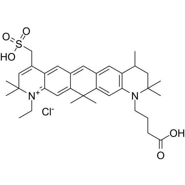
ATTO 647 is a carborhodamine fluorophore and imaging tracer with photostable properties. ATTO 647 serves as a fluorescent probe to investigate cell membrane structure and diffusion characteristics. When conjugated with wheat germ agglutinin, ATTO 647 specifically binds to N-acetyl-β-D-glucosamine and sialic acid residues on membrane glycoproteins, enabling single-molecule tracing of glycoprotein diffusion. ATTO 647 exhibits highly stable fluorescence properties with significantly reduced blinking in mounting media such as ROXS (AA/MV) and ROXS (TX/TQ), whereas its brightness properties vary in Ibidi-MM and Vectashield. ATTO 647 can also be used to label histone H2B-GFP in fixed cells for confocal microscopy photobleaching experiments.
연구목적의 판매만을 진행합니다. 환자를 대상으로 한 판매는 하지 않습니다.
- CAS No.: 906664-68-4
- 화학식: C34H45ClN2O9S
- 분자량:693.25
-
보관:
Please store the product under the recommended conditions in the Certificate of Analysis.

To place orders, for customer services and technical support, please contact: MedChemExpress USA
Tel: 609-228-6898 E-mail: [email protected] [email protected]


-
 Biological Activity
-
Chemical Information
- 순도&문서
- References
-
 Help & FAQs




Biological Activity
제품 설명
In Vitro
ATTO 647 (0.3 mg; 2 h) can covalently label the purified WGA, with an average of 1.5 ATTO 647 fluorescent groups binding to each WGA molecule[1].
ATTO 647 (100 nM) in the Atto 647-WGA conjugate will exhibit single-step or double-step photobleaching when bound to the hyaluronic acid-coated glass cover slip, which is consistent with the situation where each conjugate contains one or two fluorescent groups[1].
Guide (The following is the experimental plan we recommend. This plan serves only as a reference guide. The specific operations should be adjusted according to your actual needs.)
Application of ATTO 647 in the Stably Expressing Group Protein H2B-GFP Human HeLa Cells for Staining[2][3]:
1. Cultivate the cells until 50-80% confluence, fix with 3.7% formaldehyde.
2. Triton X-100 0.5% permeabilization, BSA/PBST 2% blocking.
3. Incubate with ATTO 647N conjugated antibody to label the intranuclear group protein H2B-GFP.
4. Seal the sections with the following media: PBS, Vectashield (VS), Ibidi-MM, ROXS (AA/MV) (1 mM ascorbic acid + 1 mM methylene blue + enzymatic deoxygenation system), ROXS (TX/TQ) (1 mM Trolox + UV pre-generated Trolox - quinone + enzymatic deoxygenation system).
5. Observe the results under a 635 nm laser.
MedChemExpress (MCE) has not independently confirmed the accuracy of these methods. They are for reference only. .


Chemical Information
-
CAS No. 906664-68-4
-
분자량 693.25
-
화학식 C34H45ClN2O9S
-
SMILES
O=C(CCCN1C2=C(C(CC1(C)C)C)C=C(C=C3C=C4C(C=C3C5(C)C)=[N+](C(C)(C)C=C4CS(=O)(O)=O)CC)C5=C2)O.O=Cl(=O)([O-])=O
-
선적
Room temperature in continental US; may vary elsewhere.
-
보관
Please store the product under the recommended conditions in the Certificate of Analysis.


순도&문서
References
[1]. Bruckbauer A, et al. Nanopipette delivery of individual molecules to cellular compartments for single-molecule fluorescence tracking. Biophys J. 2007;93(9):3120-3131. [Content Brief]
[2]. Cordes T, et al. Mechanisms and advancement of antifading agents for fluorescence microscopy and single-molecule spectroscopy. Phys Chem Chem Phys. 2011;13(14):6699-6709. [Content Brief]
[3]. Kocabey S, et al. Cellular Uptake of Tile-Assembled DNA Nanotubes. Nanomaterials (Basel). 2014;5(1):47-60. Published 2014 Dec 30. [Content Brief]

Calculators
Concentration (start) × Volume (start) = Concentration (final) × Volume (final)


ATTO 647 Related Classifications
HY-D1991 Related Classifications
 Powered by Bioz