



IP/Co-IP Kit (Protein A/G Magnetic Beads)
Based on 5 publication(s) in Google Scholar
MCE IP/Co-IP Kit (Protein A/G Magnetic Beads) can be used for protein purification, IP, Co-IP of target proteins or their protein complexes.
-
Storage :
Protein A/G Magnetic Beads: 4°C, 2 years. Do not freeze. Elution Buffer, Neutralization Buffer: 4°C, 2 years. IP Lysis Buffer, Protease Inhibitor Cocktail (100×), PMSF (100×),Protein Loading Buffer (Reducing, 4×), Protein Loading Buffer (Non-Reducing, 5×): -20°C, 2 years. TBS-T Powder: RT, 2 years.


To place orders, for customer services and technical support, please contact: MedChemExpress USA
Tel: 609-228-6898 E-mail: [email protected] [email protected]



-

HY-13259
MG-132 -

HY-K0200A
Magnetic Stand Plus -
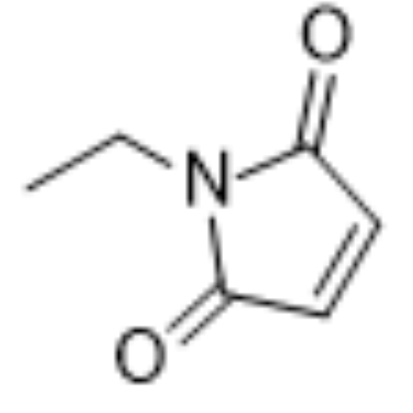
HY-D0843
N-Ethylmaleimide -

HY-P80879
Rabbit IgG Isotype Control -

HY-10071
Y-27632 -
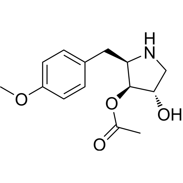
HY-18982
Anisomycin -

HY-100579
Ferrostatin-1 -
HY-K0202
Protein A/G Magnetic Beads -

HY-E70005A
Collagenase, Type I -
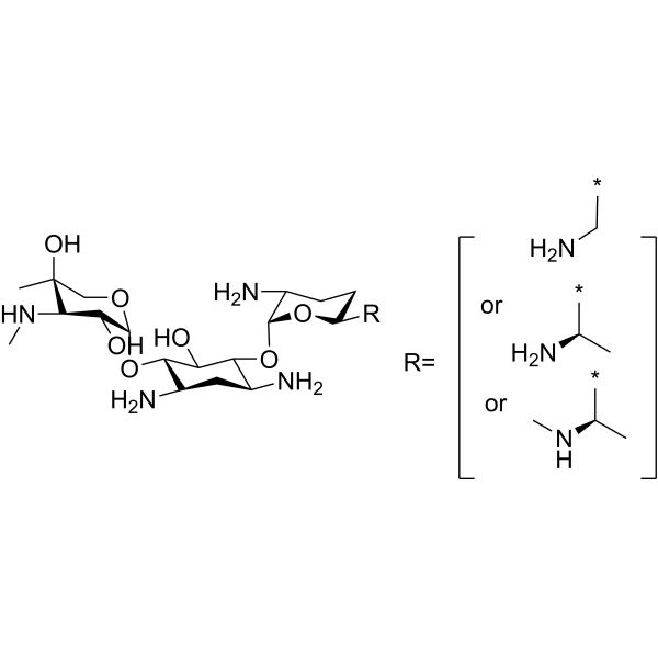
HY-A0276A
Gentamicin -
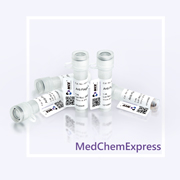
HY-K0207
Anti-Flag Magnetic Beads -
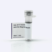
HY-K0209
Anti-His Magnetic Beads


Description & Advantages
MCE IP/Co-IP Kit (Protein A/G Magnetic Beads) is a kit developed based on Protein A/G magnetic beads. The kit includes recombinant Protein A/G magnetic beads and optimally validated reagents necessary for immunoprecipitation (IP) experiments. It can be used for protein purification, IP, Co-IP of target proteins or their protein complexes.
The kit contains sufficient reagents to perform 40 reactions using 25μL of magnetic beads.
Feature of MCE IP/Co-IP Kit (Protein A/G Magnetic Beads)
Compatible: Based on recombinant protein A/G magnetic beads, compatible with a wide range of antibody species.
Universal: Suitable for cell lysates, cell culture supernatants, serum, ascitic fluid, and other sample types.
Convenient: The kit includes magnetic beads and all necessary buffers required to complete the IP experiment.


Protocol
1. Buffer Preparation:
| Buffer | |
|---|---|
| Lysis Buffer | The IP lysis buffer is ready for use after thawing. |
| Binding/Wash Buffer | Dissolve the TBS-T instant granules in ultrapure water and bring the final volume to 1 L. |
| Elution Buffer | Use the elution buffer supplied with the kit directly. |
| Neutralization Buffer | Use the neutralization buffer supplied with the kit directly. |
| Protein Loading Buffer | Dilute the protein loading buffer supplied with the kit to 1× before use. |
Notes:
a. After thawing, it is recommended to aliquot the IP lysis buffer appropriately to minimize repeated freeze-thaw cycles.
b. Prepared TBS-T should be stored at 4°C and can be kept for at least one month.
c. Reducing or non-reducing protein loading buffer may be used according to the sample requirements.
2. Antigen Sample Preparation:
| Sample Type | Processing Method |
|---|---|
| Serum | If the target protein is highly abundant, dilute the serum sample to a final target protein concentration of 10-100 μg/mL and keep it on ice until use, or store it at -20°C for long-term preservation. |
| Suspension Cells |
1. Collect the cells by centrifugation (4°C, 500 g, 10 min). Discard the supernatant, weigh the cells, and wash twice with PBS (1×); 2. Add lysis buffer at a ratio of 5-10 μL/mg of cells. Add an appropriate amount of PMSF and protease inhibitor simultaneously, mix thoroughly, and incubate on ice for 10 min to ensure complete lysis; 3. Centrifuge at 4°C and 14000 g for 10 min. Collect the supernatant and keep it on ice until use or store it at -20°C for long-term preservation. |
| Adherent Cells |
1. Remove the culture medium and wash twice with PBS (1×); 2. Detach the cells using a cell scraper and collect them in a 1.5 mL EP tube. Add lysis buffer at a ratio of 20-30 μL per 1×10⁵ cells. Add an appropriate amount of PMSF and protease inhibitor simultaneously, mix thoroughly, and incubate on ice for 10 min to ensure complete lysis; 3. Centrifuge at 4°C and 14000 g for 10 min. Collect the supernatant and keep it on ice until use or store it at -20°C for long-term preservation. |
Note: For cell lysis in a 6-well plate, 150-250 μL of lysis buffer may also be added to each well.
3. Magnetic Bead Pretreatment:
Thoroughly resuspend the magnetic beads. Transfer 25 μL of magnetic beads into a 1.5 mL EP tube, add 200 μL of binding/wash buffer, mix thoroughly, place the tube on a magnetic stand for magnetic separation, and discard the supernatant. Repeat the washing procedure twice.
Note: To ensure an even distribution of the magnetic beads, mix them thoroughly by repeated inversion, gentle vortexing, or using a mixer.
4. Immunocomplex Preparation:
The required sample amount and incubation time depend on the specific antibody-antigen system and may need to be optimized to achieve the maximum yield. The following protocol is designed for 2-20 μg of affinity-purified antibody and may be scaled proportionally as needed.
1. In a centrifuge tube, combine the cell lysate from each sample with 2-10 μg of immunoprecipitation antibody.
The protein concentration can be measured using a Protein Concentration Assay Kit (MCE Cat. No.: HY-K2001). A total protein amount of 500-1000 μg is recommended for each immunoprecipitation reaction.
2. Dilute the antigen-antibody reaction mixture to 500 μL with binding/wash buffer, mix thoroughly, and incubate on a rotator at room temperature for 1-2 h or at 4°C overnight to form the immunocomplex.
5. Immunocomplex Precipitation:
1. Add the antigen-antibody immunocomplex prepared above to the pretreated magnetic beads and incubate on a rotator at room temperature for 1-2 h or at 4°C for 2-4 h. Perform magnetic separation and collect the magnetic beads. Transfer the supernatant to a new EP tube for subsequent analysis.
2. Add 500 μL of binding/wash buffer to the centrifuge tube, mix gently, and perform magnetic separation. Collect the magnetic beads and transfer the supernatant to a new EP tube for subsequent analysis. Repeat the washing procedure twice.
6. Antigen Elution:
Two elution methods are provided. Select the appropriate method according to the experimental requirements.
1. Acidic Elution: Samples eluted using this method retain their original biological activity and can be used for subsequent functional analysis.
Add 100 μL of elution buffer to the centrifuge tube, mix thoroughly, and incubate at room temperature for 5-10 min. Perform magnetic separation, collect the supernatant, and immediately add neutralization buffer at 1/10 of the total eluate volume to neutralize the pH. The sample can then be used for subsequent functional analysis.
Note: The eluted protein can be stored at 4°C for a short period. For long-term storage, it is recommended to store the sample at -20°C.
2. Denaturing Elution: Samples eluted using this method are suitable for SDS-PAGE analysis.
Add 100 μL of protein loading buffer (1×) to the centrifuge tube, mix thoroughly, and heat at 95°C for 5 min. Perform magnetic separation, collect the supernatant, and analyze it by SDS-PAGE using the prestained protein marker supplied with the kit (10-190 kDa).
Notes:
a. Dilute the protein loading buffer to 1× with electrophoresis running buffer or ultrapure water. Reducing or non-reducing protein loading buffer may be selected according to the electrophoresis conditions.
b. At least three protein bands should be present in reducing SDS-PAGE or Western blot results: the antibody heavy chain (50 kDa), antibody light chain (25 kDa), and the antigen.


Publications
-
Journal Impact Factor
-
Most Recent
Storage
Protein A/G Magnetic Beads: 4°C, 2 years. Do not freeze.
Elution Buffer, Neutralization Buffer: 4°C, 2 years.
IP Lysis Buffer, Protease Inhibitor Cocktail (100×), PMSF (100×),Protein Loading Buffer (Reducing, 4×), Protein Loading Buffer (Non-Reducing, 5×): -20°C, 2 years.
TBS-T Powder: RT, 2 years.


Attention
1. Do not centrifuge, dry, or freeze the magnetic beads. These operations may cause the beads to aggregate and reduce their binding capacity.
2. Before use, confirm the antibody subclass and its affinity with Protein A/G by referring to the appendix. If the affinity is poor, you can improve the binding efficiency by increasing the incubation time of the antibody with the magnetic beads (30 - 120 min), raising the pH of the binding buffer (8 - 9), and reducing the ionic strength (25 - 100 mM NaCl).
3. Ultrasonication will cause the antibody captured by the magnetic beads to detach from the beads. Therefore, after capturing the antibody, avoid using this method to resuspend the magnetic beads.
4. This product is for R&D use only, not for drug, household, or other uses.
5. For your safety and health, please wear a lab coat and disposable gloves to operate.


Components
| Cat. No. | Product List | Components | HY-K0202K-40 T | Storage |
|---|---|---|---|---|
| HY-K0202K-A (1 Kit) |
IP/Co-IP reagent A |
Protein A/G IP Magnetic Beads Elution Buffer Neutralization Buffer |
1 mL 5 mL 1 mL |
4°C, 2 years. Do not freeze the magnetic beads. |
| HY-K0202K-B (1 Kit) |
IP/Co-IP reagent B |
IP Lysis Buffer Protease Inhibitor Cocktail (100×) PMSF (100×) Protein Loading Buffer (Reducing, 4×) Protein Loading Buffer (Non-Reducing, 5×) Prestained Protein Marker |
50 mL 1 mL 1 mL 1 mL 1 mL 250 μL |
-20°C, 2 years. |
| HY-K0202K-C (1 Kit) |
IP/Co-IP reagent C |
TBS-T Powder | 1 pouch | RT, 2 years. |


 Please enter the correct Lot Number
