From 11:00 pm to 12:00 pm EST ( 8:00 pm to 9:00 pm PST ) on January 6th, the website will be under maintenance. We are sorry for the inconvenience. Please arrange your schedule properly.
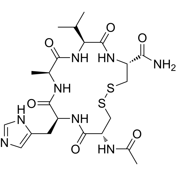
Adrixetinib (Q702) is an orally active triple inhibitor against CSF1R, Mer, and Axl, with Kd values of 8.7 nM, 0.8 nM, and 0.3 nM, respectively. Adrixetinib acts as a potent immune modulator that remodels the tumor microenvironment. Adrixetinib increases the abundance of M1 macrophages and CD8⁺ T cells, while decreasing the levels of M2 macrophages and myeloid-derived suppressor cells (MDSCs). Adrixetinib upregulates the expression of MHC class I and E-cadherin in tumor cells. Adrixetinib shows remarkable antitumor efficacy in syngeneic mouse tumor models. Adrixetinib is suitable for the research of breast cancer, renal adenocarcinoma, colon carcinoma, and melanoma .
Zarutatug (TORL-3-600 antibody) is an IgG1κ humanized antibody targeting cadherin 17 (CDH17). It selectively binds to cell-surface CDH17, triggering endocytosis and trafficking to lysosomes. Zarutatug can be used to construct ADCs, such as TORL-3-600 .
Aristolactam I is an AQP1 inhibitor and Aristolochic acid I metabolite. Aristolactam I can be isolated from Aristolochia plants. Aristolactam I downregulates Twist1 expression, increases E-cadherin expression, and activates the TGF-β/Smad signaling pathway. Aristolactam I has anticancer activity against breast cancer. Aristolactam I is nephrotoxic. Aristolactam I is mainly used in the study of breast cancer and kidney diseases such as renal interstitial fibrosis .
The Anti-CDH17/Cadherin-17 Antibody (PTA001_A4) is a humanized antibody expressed in CHO cells, targeting CDH17/Cadherin-17. The Anti-CDH17/Cadherin-17 Antibody (PTA001_A4) features an IgG1 heavy chain and a huκ light chain, with a predicted molecular weight (MW) of 149.12 kDa. The isotype control for the Anti-CDH17/Cadherin-17 Antibody (PTA001_A4) can be referenced as Human IgG1 kappa, Isotype Control (HY-P99001).
Murine Fibrinogen is a native fibrinogen derived from mouse plasma. Murine Fibrinogen acts as a cerebrovascular permeability enhancer. Murine Fibrinogen activates matrix metalloproteinase-9 (MMP-9), downregulates the expression of vascular endothelial cadherin(VE-cadherin), and upregulates the expression of plasmalemmal vesicle-associated protein-1 (PV-1). Murine Fibrinogen increases macromolecular leakage from pial veins, thereby disrupting the microvascular integrity of cerebral blood vessels. Murine Fibrinogen can be used in studies related to cerebrovascular dysfunction .
LNSMGQD is a cyclic peptide fragment derived from desmoglein 1 (amino acids 81-86), which mimics trans-interactions and acts as part of the tandem peptide binding interface of desmoglein 2. LNSMGQD not only binds to desmoglein 1 and 3, but also effectively inhibits their homophilic trans-interactions, while reducing the probability of homophilic or heterophilic binding between desmoglein 2 and Dsc2, N-cadherin and E-cadherin. LNSMGQD is applicable to the research on disease mechanisms such as Crohn's disease and pemphigus vulgaris .
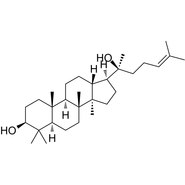
Dammarenediol II is a ginsenoside precursor . Dammarenediol II reduces the activity of O-GlcNAc transferase (OGT) and downregulates the global O-GlcNAcylation level. Dammarenediol II inhibits the phosphorylation of Akt, mTOR and GSK3β. Dammarenediol II inhibits human carboxylesterase activity, VEGF-induced ROS production, stress fiber formation and vascular endothelial cadherin disruption. Dammarenediol II promotes cell apoptosis (apoptosis), increases the levels of cleaved PARP1 and p53, and inhibits retinal microvascular leakage. Dammarenediol II can be used in studies related to liver cancer and diabetic retinopathy .
Fibrin is an insoluble protein found in blood that is produced in response to bleeding. As the main component of blood clots, fibrin functions in blood coagulation. Fibrin binds to the integrins ICAM-1, VE-cadherin, αIIbβ3, αMβ2, αvβ3 and α5β1. Fibrin is used in research related to chronic wounds .
Vasculotide is a blood-brain barrier (BBB)-penetrant Tie2 agonist. Vasculotide binds to a unique domain of Tie2, induces receptor clustering to drive phosphorylation, activates downstream PI3K/Akt and eNOS pathways, enhances inter-endothelial cell junctions (such as VE-cadherin and claudin-5), and inhibits inflammatory adhesion molecules, ultimately stabilizing the vascular endothelial barrier and reducing its permeability . Vasculotide alleviates pulmonary microvascular leakage and microcirculatory dysfunction caused by cardiopulmonary bypass, acts as an adjuvant radioprotective agent to reduce acute radiation dermatitis, and promotes BBB recovery after focused ultrasound (FUS). Combination of Vasculotide with antibiotics reduces lung injury .
Anti-CDH17/Cadherin-17 Antibody (10C12) is a human antibody expressed in CHO cells, targeting CDH17/Cadherin-17. Anti-CDH17/Cadherin-17 Antibody (10C12) can be referenced as Human IgG1 kappa, Isotype Control (HY-P99001). Anti-CDH17/Cadherin-17 Antibody (10C12) can be used in the study of cancer .
BAS00602705 is an E-cadherin inhibitor. BAS00602705 exerts its function by blocking the trans-interactions of E-cadherin molecules within junctional complexes, without permanently altering E-cadherin expression levels. BAS00602705 significantly impairs invadopodia formation in pancreatic cancer cells. BAS00602705 can be used for the study of anti-invasive therapeutic strategies in cancers characterized by dysregulated E-cadherin-mediated invadopodia activity, such as pancreatic ductal adenocarcinoma (PDAC) .
FX-06 (Fibrin-derived peptide Bβ15-42) is a fibrin Bbeta chain-derived peptide. FX-06 binds to VE-cadherin and inhibits leukocyte transmigration and initiates VE-cadherin-mediated signaling. FX-06 can be used in the research of ischemia/reperfusion injury, Dengue shock syndrome (DSS) .
PRMT7-IN-2 (A33) is a selective PRMT7 inhibitor with an IC50 of 0.50 μM. PRMT7-IN-2 arrests cell cycle at G0/G1 phase, induces cell apoptosis, and inhibits cell growth in vivo and in vitro. PRMT7-IN-2 decreases the monomethylarginine level of PRMT7, increases expression of epithelial marker (E-cadherin, and reduces expression of mesenchymal markers such as N-cadherin, Vimentin, and ZEB2 .
ANT308 is a vasoactive intestinal polypeptide (VIP receptor) antagonist. ANT308 significantly enhances the activation and proliferation of T cells. ANT308 inhibits the migration and metastasis, induces apoptosis of melanoma tumor cells by inhibiting VIP-VPAC2 signaling and reducing the expression of MCAM and N-cadherin. ANT308 can be used for the studies of acute myeloid leukemia (AML) and uveal melanoma (UVM) .
ANT308 TFA is a vasoactive intestinal polypeptide (VIP receptor) antagonist. ANT308 TFA significantly enhances the activation and proliferation of T cells. ANT308 TFA inhibits the migration and metastasis, induces apoptosis of melanoma tumor cells by inhibiting VIP-VPAC2 signaling and reducing the expression of MCAM and N-cadherin. ANT308 TFA can be used for the study of acute myeloid leukemia (AML) and uveal melanoma (UVM) .
(R)-MMP408 is an isomer of MMP408 (HY-12093). MMP408 is an orally active MMP-12 inhibitor (IC50=2.0 nM for hMMP-12) that effectively interferes with the epithelial-mesenchymal transition (EMT) process. MMP408 significantly upregulates the expression of E-cadherin in nasal epithelial cells, while inhibiting mesenchymal markers such as vimentin, α-smooth muscle actin and fibronectin, thereby reversing the EMT phenotype. MMP408 is used in studies of airway remodeling-related diseases, including chronic rhinosinusitis with nasal polyps, chronic obstructive pulmonary disease (COPD) and asthma .
Anti-CDH6/K-Cadherin Antibody (HKT288 antibody) is a human antibody expressed in CHO cells, targeting CDH6. Anti-CDH6/K-Cadherin Antibody (HKT288 antibody) can be referenced as Human IgG1 kappa, Isotype Control (HY-P99001).
Hakin-1 is a E3 Ubiquitin-Ligase Hakai inhibitor. Hakin-1 blocks Hakai-mediated global ubiquitination and specific ubiquitination of E-cadherin and inhibits epithelial-mesenchymal transition (EMT) progression. Hakan-1 inhibits tumor progression and cancer metastasis. Hakin-1 can be used for the study of carcinoma such as colorectal cancer .
PF-03732010 is a humanized antibody expressed in CHO, targeting CDH3/P-cadherin. PF-03732010 has a huIgG1 type heavy chain and a huλ type light chain, with a predicted molecular weight (MW) of 142.7 kDa. The isotype control for PF-03732010 can refer to Human IgG1 kappa, Isotype Control (HY-P99001).
VSLRGDTRG acetate is a synthetic peptide containing the RGD motif from cadherin 17 (CDH17), which binds to α2β1 integrin and activates its signaling pathway. VSLRGDTRG acetate promotes the high-affinity conformational change of β1 integrin through the RGD motif, enhancing cell adhesion and phosphorylation of FAK and ERK1/2, thereby driving tumor proliferation and metastasis. VSLRGDTRG acetate can be used in research on cancers expressing CDH17, such as colon cancer and pancreatic cancer .
TRPM7-IN-1 (compound SUD), a benzoylurea derivative, is an effective TRPM7 inhibitor. TRPM7-IN-1 induces cell cycle arrest and apoptosis, decreases the migration of MCF-7 and BGC-823 cells. TRPM7-IN-1 decreases vimentin expression and increases E-cadherin expression. TRPM7-IN-1 potentially reduces the TRPM7-like current and decreases TRPM7 expression through the PI3K/Akt signaling pathway. TRPM7-IN-1 is a potential agent to suppress the metastasis of breast and gastric cancer by inhibiting TRPM7 expression and function .
(Rac)-AAA is a regulator and inhibitor targeting GPR75. By blocking the 20-HETE-induced downregulation of GPR75 expression, (Rac)-AAA effectively inhibits the activation of key downstream signaling pathways including EGFR, AKT, NF-κB and FAK. (Rac)-AAA reverses 20-HETE-mediated epithelial-mesenchymal transition, which is specifically characterized by downregulating vimentin (vimentin), upregulating E-Cadherin, as well as reducing MMP-2 activity and cancer cell migration ability. (Rac)-AAA also abolishes the 20-HETE-induced upregulation of HIC-5 expression and anchorage-independent growth, and modulates the subcellular localization of PKC-α and phosphorylated AKT. (Rac)-AAA is investigated in androgen-independent prostate cancer (castration-resistant prostate cancer) .
Vasculotide TFA is a blood-brain barrier (BBB)-penetrant Tie2 agonist. Vasculotide TFA binds to a unique domain of Tie2, induces receptor clustering to drive phosphorylation, activates downstream PI3K/Akt and eNOS pathways, enhances inter-endothelial cell junctions (such as VE-cadherin and claudin-5), and inhibits inflammatory adhesion molecules, ultimately stabilizing the vascular endothelial barrier and reducing its permeability . Vasculotide TFA alleviates pulmonary microvascular leakage and microcirculatory dysfunction caused by cardiopulmonary bypass, acts as an adjuvant radioprotective agent to reduce acute radiation dermatitis, and promotes BBB recovery after focused ultrasound (FUS). Combination of Vasculotide TFA with antibiotics reduces lung injury .
Anti-CDH1/E-cadherin/CD324 Antibody is a humanized antibody expressed in CHO, targeting CDH1/E-cadherin/CD324. The Anti-CDH1/E-cadherin/CD324 Antibody contains a huIgG1 heavy chain and a huκ light chain, with a predicted molecular weight (MW) of 146.6 kDa. The isotype control for Anti-CDH1/E-cadherin/CD324 Antibody can be referenced as Human IgG1 kappa, Isotype Control (HY-P99001).
CQY684 (PCA062 antibody) is a monoclonal antibody targeting CDH3/P-cadherin, which locks P-cadherin in an X-dimer conformation and enhances the stability of this adhesion structure. CQY684 induces P-cadherin phosphorylation and promotes its dissociation from the cytoplasmic region of P-cadherin. As an endocytosis inducer and lysosome-targeting agent, CQY684 facilitates the internalization of the P-cadherin-CQY684 complex and improves the turnover efficiency of P-cadherin. CQY684 serves as a platform for the delivery of intracellular anticancer compounds, which is achieved through the targeted lysosomal transport of the antibody-P-cadherin complex. CQY684 is applicable to the research of breast cancer, esophageal cancer, and head and neck cancer .
VSLRGDTRG is a synthetic peptide containing the RGD motif from cadherin 17 (CDH17), which binds to α2β1 integrin and activates its signaling pathway. VSLRGDTRG promotes the high-affinity conformational change of β1 integrin through the RGD motif, enhancing cell adhesion and phosphorylation of FAK and ERK1/2, thereby driving tumor proliferation and metastasis. VSLRGDTRG can be used in research on cancers expressing CDH17, such as colon cancer and pancreatic cancer .
BAS00093476 is an E-cadherin inhibitor. BAS00093476 exhibits inhibitory activity against E-cadherin-mediated cell-cell adhesion in human pancreatic tumor BxPC-3 cells. BAS00093476 can be applied to the research on the modulation of E-cadherin-mediated cell-cell adhesion in solid tumors expressing cadherins .
FF-21101 is a human IgG1 monoclonal antibody (mAb) targeting CDH3/P-cadherin. Recommend Isotype Controls: Human IgG1 kappa, Isotype Control (HY-P99001) .
N-Cadherin mimic peptide is a N-cadherin agonist. N-Cadherin mimic peptide promotes N-cadherin homodimerization via enhancing β-catenin signaling, inducing early chondrogenesis and cartilage matrix production in mesenchymal stem cells (MSCs). N-Cadherin mimic peptide is promising for research of MSC-based cartilage regeneration .
Anti-Mouse E-Cadherin/CD324 Antibody (DECMA-1) is an anti-mouse E-Cadherin/CD324 IgG1 monoclonal antibody. Anti-Mouse E-Cadherin/CD324 Antibody (DECMA-1) can downregulate the HER signaling axis and PI3K/Akt/mTOR signaling pathway. Anti-Mouse E-Cadherin/CD324 Antibody (DECMA-1) can inhibit the proliferation of tumor cells and induce their apoptosis. Anti-Mouse E-Cadherin/CD324 Antibody (DECMA-1) can be used for researches on cancer and inflammation conditions such as breast cancer, chronic compression injury (CCI) and asthma .
BAS00132635 (Compound AS9) is an E-cadherin inhibitor. BAS00132635 can block trans-interactions of E-cadherin molecules in junctional complexes and reduce the number of cancer cells exhibiting invadopodia. BAS00132635 can be used for the research of cancer, such as Pancreatic cancer .
SLEC-11 is a CDH1/E-cadherin modulator that potently inhibits cell death in E-cadherin-deficient cells (EC50=8.2 μM). SLEC-11 can be used to study potential synthetic lethal therapies for gastric cancer .
Ovalitenone is a flavonoid compound that can be isolated from the plant Millettia peguensis. It shows no cytotoxic effects on lung cancer H460 and A549 cells, but it significantly inhibits anchorage-independent growth, CSC-like phenotypes, colony formation, and the migration and invasion capabilities of cancer cells. Ovalitenone can significantly reduce the levels of N-cadherin, snail, and slug, while increasing E-cadherin, thus inhibiting the EMT pathway. Additionally, Ovalitenone suppresses the signaling pathways regulated by focal adhesion kinase (FAK), ATP-dependent tyrosine kinase (AKT), mammalian target of rapamycin (mTOR), and cell division cycle 42 (Cdc42) .
SHP2 ATTEC degrader-1 is a SHP2ATTEC degrader. SHP2 ATTEC degrader-1 has degradation rate of 83.31 at 1.0 μM for 24 h in the PANC-1 cell line. SHP2 ATTEC degrader-1 inhibits cell growth in vivo and in vitro. SHP2 ATTEC degrader-1 induces apoptosis and increases the expression of the epithelial marker (E-cadherin), and reduces the expression of interstitial markers (such as N-cadherin, Vimentin) (Pink: LC3 ligand (HY-174085); Black :linker HY-140468; Blue: SHP2 ligand (HY-174084) .
Theophylline-platinum(IV) prodrug-1 is a PARP-1 inhibitor. Theophylline-platinum(IV) prodrug-1 enhances DNA
damage, ROS production, mitochondrial dysfunction, apoptosis and S-phase arrest, along with reducing invasion and metastasis in cells. Theophylline-platinum(IV) prodrug-1 exhibits superior antitumor activity in the xenograft SKOV3-BRCA1-KD tumor model. Theophylline-platinum(IV) prodrug-1 can be used for the study of ovarian cancer .
Apoptosis inducer 44 is an apoptosis inducer. Apoptosis inducer 44 triggers apoptosis in MDA-MB-231 cells by increasing the levels of Bax and Cyt C, reducing Bcl-2, and initiating caspase-3 cleavage. Apoptosis inducer 44 suppresses the invasion and migration of MDA-MB-231 cells by down-regulating MMP-2 and MMP-9 expression and up-regulating E-cadherin protein levels. Apoptosis inducer 44 can be used for the study of breast cancer .
Nef-M1 (Nef-Motif-1) is an antagonist peptide targeting CXCR4 and an apoptosis inducer derived from a myristoylated protein encoded by the nef gene in HIV. Nef-M1 inhibits tumor angiogenesis and epithelial-mesenchymal transition (EMT). Nef-M1 activates the apoptosis pathway by increasing the level of caspase-3 in cancer cells. Nef-M1 simultaneously inhibits VEGF-A, p-GSK-3β and vimentin, and enhances E-cadherin, thereby inhibiting angiogenesis and EMT processes. Nef-M1 can be used in the study of colorectal cancer and breast cancer .
Adrixetinib (Q702) TFA is an orally active triple inhibitor against CSF1R, Mer, and Axl, with Kd values of 8.7 nM, 0.8 nM, and 0.3 nM, respectively. Adrixetinib TFA acts as a potent immune modulator that remodels the tumor microenvironment. Adrixetinib TFA increases the abundance of M1 macrophages and CD8⁺ T cells, while decreasing the levels of M2 macrophages and myeloid-derived suppressor cells (MDSCs). Adrixetinib TFA upregulates the expression of MHC class I and E-cadherin in tumor cells. Adrixetinib TFA shows remarkable antitumor efficacy in syngeneic mouse tumor models. Adrixetinib TFA is suitable for the research of breast cancer, renal adenocarcinoma, colon carcinoma, and melanoma .
Human CDH5 mRNA encodes the human cadherin 5 (CDH5) protein, a classical cadherin of the cadherin superfamily. CDH5 functioning as a classical cadherin by imparting to cells the ability to adhere in a homophilic manner. It plays a role in endothelial adherens junction assembly and maintenance.
Ecad saRNA is a small activating RNA (saRNA) targeting the E-cadherin genes. Ecad saRNA induce expression of the E-cadherin genes, and targets the E-cadherin promoters at ?215 relative to gene's transcription start site .
AW01178 is a Class I HDAC inhibitor. AW01178 induces the upregulation of E-cadherin at both mRNA and protein levels and inhibits the EMT of breast cancer cells .
anti-TNBC agent-9 (Compound 3as) is an anti-cancer agent for triple-negative breast cancer (TNBC). anti-TNBC agent-9 exhibits significant inhibitory activity against MDA-MB-453 cells with an IC50 value of 8.5 μM. anti-TNBC agent-9 inhibits tumor cell migration by upregulating E-cadherin and downregulating N-cadherin, matrix metalloproteinase 2 (MMP2), and MMP9. anti-TNBC agent-9 induces apoptosis by increasing the expression of the pro-apoptotic protein BAX and decreasing the expression of the anti-apoptotic protein BCL-2, thereby inhibiting tumor cell proliferation .
CT1-3 is a potent anticancer agent. CT1-3 induces mitochondria-mediated apoptosis by regulating JNK/Bcl-2/Bax/XIAP pathway. CT1-3 suppresses the epithelial mesenchymal transition (EMT) potential of human cancer cells (HCCs) via regulating the E-cadherin/Snail axis, thus inhibits tumorigenesis. CT1-3 has a strong antitumor effect in mice model and exhibits no significant hepatic and renal toxicity .
Aristolactam I (Standard) is the analytical standard of Aristolactam I. This product is intended for research and analytical applications. Aristolactam I is an AQP1 inhibitor and Aristolochic acid I metabolite. Aristolactam I can be isolated from Aristolochia plants. Aristolactam I downregulates Twist1 expression, increases E-cadherin expression, and activates the TGF-β/Smad signaling pathway. Aristolactam I has anticancer activity against breast cancer. Aristolactam I is nephrotoxic. Aristolactam I is mainly used in the study of breast cancer and kidney diseases such as renal interstitial fibrosis .
AL-GDa62 is a derivative of the CDH1/E-cadherin modulator SLEC-11 (HY-145268) and induces apoptosis in CDH1 -/- cells. AL-GDa62 has an EC50 of 3.2 μM and 2 μM for isogenic mammary epithelial cells MCF10A-WT (wild type) and mutant MCF10A-CDH1 -/-, respectively. AL-GDa62 specifically inhibits TCOF1, ARPC5, and UBC9, and suppresses SUMOylation at low micromolar concentrations .
TKL002 is a blood-brain barrier-permeable inhibitor of the CTH/H2S/NF-κB/EMT signaling axis. TKL002 induces G2/M phase cell cycle arrest and apoptosis in glioblastoma cells. TKL002 inhibits the migration and invasion of glioblastoma cells by upregulating E-cadherin and downregulating N-cadherin and vimentin. TKL002 is applicable to relevant research on glioblastoma .
OSU-03013 is a Celecoxib (HY-14398) analog. OSU-03013 can promote apoptosis, up-regulate E-cadherin, and down-regulate β-catenin, c-myc, Wnt1, and N-cadherin. OSU-03013 reduces cell migration and invasion. OSU-03013 regulates both Wnt and mTOR expression to inhibit colon cancer (CC) cell proliferation. OSU-03013 can be used for CC cancer research .
ML327 (Standard) is the analytical standard of ML327 (HY-103038). This product is intended for research and analytical applications. ML327 is a blocker of MYC which can also de-repress E-cadherin transcription and reverse Epithelial-to-Mesenchymal Transition (EMT).
Salviamarinic acid A is a water-soluble phenolic acid that can be extracted from Salvia miltiorrhiza with potent anti-pulmonary fibrosis activity. Salviamarinic acid A significantly increases cell viability, cell index, cell motility and E-cadherin expression, and reduces TGF-β1, α-SMA and Collagen I levels. Salviamarinic acid A can be used for pulmonary fibrosis research .
NUAK1-IN-3 is a potent and selective NUAK1 inhibitor with an IC50 of 0.49 nM. NUAK1-IN-3 also inhibits NUAK2 and JAK3 with IC50 values of265 and 225 nM. NUAK1-IN-3 engages Glu139 of NUAK1, forms a salt bridge between its bicyclic ring nitrogen and Asp142, and uses a fluorine atom to enhance hydrophobic binding interactions. NUAK1-IN-3 attenuates MYPT1 phosphorylation, suppresses the NUAK1-MYPT1 signaling axis, and inhibits proliferation, migration, and invasion of triple-negative breast cancer cells. NUAK1-IN-3 reverses TGF-β1-induced epithelial-mesenchymal transition (EMT) marker alterations, downregulates Snail and N-cadherin, and upregulates E-cadherin in tumor tissues. NUAK1-IN-3 suppresses tumor growth in triple-negative breast cancer xenograft models. NUAK1-IN-3 can be used for the research of triple-negative breast cancer .
Picrasidine J is a selective inhibitor targeting the KLK-10 protease and the ERK signaling pathway. Picrasidine J inhibits epithelial-mesenchymal transition (EMT) by upregulating E-Cadherin and ZO-1 and downregulating β-catenin and Snail, while simultaneously reducing KLK-10 expression and inhibiting ERK phosphorylation, thereby exhibiting significant anti-migratory and anti-invasive activity. Picrasidine J can inhibit the metastasis of head and neck squamous cell carcinoma (HNSCC) and is primarily used in anti-metastasis research for head and neck tumors .
JNK3-IN-11 is a selective JNK3 inhibitor with an IC50 of 2.08 nM. JNK3-IN-11 binds to the JNK3 ATP-binding pocket, forming conserved hydrogen bonds with Met149 and a water-mediated hydrogen bond with Lys93. JNK3-IN-11 suppresses TGF-β1-induced c-Jun phosphorylation, reduces profibrotic markers COL1A1 and PAI-1, restores E-cadherin expression, and has protection against podocyte injure. JNK3-IN-11 can be used for the research of chronic kidney disease .
JNK3-IN-10 is a blood-brain barrier-impermeable JNK3 inhibitor (IC50=0.257 nM) with over 400-fold selectivity over JNK1. JNK3-IN-10 blocks the JNK3-mediated signaling pathway downstream of TGF-β1, inhibits TGF-β1-induced phosphorylation of c-Jun, reduces the expression of pro-fibrotic markers, and restores the expression of the epithelial protein E-cadherin. JNK3-IN-10 exhibits low cytotoxicity, anti-fibrotic, cytoprotective and renoprotective effects, and alleviates albuminuria, glomerulosclerosis and podocyte foot process fusion. JNK3-IN-10 can be used for the research of chronic kidney disease, glomerulosclerosis and adriamycin-induced nephropathy .
LNSMGQD is a cyclic peptide fragment derived from desmoglein 1 (amino acids 81-86), which mimics trans-interactions and acts as part of the tandem peptide binding interface of desmoglein 2. LNSMGQD not only binds to desmoglein 1 and 3, but also effectively inhibits their homophilic trans-interactions, while reducing the probability of homophilic or heterophilic binding between desmoglein 2 and Dsc2, N-cadherin and E-cadherin. LNSMGQD is applicable to the research on disease mechanisms such as Crohn's disease and pemphigus vulgaris .
Vasculotide is a blood-brain barrier (BBB)-penetrant Tie2 agonist. Vasculotide binds to a unique domain of Tie2, induces receptor clustering to drive phosphorylation, activates downstream PI3K/Akt and eNOS pathways, enhances inter-endothelial cell junctions (such as VE-cadherin and claudin-5), and inhibits inflammatory adhesion molecules, ultimately stabilizing the vascular endothelial barrier and reducing its permeability . Vasculotide alleviates pulmonary microvascular leakage and microcirculatory dysfunction caused by cardiopulmonary bypass, acts as an adjuvant radioprotective agent to reduce acute radiation dermatitis, and promotes BBB recovery after focused ultrasound (FUS). Combination of Vasculotide with antibiotics reduces lung injury .
pVEC (Cadherin-5) is a cell-penetrating 18-amino acid-long peptide derived from the murine sequence of the cell adhesion molecule vascular endothelial cadherin. pVEC (Cadherin-5) is efficiently and rapidly taken up into cells, it can be used as a carrier peptide .
FX-06 (Fibrin-derived peptide Bβ15-42) is a fibrin Bbeta chain-derived peptide. FX-06 binds to VE-cadherin and inhibits leukocyte transmigration and initiates VE-cadherin-mediated signaling. FX-06 can be used in the research of ischemia/reperfusion injury, Dengue shock syndrome (DSS) .
ANT308 is a vasoactive intestinal polypeptide (VIP receptor) antagonist. ANT308 significantly enhances the activation and proliferation of T cells. ANT308 inhibits the migration and metastasis, induces apoptosis of melanoma tumor cells by inhibiting VIP-VPAC2 signaling and reducing the expression of MCAM and N-cadherin. ANT308 can be used for the studies of acute myeloid leukemia (AML) and uveal melanoma (UVM) .
ANT308 TFA is a vasoactive intestinal polypeptide (VIP receptor) antagonist. ANT308 TFA significantly enhances the activation and proliferation of T cells. ANT308 TFA inhibits the migration and metastasis, induces apoptosis of melanoma tumor cells by inhibiting VIP-VPAC2 signaling and reducing the expression of MCAM and N-cadherin. ANT308 TFA can be used for the study of acute myeloid leukemia (AML) and uveal melanoma (UVM) .
SWELYYPLRANL-NH2 is an E-cadherin and N-cadherin antagonist. SWELYYPLRANL-NH2 inhibits phage clone binding to E- or N-cad/Fc chimeric protein (IC50: 0.7 and 0.09 μM respectively). SWELYYPLRANL-NH2 inhibits cell aggregation. SWELYYPLRANL-NH2 can be used to promote drug delivery through epithelial and endothelial permeability barriers .
VSLRGDTRG acetate is a synthetic peptide containing the RGD motif from cadherin 17 (CDH17), which binds to α2β1 integrin and activates its signaling pathway. VSLRGDTRG acetate promotes the high-affinity conformational change of β1 integrin through the RGD motif, enhancing cell adhesion and phosphorylation of FAK and ERK1/2, thereby driving tumor proliferation and metastasis. VSLRGDTRG acetate can be used in research on cancers expressing CDH17, such as colon cancer and pancreatic cancer .
Vasculotide TFA is a blood-brain barrier (BBB)-penetrant Tie2 agonist. Vasculotide TFA binds to a unique domain of Tie2, induces receptor clustering to drive phosphorylation, activates downstream PI3K/Akt and eNOS pathways, enhances inter-endothelial cell junctions (such as VE-cadherin and claudin-5), and inhibits inflammatory adhesion molecules, ultimately stabilizing the vascular endothelial barrier and reducing its permeability . Vasculotide TFA alleviates pulmonary microvascular leakage and microcirculatory dysfunction caused by cardiopulmonary bypass, acts as an adjuvant radioprotective agent to reduce acute radiation dermatitis, and promotes BBB recovery after focused ultrasound (FUS). Combination of Vasculotide TFA with antibiotics reduces lung injury .
VSLRGDTRG is a synthetic peptide containing the RGD motif from cadherin 17 (CDH17), which binds to α2β1 integrin and activates its signaling pathway. VSLRGDTRG promotes the high-affinity conformational change of β1 integrin through the RGD motif, enhancing cell adhesion and phosphorylation of FAK and ERK1/2, thereby driving tumor proliferation and metastasis. VSLRGDTRG can be used in research on cancers expressing CDH17, such as colon cancer and pancreatic cancer .
N-Cadherin mimic peptide is a N-cadherin agonist. N-Cadherin mimic peptide promotes N-cadherin homodimerization via enhancing β-catenin signaling, inducing early chondrogenesis and cartilage matrix production in mesenchymal stem cells (MSCs). N-Cadherin mimic peptide is promising for research of MSC-based cartilage regeneration .
Nef-M1 (Nef-Motif-1) is an antagonist peptide targeting CXCR4 and an apoptosis inducer derived from a myristoylated protein encoded by the nef gene in HIV. Nef-M1 inhibits tumor angiogenesis and epithelial-mesenchymal transition (EMT). Nef-M1 activates the apoptosis pathway by increasing the level of caspase-3 in cancer cells. Nef-M1 simultaneously inhibits VEGF-A, p-GSK-3β and vimentin, and enhances E-cadherin, thereby inhibiting angiogenesis and EMT processes. Nef-M1 can be used in the study of colorectal cancer and breast cancer .
SWELYYPLRANL-NH2 TFA is an E-cadherin and N-cadherin antagonist. SWELYYPLRANL-NH2 TFA inhibits phage clone binding to E- or N-cad/Fc chimeric protein (IC50: 0.7 and 0.09 μM respectively). SWELYYPLRANL-NH2 TFA inhibits cell aggregation. SWELYYPLRANL-NH2 can be used to promote drug delivery through epithelial and endothelial permeability barriers .
Zarutatug (TORL-3-600 antibody) is an IgG1κ humanized antibody targeting cadherin 17 (CDH17). It selectively binds to cell-surface CDH17, triggering endocytosis and trafficking to lysosomes. Zarutatug can be used to construct ADCs, such as TORL-3-600 .
The Anti-CDH17/Cadherin-17 Antibody (PTA001_A4) is a humanized antibody expressed in CHO cells, targeting CDH17/Cadherin-17. The Anti-CDH17/Cadherin-17 Antibody (PTA001_A4) features an IgG1 heavy chain and a huκ light chain, with a predicted molecular weight (MW) of 149.12 kDa. The isotype control for the Anti-CDH17/Cadherin-17 Antibody (PTA001_A4) can be referenced as Human IgG1 kappa, Isotype Control (HY-P99001).
Anti-CDH17/Cadherin-17 Antibody (10C12) is a human antibody expressed in CHO cells, targeting CDH17/Cadherin-17. Anti-CDH17/Cadherin-17 Antibody (10C12) can be referenced as Human IgG1 kappa, Isotype Control (HY-P99001). Anti-CDH17/Cadherin-17 Antibody (10C12) can be used in the study of cancer .
Anti-CDH6/K-Cadherin Antibody (HKT288 antibody) is a human antibody expressed in CHO cells, targeting CDH6. Anti-CDH6/K-Cadherin Antibody (HKT288 antibody) can be referenced as Human IgG1 kappa, Isotype Control (HY-P99001).
PF-03732010 is a humanized antibody expressed in CHO, targeting CDH3/P-cadherin. PF-03732010 has a huIgG1 type heavy chain and a huλ type light chain, with a predicted molecular weight (MW) of 142.7 kDa. The isotype control for PF-03732010 can refer to Human IgG1 kappa, Isotype Control (HY-P99001).
Anti-CDH1/E-cadherin/CD324 Antibody is a humanized antibody expressed in CHO, targeting CDH1/E-cadherin/CD324. The Anti-CDH1/E-cadherin/CD324 Antibody contains a huIgG1 heavy chain and a huκ light chain, with a predicted molecular weight (MW) of 146.6 kDa. The isotype control for Anti-CDH1/E-cadherin/CD324 Antibody can be referenced as Human IgG1 kappa, Isotype Control (HY-P99001).
CQY684 (PCA062 antibody) is a monoclonal antibody targeting CDH3/P-cadherin, which locks P-cadherin in an X-dimer conformation and enhances the stability of this adhesion structure. CQY684 induces P-cadherin phosphorylation and promotes its dissociation from the cytoplasmic region of P-cadherin. As an endocytosis inducer and lysosome-targeting agent, CQY684 facilitates the internalization of the P-cadherin-CQY684 complex and improves the turnover efficiency of P-cadherin. CQY684 serves as a platform for the delivery of intracellular anticancer compounds, which is achieved through the targeted lysosomal transport of the antibody-P-cadherin complex. CQY684 is applicable to the research of breast cancer, esophageal cancer, and head and neck cancer .
RG-6125 is a humanized antibody expressed in CHO, targeting CDH11/Cadherin-11. RG-6125 has a huIgG2 type heavy chain and a huκ type light chain, with a predicted molecular weight (MW) of 145 kDa. The isotype control for RG-6125 can refer to Human IgG2 kappa, Isotype Control (HY-P99002).
FF-21101 is a human IgG1 monoclonal antibody (mAb) targeting CDH3/P-cadherin. Recommend Isotype Controls: Human IgG1 kappa, Isotype Control (HY-P99001) .
Anti-Mouse E-Cadherin/CD324 Antibody (DECMA-1) is an anti-mouse E-Cadherin/CD324 IgG1 monoclonal antibody. Anti-Mouse E-Cadherin/CD324 Antibody (DECMA-1) can downregulate the HER signaling axis and PI3K/Akt/mTOR signaling pathway. Anti-Mouse E-Cadherin/CD324 Antibody (DECMA-1) can inhibit the proliferation of tumor cells and induce their apoptosis. Anti-Mouse E-Cadherin/CD324 Antibody (DECMA-1) can be used for researches on cancer and inflammation conditions such as breast cancer, chronic compression injury (CCI) and asthma .
Aristolactam I is an AQP1 inhibitor and Aristolochic acid I metabolite. Aristolactam I can be isolated from Aristolochia plants. Aristolactam I downregulates Twist1 expression, increases E-cadherin expression, and activates the TGF-β/Smad signaling pathway. Aristolactam I has anticancer activity against breast cancer. Aristolactam I is nephrotoxic. Aristolactam I is mainly used in the study of breast cancer and kidney diseases such as renal interstitial fibrosis .
Dammarenediol II is a ginsenoside precursor . Dammarenediol II reduces the activity of O-GlcNAc transferase (OGT) and downregulates the global O-GlcNAcylation level. Dammarenediol II inhibits the phosphorylation of Akt, mTOR and GSK3β. Dammarenediol II inhibits human carboxylesterase activity, VEGF-induced ROS production, stress fiber formation and vascular endothelial cadherin disruption. Dammarenediol II promotes cell apoptosis (apoptosis), increases the levels of cleaved PARP1 and p53, and inhibits retinal microvascular leakage. Dammarenediol II can be used in studies related to liver cancer and diabetic retinopathy .
Fibrin is an insoluble protein found in blood that is produced in response to bleeding. As the main component of blood clots, fibrin functions in blood coagulation. Fibrin binds to the integrins ICAM-1, VE-cadherin, αIIbβ3, αMβ2, αvβ3 and α5β1. Fibrin is used in research related to chronic wounds .
Ovalitenone is a flavonoid compound that can be isolated from the plant Millettia peguensis. It shows no cytotoxic effects on lung cancer H460 and A549 cells, but it significantly inhibits anchorage-independent growth, CSC-like phenotypes, colony formation, and the migration and invasion capabilities of cancer cells. Ovalitenone can significantly reduce the levels of N-cadherin, snail, and slug, while increasing E-cadherin, thus inhibiting the EMT pathway. Additionally, Ovalitenone suppresses the signaling pathways regulated by focal adhesion kinase (FAK), ATP-dependent tyrosine kinase (AKT), mammalian target of rapamycin (mTOR), and cell division cycle 42 (Cdc42) .
Aristolactam I (Standard) is the analytical standard of Aristolactam I. This product is intended for research and analytical applications. Aristolactam I is an AQP1 inhibitor and Aristolochic acid I metabolite. Aristolactam I can be isolated from Aristolochia plants. Aristolactam I downregulates Twist1 expression, increases E-cadherin expression, and activates the TGF-β/Smad signaling pathway. Aristolactam I has anticancer activity against breast cancer. Aristolactam I is nephrotoxic. Aristolactam I is mainly used in the study of breast cancer and kidney diseases such as renal interstitial fibrosis .
Salviamarinic acid A is a water-soluble phenolic acid that can be extracted from Salvia miltiorrhiza with potent anti-pulmonary fibrosis activity. Salviamarinic acid A significantly increases cell viability, cell index, cell motility and E-cadherin expression, and reduces TGF-β1, α-SMA and Collagen I levels. Salviamarinic acid A can be used for pulmonary fibrosis research .
Picrasidine J is a selective inhibitor targeting the KLK-10 protease and the ERK signaling pathway. Picrasidine J inhibits epithelial-mesenchymal transition (EMT) by upregulating E-Cadherin and ZO-1 and downregulating β-catenin and Snail, while simultaneously reducing KLK-10 expression and inhibiting ERK phosphorylation, thereby exhibiting significant anti-migratory and anti-invasive activity. Picrasidine J can inhibit the metastasis of head and neck squamous cell carcinoma (HNSCC) and is primarily used in anti-metastasis research for head and neck tumors .
Cadherin-6/KCAD Proteinas are calcium-dependent cell adhesion proteins that facilitate cell-to-cell interactions. They preferentially bind to other cadherins of the same type, promoting homophilic interactions. This aids in the organization and sorting of various cell types within tissues. Cadherin-6/KCAD Protein, Human (562a.a, HEK293, His) is the recombinant human-derived Cadherin-6/KCAD protein, expressed by HEK293 , with C-His labeled tag.
CDH16, a calcium-dependent cell adhesion protein, belongs to the cadherin family. It exhibits homophilic interactions, potentially contributing to cell-type sorting and regulating cell-cell adhesions for cellular organization. CDH16 Protein, Human (HEK293, His) is the recombinant human-derived CDH16 protein, expressed by HEK293 , with N-His labeled tag.
CDH16, a calcium-dependent cell adhesion protein, belongs to the cadherin family. It exhibits homophilic interactions, potentially contributing to cell-type sorting and regulating cell-cell adhesions for cellular organization. CDH16 Protein, Human (HEK293, C-His) is the recombinant human-derived CDH16 protein, expressed by HEK293 , with C-His labeled tag.
Cadherin-12/CDH12 is a calcium-dependent cell adhesion protein in the cadherin family known for its preferential homophilic interactions (binding to itself between connecting cells). This interaction is shown to play a crucial role in mediating cell adhesion and may play a key role in sorting heterogeneous cell types. Cadherin-12/CDH12 Protein, Human (HEK293, His) is the recombinant human-derived Cadherin-12/CDH12 protein, expressed by HEK293 , with C-His labeled tag.
Cadherin-9 Protein, a calcium-dependent cell adhesion molecule, functions within cellular interactions. Exhibiting calcium-dependent adhesion, CDH9 prefers homophilic interactions, connecting cells. This unique feature suggests a potential role in sorting heterogeneous cell types. The calcium-dependent mechanism underscores cadherins' regulatory role in cellular cohesion within intercellular junctions, maintaining tissue integrity and function. Cadherin-3 Protein, Pig (P.pastoris) is the recombinant pig-derived Cadherin-3 protein, expressed by P. pastoris , with tag free.
Cadherin-13 Protein is an atypical member of the cadherin family, devoid of a transmembrane domain and anchored to the exterior surface of the plasma membrane via a glycosylphosphatidylinositol anchor. And it exhibits a preference for homophilic interactions, fostering connections between neighboring cells and potentially playing a role in cell type sorting. Cadherin-13 may act as negative regulators of neural cell growth. Moreover, it may act rather as a signalling receptor participating in recognition of the environment and regulation of cell motility, proliferation and phenotype. Cadherin-13 Protein, Rat (HEK293) is the recombinant rat-derived Cadherin-13 protein, expressed by HEK293 , with tag free.
Cadherin-6/KCAD Protein, a member of the calcium-dependent cell adhesion protein family, assumes a crucial role in mediating cellular adhesion. It exhibits a preference for interacting with other cadherin molecules of the same type in a homophilic manner, facilitating the adhesion between connecting cells. As a calcium-dependent adhesion protein, Cadherin-6/KCAD participates in maintaining the structural integrity of tissues and regulating cellular processes such as development, differentiation, and morphogenesis. Cadherin-6/KCAD Protein, Rat (HEK293, His) is the recombinant rat-derived Cadherin-6/KCAD protein, expressed by HEK293 , with C-His labeled tag.
Cadherin-6/KCAD protein acts as a calcium-dependent cell adhesion protein and mediates cell-cell interactions. They preferentially interact in the same way with other cadherin molecules of the same type, promoting adhesion between cells. Cadherin-6/KCAD Protein, Mouse (HEK293, His) is the recombinant mouse-derived Cadherin-6/KCAD protein, expressed by HEK293 , with C-His labeled tag.
Cadherin-3 protein mediates cell-cell interactions by selectively binding to other cadherin molecules in a homophilic manner. This promotes adhesion between cells, aiding in the sorting and organization of different cell types. Cadherin-3 also interacts with CDCP1 and CTNNB1, regulating cellular processes and signaling pathways. Cadherin-3 Protein, Mouse (HEK293, His) is the recombinant mouse-derived Cadherin-3 protein, expressed by HEK293 , with C-His labeled tag.
Cadherin-9 Protein, a calcium-dependent cell adhesion molecule, functions within cellular interactions. Exhibiting calcium-dependent adhesion, CDH9 prefers homophilic interactions, connecting cells. This unique feature suggests a potential role in sorting heterogeneous cell types. The calcium-dependent mechanism underscores cadherins' regulatory role in cellular cohesion within intercellular junctions, maintaining tissue integrity and function. Cadherin-3 Protein, Cynomolgus (HEK293, His) is the recombinant cynomolgus-derived Cadherin-3 protein, expressed by HEK293 , with C-His labeled tag.
Cadherin-13 proteins are calcium-dependent cell adhesion molecules that exhibit a preference for homogeneous interactions, promote connections between adjacent cells, and may play a role in cell type sorting. Cadherin-13 Protein, Mouse (HEK293, His) is the recombinant mouse-derived Cadherin-13 protein, expressed by HEK293 , with C-His labeled tag.
K-Cadherin/CDH6, a calcium-dependent cell adhesion protein, shows significant transcriptional expression in hepatocellular and renal carcinoma cell lines, hinting at potential involvement in metastasis and invasion. Cadherin-6/CDH6 Protein, Cynomolgus (HEK293, His) is the recombinant cynomolgus-derived K-Cadherin/CDH6 protein, expressed by HEK293 , with C-His labeled tag.
The R-Cadherin/CDH4 protein facilitates cell adhesion through homophilic interactions, aiding in cell connections and potential cell type sorting. It may play a crucial role in retinal development, indicating its significance in orchestrating cellular processes within the context of eye development and function. R-Cadherin/CDH4 Protein, Human (HEK293, His) is the recombinant human-derived R-Cadherin/CDH4 protein, expressed by HEK293 , with C-His labeled tag.
Cadherin-13 Protein is an atypical member of the cadherin family, devoid of a transmembrane domain and anchored to the exterior surface of the plasma membrane via a glycosylphosphatidylinositol anchor. And it exhibits a preference for homophilic interactions, fostering connections between neighboring cells and potentially playing a role in cell type sorting. Cadherin-13 may act as negative regulators of neural cell growth. Moreover, it may act rather as a signalling receptor participating in recognition of the environment and regulation of cell motility, proliferation and phenotype. Cadherin-13 Protein, Rat (HEK293, His) is the recombinant rat-derived Cadherin-13 protein, expressed by HEK293 , with C-His labeled tag.
Cadherin-13 Protein is an atypical member of the cadherin family, devoid of a transmembrane domain and anchored to the exterior surface of the plasma membrane via a glycosylphosphatidylinositol anchor. And it exhibits a preference for homophilic interactions, fostering connections between neighboring cells and potentially playing a role in cell type sorting. Cadherin-13 may act as negative regulators of neural cell growth. Moreover, it may act rather as a signalling receptor participating in recognition of the environment and regulation of cell motility, proliferation and phenotype. Cadherin-13 Protein, Rat (HEK293, Fc) is the recombinant rat-derived Cadherin-13 protein, expressed by HEK293 , with C-hFc labeled tag.
Cadherin-13 proteins are calcium-dependent cell adhesion molecules that exhibit a preference for homogeneous interactions, promote connections between adjacent cells, and may play a role in cell type sorting. Cadherin-13 Protein, Mouse (HEK293, Fc) is the recombinant mouse-derived Cadherin-13 protein, expressed by HEK293 , with C-hFc labeled tag.
Cadherin-11 Protein, Human (HEK293, His) is a recombinant human Cadherin-11 produced in HEK293 cells, with His tag. Cadherin-11 is a classical cadherin adhesion molecule that mediates homophilic cell-to-cell adhesion.
Cadherin-3 protein, a calcium-dependent adhesion molecule, facilitates cell interactions. It prefers homophilic interaction with other cadherin-3 proteins, aiding cell connection. This interaction helps sort and organize cell types. Cadherin-3 also interacts with CDCP1 and CTNNB1, impacting cellular processes. Cadherin-3 Protein, Human (547a.a, HEK293, His) is the recombinant human-derived Cadherin-3 protein, expressed by HEK293 , with C-His labeled tag.
The Cadherin-1/CD324 protein is a calcium-dependent receptor on myeloid cells that binds to carbohydrates on neutrophils and monocytes. It promotes interactions between activated endothelial cells or platelets and leukocytes. Cadherin-1/CD324 Protein, Cynomolgus (HEK293, His) is the recombinant cynomolgus-derived Cadherin-1/CD324 protein, expressed by HEK293 , with C-His labeled tag.
VE-cadherin is critical for cellular functions such as β-catenin binding and signaling receptor activity, and is involved in processes such as cell-cell junction organization and vascular maturation. It is predicted to localize in cell-cell junctions and nuclei, serving as a biomarker for pulmonary hypertension. VE-Cadherin Protein, Rat (HEK293, His) is the recombinant rat-derived VE-Cadherin protein, expressed by HEK293 , with C-His labeled tag.
VE-cadherin is a calcium-dependent glycoprotein critical for mediating cell adhesion and coordinating morphogenetic events during development. Lack of VE-cadherin in mice leads to uterine death due to increased endothelial cell apoptosis leading to vascular insufficiency. VE-Cadherin Protein, Mouse (HEK293, His) is the recombinant mouse-derived VE-Cadherin protein, expressed by HEK293 , with C-10*His labeled tag.
Cadherin-17 Protein, Human (HEK293, His) is a recombinant human Cadherin-17 produced in HEK293 cells, with His tag. Cadherin-17 is a calcium-dependent transmembrane glycoprotein that mediates cell-cell adhesion in intestinal epithelium.
Cadherin-17 protein mediates cell-cell interactions, sorting and organizing heterogeneous cell types by preferentially binding to other Cadherin-17 molecules. LI-cadherin, a subtype of Cadherin-17, is implicated in liver and intestinal morphological organization. Cadherin-17 also facilitates intestinal peptide transport, emphasizing its functional significance. Cadherin-17 Protein, Human (HEK293, hFc) is the recombinant human-derived Cadherin-17 protein, expressed by HEK293 , with C-hFc labeled tag.
Cadherin-17 Protein, a member of the calcium-dependent cell adhesion protein family, assumes a crucial role in mediating cellular adhesion. Employing preferential homophilic interactions, Cadherin-17 facilitates self-binding between connecting cells. With its calcium-dependent adhesion properties, Cadherin-17 actively participates in establishing selective connections between cells, influencing the dynamic regulation of cellular interactions and contributing to the overall structural integrity of tissues, particularly in the liver and intestine. Cadherin-17 Protein, Cynomolgus (HEK293, His) is the recombinant cynomolgus-derived Cadherin-17 protein, expressed by HEK293 , with C-His labeled tag.
Cadherin-17 protein mediates cell-cell interactions by selectively binding to other cadherin-17 proteins in a homophilic manner. This interaction promotes connections between adjacent cells and helps sort and organize different cell types within the tissue. Cadherin-17 Protein, Mouse (HEK293, His) is the recombinant mouse-derived Cadherin-17 protein, expressed by HEK293 , with C-His labeled tag.
Cadherin-17 protein mediates cell-cell interactions by selectively binding to other cadherin-17 proteins in a homophilic manner. This interaction promotes connections between adjacent cells and helps sort and organize different cell types within the tissue. Cadherin-17 Protein, Mouse (HEK293, hFc) is the recombinant mouse-derived Cadherin-17 protein, expressed by HEK293, with C-hFc labeled tag.
The Cadherin-1/CD324 protein is a calcium-dependent cell adhesion protein that interacts with homology in junctional cells to facilitate cell type sorting. CDH1 is involved in regulating intercellular adhesion, mobility, and epithelial cell proliferation. Cadherin-1/CD324 Protein, Human (Biotinylated, HEK293, His, Avi) is the recombinant human-derived Cadherin-1/CD324 protein, expressed by HEK293, with C-Avi and C-His labeled tag.
Cadherin-17 protein mediates cell-cell interactions, sorting and organizing heterogeneous cell types by preferentially binding to other Cadherin-17 molecules. LI-cadherin, a subtype of Cadherin-17, is implicated in liver and intestinal morphological organization. Cadherin-17 also facilitates intestinal peptide transport, emphasizing its functional significance. Cadherin-17 Protein, Human (106a.a, HEK293, mFc) is the recombinant human-derived Cadherin-17 protein, expressed by HEK293, with C-mFc labeled tag.
Cadherin-17 protein is a calcium-dependent cell adhesion member that plays a crucial role in cell adhesion through self-association. Cadherin-17 Protein, Rhesus macaque (HEK293, His) is the recombinant Rhesus Macaque-derived Cadherin-17 protein, expressed by HEK293 , with C-His labeled tag.
N-Cadherin Protein forms dimers with CDH2 chains, promoting cell-cell adhesion and sorting of cell types. It regulates neural stem cell quiescence by anchoring them to ependymocytes, disrupted by MMP24 cleavage. It also promotes cell-to-cell junctions and neurite branching, and regulates dendritic spine density. N-Cadherin Protein interacts with CDCP1, PCDH8, OBSCN, FGFR4, NCAM1, CDH2, PLCG1, FRS2, SRC, SHC1, GAP43, CTTN, and FBXO45. N-Cadherin Protein, Human (699a.a, HEK293, His) N-Cadherin Protein, Human (HEK293, His) is the recombinant human-derived N-Cadherin protein, expressed by HEK293 , with C-His labeled tag.
Cadherin-18, a calcium-dependent cell adhesion protein and member of the cadherin family, regulates cellular adhesion processes. Cadherin-18 Protein, Rhesus Macaque (HEK293, His) is the recombinant Rhesus Macaque-derived Cadherin-18 protein, expressed by HEK293 , with C-His labeled tag.
Cadherin-10/CDH10 is a calcium-dependent cell adhesion molecule in the cadherin family that participates in preferential homophilic interactions and promotes self-association between adjacent cells. This suggests a key role in mediating cell adhesion and may aid in sorting heterogeneous cell types. Cadherin-10/CDH10 Protein, Human (HEK293, His) is the recombinant human-derived Cadherin-10/CDH10 protein, expressed by HEK293 , with C-His labeled tag.
The FGFR-4 protein binds to its ligands and activates signaling pathways involved in cell growth, differentiation, and survival. It plays a role in various developmental processes, including skeletal development, liver metabolism, and cancer progression. In the absence of ligand binding, FGFR-4 remains inactive and does not initiate downstream signaling cascades. VE-Cadherin Protein, Human (HEK293, His-Fc) is the recombinant human-derived VE-Cadherin protein, expressed by HEK293 , with C-hFc, C-6*His labeled tag.
Cadherin-6/KCAD Proteinas are calcium-dependent cell adhesion proteins that facilitate cell-to-cell interactions. They preferentially bind to other cadherins of the same type, promoting homophilic interactions. This aids in the organization and sorting of various cell types within tissues. Cadherin-6/KCAD Protein, Human (Biotinylated, HEK293, His, Avi) is the recombinant human-derived Cadherin-6/KCAD protein, expressed by HEK293, with C-His and C-Avi labeled tag.
Cadherin-17 protein mediates cell-cell interactions, sorting and organizing heterogeneous cell types by preferentially binding to other Cadherin-17 molecules. LI-cadherin, a subtype of Cadherin-17, is implicated in liver and intestinal morphological organization. Cadherin-17 also facilitates intestinal peptide transport, emphasizing its functional significance. Cadherin-17 Protein, Human (Biotinylated, HEK293, His-Avi) is the recombinant human-derived Cadherin-17 protein, expressed by HEK293 , with C-Avi, C-His labeled tag.
Cadherin-3 Protein, Mouse (HEK293, Fc) is a recombinant mouse Cadherin-3 (P-cadherin) produced in HEK293 cells, with an Fc fragment at the C-terminus. Cadherin-3 is a classical cell-to-cell adhesion molecule with a homeostatic function in several normal tissues.
Cadherin-17 protein mediates cell-cell interactions, sorting and organizing heterogeneous cell types by preferentially binding to other Cadherin-17 molecules. LI-cadherin, a subtype of Cadherin-17, is implicated in liver and intestinal morphological organization. Cadherin-17 also facilitates intestinal peptide transport, emphasizing its functional significance. Cadherin-17 Protein, Human (328a.a, HEK293, His) is the recombinant human-derived Cadherin-17 protein, expressed by HEK293 , with C-His labeled tag.
Cadherin-17 protein mediates cell-cell interactions, sorting and organizing heterogeneous cell types by preferentially binding to other Cadherin-17 molecules. LI-cadherin, a subtype of Cadherin-17, is implicated in liver and intestinal morphological organization. Cadherin-17 also facilitates intestinal peptide transport, emphasizing its functional significance. Cadherin-17 Protein, Human (215a.a, HEK293, His) is the recombinant human-derived Cadherin-17 protein, expressed by HEK293 , with C-His labeled tag.
Cadherin-17 protein mediates cell-cell interactions, sorting and organizing heterogeneous cell types by preferentially binding to other Cadherin-17 molecules. LI-cadherin, a subtype of Cadherin-17, is implicated in liver and intestinal morphological organization. Cadherin-17 also facilitates intestinal peptide transport, emphasizing its functional significance. Cadherin-17 Protein, Human (765a.a, HEK293, mFc) is the recombinant human-derived Cadherin-17 protein, expressed by HEK293, with C-mFc labeled tag.
N-Cadherin Protein is a calcium-dependent cell adhesion protein that mediates homotypic cell-cell adhesion by dimerizing with a CDH2 chain from another cell. N-Cadherin Protein plays a role in cell adhesion, cell sorting, and neural development. N-Cadherin Protein, Mouse (HEK293) is the recombinant mouse-derived N-Cadherin protein, expressed by HEK293 , with tag free.
Cadherin-6/KCAD Protein, Human (HEK293, Fc), a recombinant human Cadherin-6 produced in HEK293 cells, has an Fc fragment at the C-terminus. Cadherin-6 (CDH6) is a type 2 cadherin, which drives epithelial cells toward a mesenchymal condition (EMT) during embryonic development and it is aberrantly re-activated in cancer.
Cadherin-11, a calcium-dependent cell adhesion protein, belongs to the cadherin family. Cadherin-11 Protein, Human (564a.a, HEK293, His) is the recombinant human-derived Cadherin-11 protein, expressed by HEK293 , with C-His labeled tag.
Cadherin-17 protein mediates cell-cell interactions, sorting and organizing heterogeneous cell types by preferentially binding to other Cadherin-17 molecules. LI-cadherin, a subtype of Cadherin-17, is implicated in liver and intestinal morphological organization. Cadherin-17 also facilitates intestinal peptide transport, emphasizing its functional significance. Cadherin-17 Protein, Human (Biotinylated, 328a.a, HEK293, His) is the recombinant human-derived Cadherin-17 protein, expressed by HEK293 , with C-His labeled tag.
Cadherin-3 Protein, Human (630a.a, HEK293, His) is a recombinant human Cadherin-3 (P-cadherin) produced in HEK293 cells, with His tag. Cadherin-3 is a classical cell-to-cell adhesion molecule with a homeostatic function in several normal tissues.
Cadherin-1/CD324 protein, a calcium-dependent adhesion molecule, mediates cell processes. It interacts homophilically with other cadherin molecules, sorting and organizing cell types. CDH1 regulates adhesion, mobility, and proliferation of epithelial cells. It serves as an integrin ligand, suppressing invasion and promoting non-amyloidogenic degradation. CDH1 inhibits APP C99 and C83 production. Cadherin-1/CD324 Protein, Rat (HEK293, His) is the recombinant rat-derived Cadherin-1/CD324 protein, expressed by HEK293 , with C-His labeled tag.
Cadherin-1/CD324 protein, a calcium-dependent adhesion molecule, mediates cell processes. It interacts homophilically with other cadherin molecules, sorting and organizing cell types. CDH1 regulates adhesion, mobility, and proliferation of epithelial cells. It serves as an integrin ligand, suppressing invasion and promoting non-amyloidogenic degradation. CDH1 inhibits APP C99 and C83 production. Cadherin-1/CD324 Protein, Rat (HEK293, Fc) is the recombinant rat-derived Cadherin-1/CD324 protein, expressed by HEK293 , with C-hFc labeled tag.
The Cadherin-1/CD324 protein is a calcium-dependent cell adhesion protein that interacts with homology in junctional cells to facilitate cell type sorting. CDH1 is involved in regulating intercellular adhesion, mobility, and epithelial cell proliferation. Cadherin-1/CD324 Protein, Human (HEK293, Fc) is the recombinant human-derived Cadherin-1/CD324 protein, expressed by HEK293 , with C-hFc labeled tag.
Cadherin-1/CD324 is a calcium-dependent cell adhesion protein that orchestrates homogeneous interactions, promoting cell cohesion and potential cell type sorting. CDH1 regulates epithelial cell adhesion, mobility, and proliferation and acts as an invasive suppressor. Cadherin-1/CD324 Protein, Mouse (HEK293, His) is the recombinant mouse-derived Cadherin-1/CD324 protein, expressed by HEK293 , with C-6*His labeled tag.
Cadherin-6 Protein, Human (594a.a, HEK293, His), a recombinant human Cadherin-6 produced in HEK293 cells, has a His tag at the N-terminus. Cadherin-6 (CDH6) is a type 2 cadherin, which drives epithelial cells toward a mesenchymal condition (EMT) during embryonic development and it is aberrantly re-activated in cancer.
Cadherin-8, a calcium-dependent cell adhesion protein, participates in homophilic interactions, potentially aiding the sorting of diverse cell types. Its ability for preferential homophilic interaction underscores its role in mediating cell-cell adhesions and implies involvement in cellular organization and coordination. Cadherin-8 Protein, Rat (HEK293) is the recombinant rat-derived Cadherin-8 protein, expressed by HEK293 , with tag free.
The Cadherin-1/CD324 protein is a calcium-dependent cell adhesion protein that interacts with homology in junctional cells to facilitate cell type sorting. CDH1 is involved in regulating intercellular adhesion, mobility, and epithelial cell proliferation. Cadherin-1/CD324 Protein, Human (Biotinylated, HEK293, Fc) is the recombinant human-derived Cadherin-1/CD324 protein, expressed by HEK293 , with C-hFc labeled tag.
Cadherin-9/CDH9 is a calcium-dependent cell adhesion protein that plays a key role in mediating cell adhesion through preferential homologous interactions, promoting self-association between adjacent cells. This emphasizes its importance in cell adhesion and implies a significant contribution to the sorting of heterogeneous cell types. Cadherin-9/CDH9 Protein, Human (HEK293, His) is the recombinant human-derived Cadherin-9/CDH9 protein, expressed by HEK293 , with C-His labeled tag.
Cadherin-8, a calcium-dependent cell adhesion protein, participates in homophilic interactions, potentially aiding the sorting of diverse cell types. Its ability for preferential homophilic interaction underscores its role in mediating cell-cell adhesions and implies involvement in cellular organization and coordination. Cadherin-8 Protein, Rat (HEK293, His) is the recombinant rat-derived Cadherin-8 protein, expressed by HEK293 , with C-His labeled tag.
Cadherin-8 Protein, Human (HEK293, His), a recombinant human Cadherin-8 produced in HEK293 cells, has a His tag at the N-terminus. Cadherin-8 (Cdh8) is a classical type II cadherin that can bind β-catenin and has a role in the migration and growth of other neuronal cell types.
Cadherin-1/CD324 Protein, Human (HEK293, His) is a recombinant human E-cadherin produced in HEK293 cells, with His tag. Cadherin-1/CD324/E-cadherin is one of the most important molecules in cell-cell adhesion in epithelial tissues.
CDH19 Protein, a calcium-dependent cell adhesion protein, engages in homophilic interactions, primarily connecting cells. This adhesive property suggests a potential role for cadherins, including CDH19, in facilitating the sorting of diverse cell types. CDH19 Protein, Human (Flag-MBP) is the recombinant human-derived CDH19 protein, expressed by E. coli , with N-MBP, N-Flag labeled tag.
Cadherin-17 protein is a calcium-dependent cell adhesion member that plays a crucial role in cell adhesion through self-association. Cadherin-17 Protein, Rat (HEK293, His) is the recombinant rat-derived Cadherin-17 protein, expressed by HEK293 , with C-His labeled tag.
Cadherin-17 Protein, a member of the calcium-dependent cell adhesion protein family, assumes a crucial role in mediating cellular adhesion. Employing preferential homophilic interactions, Cadherin-17 facilitates self-binding between connecting cells. With its calcium-dependent adhesion properties, Cadherin-17 actively participates in establishing selective connections between cells, influencing the dynamic regulation of cellular interactions and contributing to the overall structural integrity of tissues, particularly in the liver and intestine. Cadherin-17 Protein, Cynomolgus (HEK293, Fc) is the recombinant cynomolgus-derived Cadherin-17 protein, expressed by HEK293 , with C-hFc labeled tag.
The PCDH1 protein has been implicated in cell-cell interactions and adhesion, suggesting a role in important cellular processes. Its involvement in cell adhesion highlights its importance in forming and maintaining cell junctions. PCDH1 Protein, Human (HEK293, His) is the recombinant human-derived PCDH1 protein, expressed by HEK293 , with C-6*His labeled tag.
DSC2 (or Desmocollin-2) is an important component of desmosome connections between cells and plays a key role in the complex interaction network between plaque proteins and intermediate filaments. At these junctions, DSC2 contributes to cell-cell adhesion, promoting tissue cohesion and stability. DSC2/Desmocollin-2 Protein, Rat (HEK293, His) is the recombinant rat-derived DSC2/Desmocollin-2 protein, expressed by HEK293 , with C-His labeled tag.
DSC2 (or Desmocollin-2) is an important component of desmosome connections between cells and plays a key role in the complex interaction network between plaque proteins and intermediate filaments. At these junctions, DSC2 contributes to cell-cell adhesion, promoting tissue cohesion and stability. DSC2/Desmocollin-2 Protein, Rat (HEK293, Fc) is the recombinant rat-derived DSC2/Desmocollin-2 protein, expressed by HEK293 , with C-hFc labeled tag.
DSC2/Desmocollin-2 protein is vital for intercellular desmosome junctions, mediating adhesion by interacting with plaque proteins and intermediate filaments. It also regulates cell positioning and interacts with DSP, PKP2, and JUP, strengthening junction stability. Overall, DSC2/Desmocollin-2 protein maintains tissue organization and functionality. DSC2/Desmocollin-2 Protein, Human (HEK293, His, solution) is the recombinant human-derived DSC2/Desmocollin-2 protein, expressed by HEK293 , with C-His labeled tag.
DSC2/Desmocollin-2 protein is vital for intercellular desmosome junctions, mediating adhesion by interacting with plaque proteins and intermediate filaments. It also regulates cell positioning and interacts with DSP, PKP2, and JUP, strengthening junction stability. Overall, DSC2/Desmocollin-2 protein maintains tissue organization and functionality. DSC2/Desmocollin-2 Protein, Human (HEK293, C-His) is the recombinant human-derived DSC2/Desmocollin-2, expressed by HEK293, with C-10*His labeled tag. The total length of DSC2/Desmocollin-2 Protein, Human (HEK293, C-His) is 657 a.a..
DSG3 is a component of intercellular desmosome junctions and plays a key role in promoting intercellular adhesion by participating in important interactions with plaque proteins and intermediate filaments. Carefully orchestrated interactions between DSG3 and these structural elements are critical for maintaining the integrity and strength of cell-to-cell connections. DSG3 Protein, Human (sf9, His) is the recombinant human-derived DSG3 protein, expressed by sf9 insect cells , with C-6*His labeled tag.
DSG3 is a component of intercellular desmosome junctions and plays a key role in promoting intercellular adhesion by participating in important interactions with plaque proteins and intermediate filaments. Carefully orchestrated interactions between DSG3 and these structural elements are critical for maintaining the integrity and strength of cell-to-cell connections. DSG3 Protein, Human (His-SUMO) is the recombinant human-derived DSG3 protein, expressed by E. coli , with N-6*His, N-SUMO labeled tag.
Phospho-E-Cadherin (Ser838/Ser840) Antibody (YA2579) is a Rabbit-derived and non-conjugated IgG monoclonal antibody, targeting to Phospho-E-Cadherin (Ser838/Ser840).
Phospho-E-Cadherin (Ser838/Ser840) Antibody (YA2579) is a Rabbit-derived and non-conjugated IgG monoclonal antibody, targeting to Phospho-E-Cadherin (Ser838/Ser840).
AW01178 is a Class I HDAC inhibitor. AW01178 induces the upregulation of E-cadherin at both mRNA and protein levels and inhibits the EMT of breast cancer cells .
Human CDH5 mRNA encodes the human cadherin 5 (CDH5) protein, a classical cadherin of the cadherin superfamily. CDH5 functioning as a classical cadherin by imparting to cells the ability to adhere in a homophilic manner. It plays a role in endothelial adherens junction assembly and maintenance.
Ecad saRNA is a small activating RNA (saRNA) targeting the E-cadherin genes. Ecad saRNA induce expression of the E-cadherin genes, and targets the E-cadherin promoters at ?215 relative to gene's transcription start site .
Inquiry Online
Your information is safe with us. * Required Fields.
Western blot analysis of extracts from THP-1(lane 2(20μg), Jurkat (lane 3(20μg) and NIH3T3(lane 4(20μg) using FOXO1A (HY-P80132) Rabbit mAb. Proteins were transferred
to a PVDF membrane and blocked with 5% non-fat milk in TBST for 2 hour at room temperature. The primary antibody (1/1000) and Loading control antibody (Beta Actin, HY-P80438, 1/10000) was
used in 5% non-fat milk in TBST at 4°C overnight. Goat Anti-Mouse/Rabbit IgG-HRP Secondary Antibody (1/10000) was used for 1 hour at room temperature.
Western blot analysis of extracts from THP-1(lane 2(20μg), Jurkat (lane 3(20μg) and NIH3T3(lane 4(20μg) using FOXO1A (HY-P80132) Rabbit mAb. Proteins were transferred
to a PVDF membrane and blocked with 5% non-fat milk in TBST for 2 hour at room temperature. The primary antibody (1/1000) and Loading control antibody (Beta Actin, HY-P80438, 1/10000) was
MedchemExpress Validation 03
Western blot analysis of extracts from THP-1(lane 2(20μg), Jurkat (lane 3(20μg) and NIH3T3(lane 4(20μg) using FOXO1A (HY-P80132) Rabbit mAb. Proteins were transferred
MedchemExpress Validation 04
Western blot analysis of extracts from THP-1(lane 2(20μg), Jurkat (lane 3(20μg) and NIH3T3(lane 4(20μg) using FOXO1A (HY-P80132) Rabbit mAb. Proteins were transferred
to a PVDF membrane and blocked with 5% non-fat milk in TBST for 2 hour at room temperature. The primary antibody (1/1000) and Loading control antibody (Beta Actin, HY-P80438, 1/10000) was
used in 5% non-fat milk in TBST at 4°C overnight. Goat Anti-Mouse/Rabbit IgG-HRP Secondary Antibody (1/10000) was used for 1 hour at room temperature.
MedchemExpress Validation
Western blot analysis of extracts from THP-1(lane 2(20μg), Jurkat (lane 3(20μg) and NIH3T3(lane 4(20μg) using FOXO1A (HY-P80132) Rabbit mAb. Proteins were transferred
to a PVDF membrane and blocked with 5% non-fat milk in TBST for 2 hour at room temperature. The primary antibody (1/1000) and Loading control antibody (Beta Actin, HY-P80438, 1/10000) was
used in 5% non-fat milk in TBST at 4°C overnight. Goat Anti-Mouse/Rabbit IgG-HRP Secondary Antibody (1/10000) was used for 1 hour at room temperature.
MedchemExpress Validation
Western blot analysis of extracts from THP-1(lane 2(20μg), Jurkat (lane 3(20μg) and NIH3T3(lane 4(20μg) using FOXO1A (HY-P80132) Rabbit mAb. Proteins were transferred
to a PVDF membrane and blocked with 5% non-fat milk in TBST for 2 hour at room temperature. The primary antibody (1/1000) and Loading control antibody (Beta Actin, HY-P80438, 1/10000) was
used in 5% non-fat milk in TBST at 4°C overnight. Goat Anti-Mouse/Rabbit IgG-HRP Secondary Antibody (1/10000) was used for 1 hour at room temperature.
MedChemExpress values your privacy and your trust is important to us. We use cookies to enhance your website experience. Some cookies are necessary to run the website.
Privacy and Cookie Policy