



N-Cadherin Protein, Human (HEK293, His)
Based on 1 publication(s) in Google Scholar
N-Cadherin Protein forms dimers with CDH2 chains, promoting cell-cell adhesion and sorting of cell types. It regulates neural stem cell quiescence by anchoring them to ependymocytes, disrupted by MMP24 cleavage. It also promotes cell-to-cell junctions and neurite branching, and regulates dendritic spine density. N-Cadherin Protein interacts with CDCP1, PCDH8, OBSCN, FGFR4, NCAM1, CDH2, PLCG1, FRS2, SRC, SHC1, GAP43, CTTN, and FBXO45. N-Cadherin Protein, Human (699a.a, HEK293, His) N-Cadherin Protein, Human (HEK293, His) is the recombinant human-derived N-Cadherin protein, expressed by HEK293 , with C-His labeled tag.
- Species: Human
- Source: HEK293
-
Storage:Stored at -20°C for 2 years from date of receipt. After reconstitution, it is stable at 4°C for 1 week or -20°C for longer (with carrier protein). It is recommended to freeze aliquots at -20°C or -80°C for extended storage.


To place orders, for customer services and technical support, please contact: MedChemExpress USA
Tel: 609-228-6898 E-mail: [email protected] [email protected]


-
 Biological Activity
-
Technical Parameters
-
Product Properties
- Documentation
- Help & FAQs




Biological Activity
Description
N-Cadherin Protein forms dimers with CDH2 chains, promoting cell-cell adhesion and sorting of cell types. It regulates neural stem cell quiescence by anchoring them to ependymocytes, disrupted by MMP24 cleavage. It also promotes cell-to-cell junctions and neurite branching, and regulates dendritic spine density. N-Cadherin Protein interacts with CDCP1, PCDH8, OBSCN, FGFR4, NCAM1, CDH2, PLCG1, FRS2, SRC, SHC1, GAP43, CTTN, and FBXO45. N-Cadherin Protein, Human (699a.a, HEK293, His) N-Cadherin Protein, Human (HEK293, His) is the recombinant human-derived N-Cadherin protein, expressed by HEK293 , with C-His labeled tag.
Background
N-Cadherin Protein is a calcium-dependent cell adhesion protein that plays a crucial role in homotypic cell-cell adhesion by forming dimers with CDH2 chains from neighboring cells. This interaction between cadherins contributes to the sorting of different cell types. In the adult subependymal zone, N-Cadherin Protein regulates the quiescence of neural stem cells by anchoring them to ependymocytes. However, when cleaved by MMP24, this anchorage is disrupted, leading to the modulation of neural stem cell quiescence. N-Cadherin Protein also plays a role in the formation of cell-to-cell junctions between pancreatic beta cells and neural crest stem cells, promoting the extension of processes by the latter. Furthermore, it is essential for proper neurite branching and pre- and postsynaptic organization. N-Cadherin Protein may be involved in neuronal recognition mechanisms and the regulation of dendritic spine density in hippocampal neurons. It can form homodimers or heterodimers with other cadherins, and these interactions occur in trans, meaning with cadherin chains from neighboring cells. N-Cadherin Protein interacts with various proteins, including CDCP1, PCDH8, and OBSCN, and it has been found in a complex containing FGFR4, NCAM1, CDH2, PLCG1, FRS2, SRC, SHC1, GAP43, and CTTN. Additionally, it has been shown to interact with FBXO45.
Verified Bioactivity
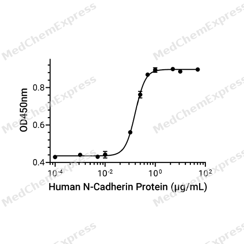
Measured by the ability of the immobilized protein to support the adhesion of MCF-7 human breast adenocarcinoma cells. The ED50 for this effect is 0.1602 μg/mL, corresponding to a specific activity is 6242.197 units/mg.
MCE Validation Data
-
Purity - SDS-PAGE
Purity - SDS-PAGE
-
Bioactivity - Cell-Based Assay
Bioactivity - Cell-Based Assay


Publications (1)
-
Journal Impact Factor
-
Most Recent


Technical Parameters
-
Species Human
-
Source HEK293
-
Tag C-10*His
-
Accession
P19022-1 (S26-A724)
-
Molecular Construction
-
N-term
-
N-Cadherin (S26-A724)
Accession # P19022-1 -
10*His
-
C-term
-
-
Protein Length
Partial Extracellular Domain (with Propeptide)
-
Synonyms
CDH2; N-Cadherin; Prev. NCAD; Calcium-Dependent Adhesion Protein, Neuronal; CDHN; CD325 Antigen; Neural Cadherin; N-Cadherin 1; Cadherin-2; ARVD14; CD325; ADHD8; Cadherin 2, Type 1, N-Cadherin (Neuronal); ACOGS; Cadherin 2; CDw325
-
AA Sequence
SGEIALCKTGFPEDVYSAVLSKDVHEGQPLLNVKFSNCNGKRKVQYESSEPADFKVDEDGMVYAVRSFPLSSEHAKFLIYAQDKETQEKWQVAVKLSLKPTLTEESVKESAEVEEIVFPRQFSKHSGHLQRQKRDWVIPPINLPENSRGPFPQELVRIRSDRDKNLSLRYSVTGPGADQPPTGIFIINPISGQLSVTKPLDREQIARFHLRAHAVDINGNQVENPIDIVINVIDMNDNRPEFLHQVWNGTVPEGSKPGTYVMTVTAIDADDPNALNGMLRYRIVSQAPSTPSPNMFTINNETGDIITVAAGLDREKVQQYTLIIQATDMEGNPTYGLSNTATAVITVTDVNDNPPEFTAMTFYGEVPENRVDIIVANLTVTDKDQPHTPAWNAVYRISGGDPTGRFAIQTDPNSNDGLVTVVKPIDFETNRMFVLTVAAENQVPLAKGIQHPPQSTATVSVTVIDVNENPYFAPNPKIIRQEEGLHAGTMLTTFTAQDPDRYMQQNIRYTKLSDPANWLKIDPVNGQITTIAVLDRESPNVKNNIYNATFLASDNGIPPMSGTGTLQIYLLDINDNAPQVLPQEAETCETPDPNSINITALDYDIDPNAGPFAFDLPLSPVTIKRNWTITRLNGDFAQLNLKIKFLEAGIYEVPIIITDSGNPPKSNISILRVKVCQCDSNGDCTDVDRIVGAGLGTGA
-
Predicted Molecular Mass
78.4 kDa
-
Molecular Weight
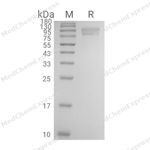
Approximately 95 kDa (Pro form) & 78 kDa (Mature form), based on SDS-PAGE under reducing conditions, due to the glycosylation.
-
Glycosylation
Yes
-
Purity
≥ 90%, as determined by reducing SDS-PAGE.


Product Properties
Lyophilized powder
Lyophilized from a 0.22 μm filtered solution of PBS, pH 7.4, 8% trehalose.
<1 EU/μg, determined by LAL method.
It is not recommended to reconstitute to a concentration less than 100 μg/mL in ddH2O.
Stored at -20°C for 2 years from date of receipt. After reconstitution, it is stable at 4°C for 1 week or -20°C for longer (with carrier protein). It is recommended to freeze aliquots at -20°C or -80°C for extended storage.
Room temperature in continental US; may vary elsewhere.


Documentation
-
Data Sheet (240 KB)
-
SDS (252 KB)
- English - EN (252 KB)
- Français - FR (252 KB)
- Deutsch - DE (252 KB)
- Norwegian - NO (252 KB)
- Español - ES (252 KB)
- Swedish - SV (252 KB)
- Italian - IT (252 KB)
- Korean - KR (252 KB)
- Portuguese - PT (252 KB)
-
Handling Instructions (2659 KB)

Calculators
Concentration (start) × Volume (start) = Concentration (final) × Volume (final)


 Powered by Bioz

