



Aristolactam I
Based on 1 publication(s) in Google Scholar
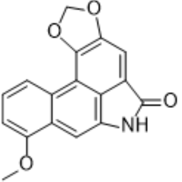
Aristolactam I is an AQP1 inhibitor and Aristolochic acid I metabolite. Aristolactam I can be isolated from Aristolochia plants. Aristolactam I downregulates Twist1 expression, increases E-cadherin expression, and activates the TGF-β/Smad signaling pathway. Aristolactam I has anticancer activity against breast cancer. Aristolactam I is nephrotoxic. Aristolactam I is mainly used in the study of breast cancer and kidney diseases such as renal interstitial fibrosis.
For research use only. We do not sell to patients.
- Purity : 99.44%
- CAS No.: 13395-02-3
- Formula: C17H11NO4
- Molecular Weight:293.27
-
Storage:
4°C, protect from light
* In solvent : -80°C, 6 months; -20°C, 1 month (protect from light)

To place orders, for customer services and technical support, please contact: MedChemExpress USA
Tel: 609-228-6898 E-mail: [email protected] [email protected]


-
 Biological Activity
-
Chemical Information
-
Solvent & Solubility
- Protocols
- Purity & Documentation
- References
-
 Help & FAQs


-
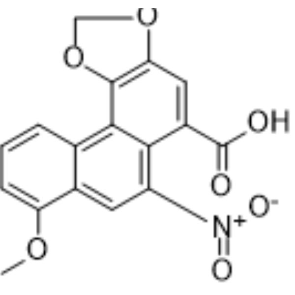
HY-N0510
Aristolochic acid ANF-κB Inhibitor -
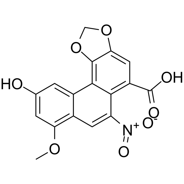
HY-N1465
Aristolochic acid DPDE2/CDK2 Inhibitor -
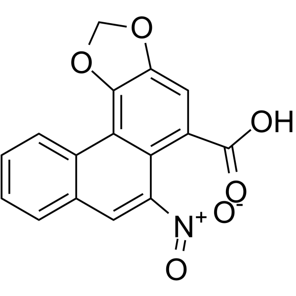
HY-N0511
Aristolochic acid BBioactive Alkaloid -
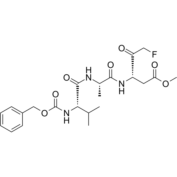
HY-16658
Z-VAD(OMe)-FMKPan-caspase Inhibitor -
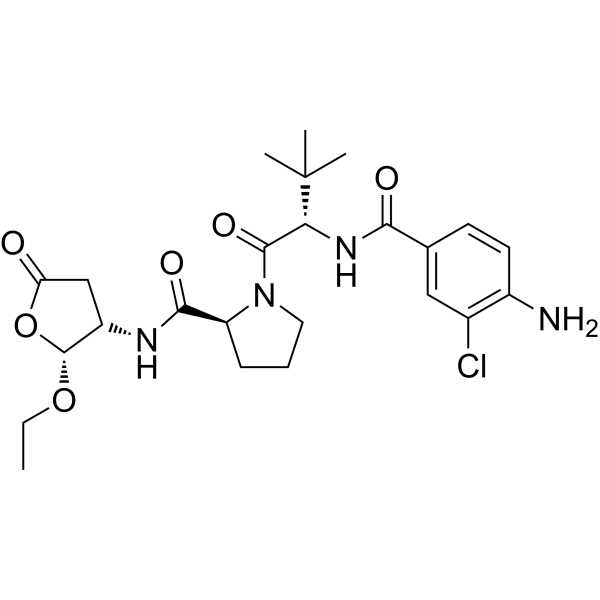
HY-13205
BelnacasanCaspase-1 Inhibitor -
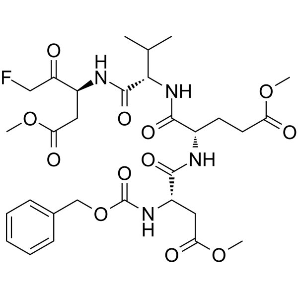
HY-12466
Z-DEVD-FMKCaspase-3 Inhibitor -
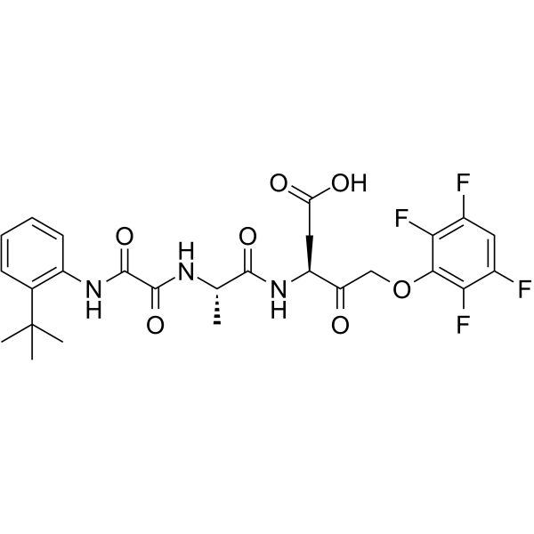
HY-10396
Emricasanpan-Caspase Inhibitor -
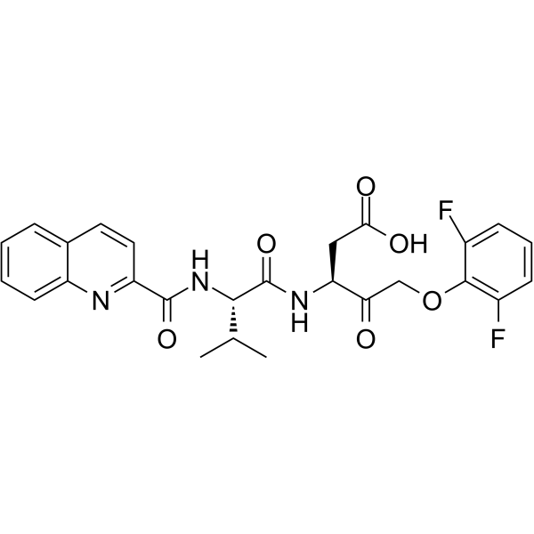
HY-12305
Q-VD-OPhPan-caspase Inhibitor -
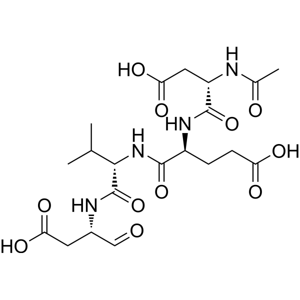
HY-P1001
Ac-DEVD-CHOCaspase-3 Inhibitor -

HY-N2013R
Aristolactam I (Standard)Aristolochic Acid I Metabolite -
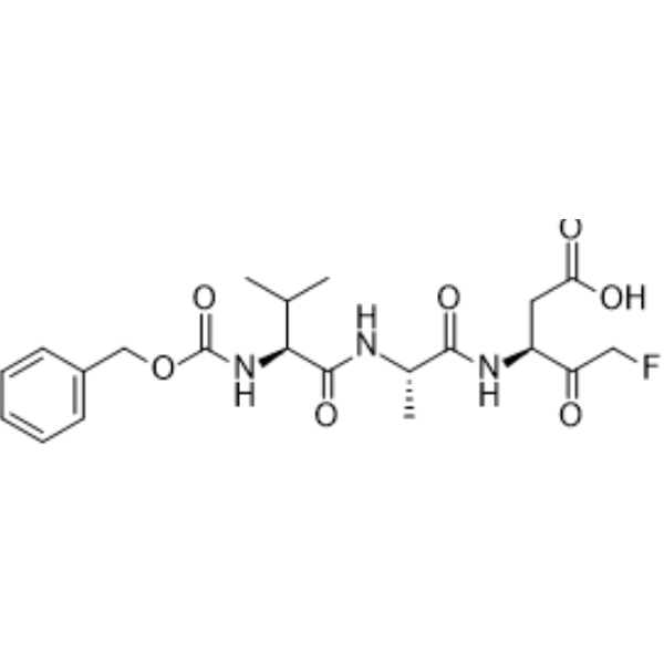
HY-16658B
Z-VAD-FMKPan-Caspase Inhibitor -
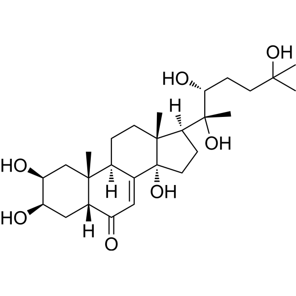
HY-N6979
CrustecdysoneCaspase Inhibitor
Publications Citing Use of MedChemExpress (MCE) Aristolactam I
More

Biological Activity
Description
In Vitro
Aristolactam I (0-40 μM; 21-48 h) inhibits cell migration and invasion through regulation of Twist1 in MDA-MB-231 breast cancer cells[1].
Aristolactam I (2.5-10 μM; 48 h) enhances the tendency of HK-2 cells to transform from epithelial cells to fibroblast-like cells[2].
MedChemExpress (MCE) has not independently confirmed the accuracy of these methods. They are for reference only.
-
Cell Line:MDA-MB-231
-
Concentration:0 μM, 40 μM
-
Incubation Time:48 h
-
Result:Upregulated mRNA expression of CDH1 and downregulated CDH2 mRNA expression.
-
Cell Line:MDA-MB-231
-
Concentration:0 μM, 20 μM, 40 μM
-
Incubation Time:48 h
-
Result:Increased protein expression of E-cadherin, decreased N-cadherin and Twist1 protein expression.
In Vivo
MedChemExpress (MCE) has not independently confirmed the accuracy of these methods. They are for reference only.
-
Animal Model:Male Sprague-Dawley rats (weighing 200-250 g)[5]
-
Dosage:2.25, 4.5, 9 mg/kg
-
Administration:Intraperitoneal injection, 5 days
-
Result:Significantly increased 24-hour urine volume.
Showed a stronger diuretic effect than Aristolochic acid I.
Caused early damage to the renal proximal tubules, with a stronger renal injury potential than Aristolochic acid I.
Inhibited the expression of renal AQP1, AQP2, and AQP4.


Chemical Information
-
CAS No. 13395-02-3
-
Appearance Solid
-
Molecular Weight 293.27
-
Formula C17H11NO4
-
Color Light yellow to yellow
-
SMILES
O=C1NC2=C(C3=C4C(C(OC)=CC=C4)=C2)C1=CC5=C3OCO5
-
Synonyms
Aristololactam; Aristolactam
-
Structure Classification
-
Initial Source
-
Shipping
Room temperature in continental US; may vary elsewhere.
-
Storage
4°C, protect from light
* In solvent : -80°C, 6 months; -20°C, 1 month (protect from light)


Publications (1)
-
Journal Impact Factor
-
Most Recent
-
Toxicol Lett
2024 Apr:394:76-91. PMID: 38428544


Solvent & Solubility
In Vitro:
DMSO : 12.5 mg/mL (42.62 mM; Need ultrasonic; Hygroscopic DMSO has a significant impact on the solubility of product, please use newly opened DMSO)
H2O : < 0.1 mg/mL (insoluble)
Please refer to the solubility information to select the appropriate solvent. Once prepared, please aliquot and store the solution to prevent product inactivation from repeated freeze-thaw cycles.
Storage method and period of stock solution: -80°C, 6 months; -20°C, 1 month (protect from light). When stored at -80°C, please use it within 6 months. When stored at -20°C, please use it within 1 month.
Please refer to the solubility information to select the appropriate solvent. Once prepared, please aliquot and store the solution to prevent product inactivation from repeated freeze-thaw cycles.
Storage method and period of stock solution: -80°C, 6 months; -20°C, 1 month (protect from light). When stored at -80°C, please use it within 6 months. When stored at -20°C, please use it within 1 month.
Concentration (start) × Volume (start) = Concentration (final) × Volume (final)


Purity & Documentation
-
Data Sheet (283 KB)
-
SDS (644 KB)
- English - EN (644 KB)
- Français - FR (644 KB)
- Deutsch - DE (644 KB)
- Norwegian - NO (644 KB)
- Español - ES (644 KB)
- Swedish - SV (644 KB)
- Italian - IT (644 KB)
- Korean - KR (644 KB)
- Portuguese - PT (644 KB)
-
Handling Instructions (2659 KB)
References
[2]. Zhang X, et al. An investigation on nephrotoxicity of Aristolactam I induced epithelial-mesenchymal transition on HK-2 cells. Toxicon. 2021 Oct 15;201:21-26. [Content Brief]
[3]. Li J , et al. Toxicities of aristolochic acid I and aristololactam I in cultured renal epithelial cells. Toxicol In Vitro. 2010 Jun;24(4):1092-7. [Content Brief]
[4]. Zhou Y , et al. Mitochondrial uptake of aristolactam I plays a critical role in its toxicity. Toxicol Lett. 2024 Apr;394:76-91. [Content Brief]
[5]. Li J, et al. Expression of Renal Aquaporins in Aristolochic Acid I and Aristolactam I-Induced Nephrotoxicity. Nephron. 2016;133(3):213-21. [Content Brief]


Complete Stock Solution Preparation Table
Please refer to the solubility information to select the appropriate solvent. Once prepared, please aliquot and store the solution to prevent product inactivation from repeated freeze-thaw cycles.
Storage method and period of stock solution: -80°C, 6 months; -20°C, 1 month (protect from light). When stored at -80°C, please use it within 6 months. When stored at -20°C, please use it within 1 month.
| Optional Solvent | Concentration Solvent Mass | 1 mg | 5 mg | 10 mg | 25 mg |
|---|---|---|---|---|---|
| DMSO | 1 mM | 3.4098 mL | 17.0491 mL | 34.0983 mL | 85.2457 mL |
| 5 mM | 0.6820 mL | 3.4098 mL | 6.8197 mL | 17.0491 mL | |
| 10 mM | 0.3410 mL | 1.7049 mL | 3.4098 mL | 8.5246 mL | |
| 15 mM | 0.2273 mL | 1.1366 mL | 2.2732 mL | 5.6830 mL | |
| 20 mM | 0.1705 mL | 0.8525 mL | 1.7049 mL | 4.2623 mL | |
| 25 mM | 0.1364 mL | 0.6820 mL | 1.3639 mL | 3.4098 mL | |
| 30 mM | 0.1137 mL | 0.5683 mL | 1.1366 mL | 2.8415 mL | |
| 40 mM | 0.0852 mL | 0.4262 mL | 0.8525 mL | 2.1311 mL |


Aristolactam I Related Classifications
HY-N2013 Related Classifications
 Powered by Bioz