



Dammarenediol II

Based on 1 Customer Validation
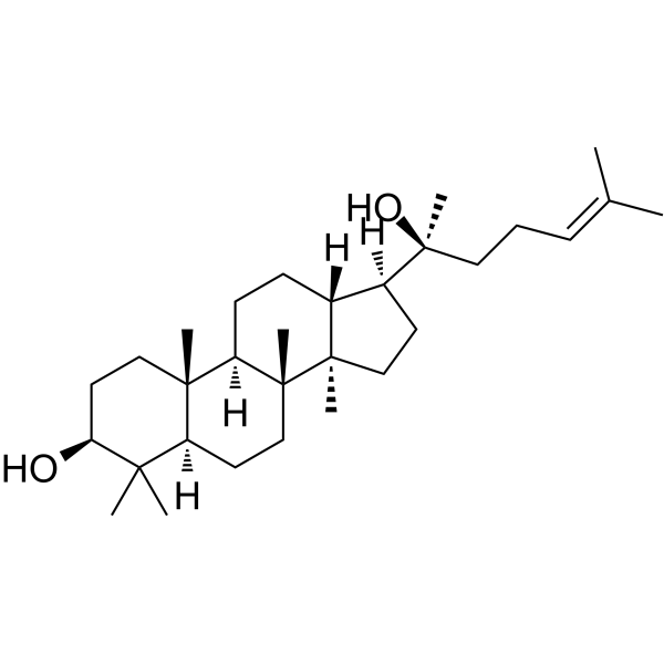
Dammarenediol II is a ginsenoside precursor. Dammarenediol II reduces the activity of O-GlcNAc transferase (OGT) and downregulates the global O-GlcNAcylation level. Dammarenediol II inhibits the phosphorylation of Akt, mTOR and GSK3β. Dammarenediol II inhibits human carboxylesterase activity, VEGF-induced ROS production, stress fiber formation and vascular endothelial cadherin disruption. Dammarenediol II promotes cell apoptosis (apoptosis), increases the levels of cleaved PARP1 and p53, and inhibits retinal microvascular leakage. Dammarenediol II can be used in studies related to liver cancer and diabetic retinopathy.
For research use only. We do not sell to patients.
- Purity : 99.81%
- CAS No.: 14351-29-2
- Formula: C30H52O2
- Molecular Weight:444.73
-
Storage:
4°C, protect from light
* In solvent : -80°C, 6 months; -20°C, 1 month (protect from light)

To place orders, for customer services and technical support, please contact: MedChemExpress USA
Tel: 609-228-6898 E-mail: [email protected] [email protected]


-
 Biological Activity
-
Chemical Information
-
Solvent & Solubility
- Protocols
- Purity & Documentation
- References
-
 Help & FAQs




Biological Activity
Description
Cellular Effect
|
Cell Line

|
Type | Value | Description | References |
|---|---|---|---|---|
| Vero | IC50 |
7 μg/mL
Compound: 2, dammarenediol-II
|
Antiviral activity against HSV1 MacIntyre replication in african green monkey Vero cells assessed as inhibition of virus-induced cytopathic effect after 48 hrs
Antiviral activity against HSV1 MacIntyre replication in african green monkey Vero cells assessed as inhibition of virus-induced cytopathic effect after 48 hrs
|
[PMID: 2828553] |
| Vero | IC50 |
7 μg/mL
Compound: 2, dammarenediol-II
|
Antiviral activity against HSV2 MS replication infected in african green monkey Vero cells assessed as inhibition of virus-induced cytopathic effect after 48 hrs
Antiviral activity against HSV2 MS replication infected in african green monkey Vero cells assessed as inhibition of virus-induced cytopathic effect after 48 hrs
|
[PMID: 2828553] |
In Vitro
Dammarenediol II (40 μM; 24 h) reduces O-GlcNAc levels in HepG2, Huh7, SK-HEP1, SNU-182 and SNU-387 cells[1].
Dammarenediol II (0.01-2.0 μg/mL; 30 min pretreatment, 10 min VEGF incubation) dose-dependently inhibits VEGF-induced intracellular ROS production in human umbilical vein endothelial cells, with the maximum inhibitory effect observed at 1.0 μg/mL[2].
Dammarenediol II (1 μg/mL; 30 min, 1 h VEGF) inhibits VEGF-induced stress fiber formation in human umbilical vein endothelial cells[2].
Dammarenediol II (1 μg/mL; 30 min preincubation, 90 min VEGF treatment) inhibits VEGF-induced VE-cadherin disruption and maintains the integrity of adherens junctions in human umbilical vein endothelial cells[2].
Dammarenediol II (0-50 μM; 24 h) significantly reduces the viability of HepG2 cells at 50 μM[1].
Dammarenediol II (0-40 μM; 24 h) significantly downregulates the expression level of OGT in HepG2 cells[1].
Dammarenediol II (0-40 μM; 24 h) reduces the EC50 of Etoposide (HY-13629) for regulating caspase-3/7 activity from 17.69 μM to 4.86 μM in HepG2 cells, upregulates the expression levels of cleaved-PARP1 and p53 proteins, induces apoptosis in HepG2, Huh7, SK-HEP1, SNU-182 and SNU-387 cells, and synergistically enhances the anticancer activity of Etoposide[1].
Dammarenediol II (40 μM; 24 h) inhibits the phosphorylation levels of Akt, GSK3β and mTOR in HepG2 cells[1].
MedChemExpress (MCE) has not independently confirmed the accuracy of these methods. They are for reference only. Further protocols information, click here.
-
Cell Line:Huh7, SK-HEP1, SNU-182, SNU-387 human liver carcinoma cells
-
Concentration:40 μM
-
Incubation Time:24 h
-
Result:Reduced O-GlcNAc levels in all tested human liver carcinoma cell lines: Huh7, SK-HEP1, SNU-182, and SNU-387.
-
Cell Line:HepG2 cells
-
Concentration:0, 2.5, 5, 10, 20, 40, 50 μM
-
Incubation Time:24 h
-
Result:Significantly reduced the viability of HepG2 cells at 50 μM
-
Cell Line:HepG2 cells
-
Concentration:0, 10, 20, 40 μM + 20 μM Etoposide
-
Incubation Time:24 h
-
Result:Significantly increased the proportion of TUNEL-positive cells in a dose-dependent manner.
In Vivo
MedChemExpress (MCE) has not independently confirmed the accuracy of these methods. They are for reference only.
-
Animal Model:C57BL/6 (6-week-old male; diabetic via single intraperitoneal injection of streptozotocin)[2]
-
Dosage:2.5 μg
-
Administration:intravitreal; single injection
-
Result:Completely blocked marked FITC-dextran extravasation in diabetic mouse retinas.
Reduced average FITC-dextran fluorescence intensity in diabetic mouse retinas to levels comparable to control mice (intensity was ~2-fold higher in untreated diabetic mice than controls).


Chemical Information
-
CAS No. 14351-29-2
-
Appearance Solid
-
Molecular Weight 444.73
-
Formula C30H52O2
-
Color White to off-white
-
SMILES
C[C@@]1(CC[C@@]2([H])C3(C)C)[C@@](CC[C@@]4([H])[C@]1(CC[C@]4([H])[C@@](C)(O)CC/C=C(C)\C)C)([H])[C@]2(CC[C@@H]3O)C
-
Structure Classification
-
Initial Source
-
Shipping
Room temperature in continental US; may vary elsewhere.
-
Storage
4°C, protect from light
* In solvent : -80°C, 6 months; -20°C, 1 month (protect from light)


Solvent & Solubility
In Vitro:
DMSO : 10 mg/mL (22.49 mM; ultrasonic and warming and heat to 60°C; Hygroscopic DMSO has a significant impact on the solubility of product, please use newly opened DMSO)
Please refer to the solubility information to select the appropriate solvent. Once prepared, please aliquot and store the solution to prevent product inactivation from repeated freeze-thaw cycles.
Storage method and period of stock solution: -80°C, 6 months; -20°C, 1 month (protect from light). When stored at -80°C, please use it within 6 months. When stored at -20°C, please use it within 1 month.
Please refer to the solubility information to select the appropriate solvent. Once prepared, please aliquot and store the solution to prevent product inactivation from repeated freeze-thaw cycles.
Storage method and period of stock solution: -80°C, 6 months; -20°C, 1 month (protect from light). When stored at -80°C, please use it within 6 months. When stored at -20°C, please use it within 1 month.
Concentration (start) × Volume (start) = Concentration (final) × Volume (final)
In Vivo:
Select the appropriate dissolution method based on your experimental animal and administration route.
- For the following dissolution methods, please ensure to first prepare a clear stock solution using an In Vitro approach and then sequentially add co-solvents:
- To ensure reliable experimental results, the clarified stock solution can be appropriately stored based on storage conditions. As for the working solution for In Vivo experiments, it is recommended to prepare freshly and use it on the same day.
- The percentages shown for the solvents indicate their volumetric ratio in the final prepared solution. If precipitation or phase separation occurs during preparation, heat and/or sonication can be used to aid dissolution.
In Vivo Dissolution Calculator
Please enter the basic information of animal experiments:
-
-
-
-
Recommended: Prepare an additional quantity of animals to account for potential losses during experiments.
Please enter your animal formula composition:
-
%DMSO +
Recommended: Keep the proportion of DMSO in working solution below 2% if your animal is weak.
-
%+
-
+%Tween-80 + +
-
%Saline +
The co-solvents required include: DMSO, . All of co-solvents are available by MedChemExpress (MCE). , Tween 80. All of co-solvents are available by MedChemExpress (MCE).
Working solution concentration: 0.22 mg/mL
Method for preparing stock solution: mg drug dissolved in μL DMSO. Stock solution concentration: mg/mL. * In solvent : -80°C, 6 months; -20°C, 1 month (protect from light)
1. Take μL DMSO stock solution;
2. Add μL .
μL , mix evenly;
3. Then add μL Tween 80, mix evenly;
4. Then add μL
Please ensure that the stock solution in the first step is dissolved to a clear state, and add co-solvents in sequence. You can use ultrasonic heating (ultrasonic cleaner, recommended frequency 20-40 kHz), vortexing, etc. to assist dissolution.

Protocols
-
Kinase activity and phosphorylation assays
Kinase activity assays measure the ability of kinases to transfer phosphate groups from ATP to specific substrates, while phosphorylation assays detect the presence and levels of phosphorylated proteins. Common methods include radiolabeled ATP incorporation (e. g. ,), ADP release detection via bioluminescence (e. g. ,[3]), enzyme-linked immunosorbent assays (ELISA) for phospho-specific epitopes (e. g. ,[6]), and microtiter-based formats for high-throughput screening (e. g. ,[8]). The ADP-Glo assay quantifies kinase activity by measuring ADP produced during phosphorylation using a luciferase-based system. Radiometric assays involve autoradiography or scintillation counting after incorporation of 32P-labeled ATP into substrate proteins. ELISA-based approaches rely on phospho-specific antibodies to detect activated kinases in cell lysates or purified samples.
-
Western Blot
Western blotting (WB) is a commonly used experimental method in molecular biology, biochemistry, and immunogenetics for identifying and quantifying target proteins. It combines gel electrophoresis with immunoassay, enabling researchers to analyze protein expression, post-translational modifications, and molecular weight.
-
Apoptosis
Apoptosis, also called programmed cell death, is generally characterized by distinct morphological characteristics.
-
TUNEL staining for apoptotic DNA fragmentation
TUNEL staining detects DNA strand breaks by using terminal deoxynucleotidyl transferase to add labeled nucleotides to exposed 3′-OH DNA termini, generating either microscopic staining in fixed cells or tissue sections, or fluorescence/cytometric signal in cell suspensions. TUNEL positivity reflects DNA fragmentation but should not be interpreted alone as definitive apoptosis, because TUNEL can also label necrotic, autolytic, mechanically damaged, or DNA-repair-associated DNA breaks.
-
Annexin V plus membrane-impermeant dye apoptosis staining
Annexin V-based apoptosis assays rely on the detection of phosphatidylserine (PS) externalization from the inner leaflet of the plasma membrane to the outer leaflet, an early biochemical hallmark of apoptosis. Fluorescently labeled Annexin V binds PS in a calcium-dependent manner, enabling identification of early apoptotic cells by flow cytometry or fluorescence microscopy. When combined with a membrane-impermeant DNA-binding dye (e. g. , propidium iodide), this approach allows discrimination between viable (Annexin V−/dye−), early apoptotic (Annexin V+/dye−), and late apoptotic or necrotic (Annexin V+/dye+) cell populations by assessing membrane integrity and PS exposure.
-
ROS/oxidative-stress fluorescent staining
ROS/oxidative-stress fluorescent staining uses cell-permeant fluorogenic probes that become fluorescent after oxidation inside cells or tissues; commonly used examples include DCFH-DA/DCFDA for broad cellular oxidant detection, DHE for superoxide-related signal detection, MitoSOX for mitochondrial superoxide-related signal detection, and CellROX probes for oxidative-stress-associated fluorescence readouts. The assay detects probe oxidation rather than a single ROS species unless the probe and analysis method have been chemically validated for that species. DCFH-DA enters cells, is deacetylated by intracellular esterases to DCFH, and produces fluorescent DCF after oxidation, so the readout is used as an operational measure of total cellular oxidative stress rather than a species-specific ROS measurement. DHE and MitoSOX can report superoxide-related oxidation, but red fluorescence alone can include non-specific ethidium-like oxidation products; HPLC or optimized spectral approaches are
-
Apoptosis Solutions
Apoptosis is a regulated, generally non-lytic cell-death pathway that removes unwanted, damaged, infected, or abnormal cells through coordinated morphological changes, caspase activation, DNA fragmentation, and membrane remodeling. The intrinsic apoptosis pathway is controlled mainly by mitochondrial outer membrane permeabilization, BCL-2 family proteins, cytochrome c release, apoptosome formation, caspase-9 activation, and downstream executioner caspase-3/7 activation. The extrinsic apoptosis pathway is initiated by death receptors such as Fas, TNFR, and TRAIL receptors, which recruit adaptor proteins and activate caspase-8 before engaging executioner caspases or mitochondrial amplification through BID cleavage. Apoptosis is linked to many phenotypes, including cancer cell killing, tissue homeostasis, immune regulation, neurodegeneration, infection response, and treatment-induced cytotoxicity; unresolved questions include how apoptosis interacts with necroptosis, pyroptosis, ferroptos
-
Protocol for Kinase activity and phosphorylation assays
Kinase activity assays measure transfer of phosphate from ATP to a protein or peptide substrate, generating phosphorylated substrate, ADP, or incorporated radiolabeled phosphate as the readout; phosphorylation assays measure site-specific phosphorylation in cells or tissues as a proxy for kinase-pathway activation, inhibition, or substrate regulation. Phosphorylation can be detected by phospho-specific Western blot, immunoprecipitation kinase assay, phospho-immunofluorescence, phospho-flow cytometry, luminescent ADP detection, radiolabeled ATP incorporation, or reporter-based pathway assays, and these readouts can be applied to cancer cells, primary neurons, mouse tumors, organoids, inflammatory macrophages, ferroptosis studies, and mitophagy studies when the kinase target is biologically relevant.
-
Liver Cancer Modeling
Liver cancer can be classified into primary liver cancer and secondary liver cancer. Secondary liver cancer is the metastatic liver cancer. Primary liver cancer includes hepatocellular carcinoma (HCC), intrahepatic cholangiocarcinoma (ICC) and fibrolamellar HCC, of which HCC is the most common form, accounting for approximately 90% of primary liver cancers[1]. HCC mouse models include chemical agent-induced models, transplanted tumor models, and genetic engineered models.


Purity & Documentation
-
Data Sheet (279 KB)
-
SDS (252 KB)
- English - EN (252 KB)
- Français - FR (252 KB)
- Deutsch - DE (252 KB)
- Norwegian - NO (252 KB)
- Español - ES (252 KB)
- Swedish - SV (252 KB)
- Italian - IT (252 KB)
- Korean - KR (252 KB)
- Portuguese - PT (252 KB)
-
Handling Instructions (2659 KB)
References
[1]. Lee J, et al. Dammarenediol II enhances etoposide-induced apoptosis by targeting O-GlcNAc transferase and Akt/GSK3β/mTOR signaling in liver cancer. Mol Oncol. Published online December 30, 2025. [Content Brief]
[2]. Kim SH, et al. Dammarenediol-II Prevents VEGF-Mediated Microvascular Permeability in Diabetic Mice. Phytother Res. 2015;29(12):1910-1916. [Content Brief]


Complete Stock Solution Preparation Table
Please refer to the solubility information to select the appropriate solvent. Once prepared, please aliquot and store the solution to prevent product inactivation from repeated freeze-thaw cycles.
Storage method and period of stock solution: -80°C, 6 months; -20°C, 1 month (protect from light). When stored at -80°C, please use it within 6 months. When stored at -20°C, please use it within 1 month.
| Optional Solvent | Concentration Solvent Mass | 1 mg | 5 mg | 10 mg | 25 mg |
|---|---|---|---|---|---|
| DMSO | 1 mM | 2.2486 mL | 11.2428 mL | 22.4856 mL | 56.2139 mL |
| 5 mM | 0.4497 mL | 2.2486 mL | 4.4971 mL | 11.2428 mL | |
| 10 mM | 0.2249 mL | 1.1243 mL | 2.2486 mL | 5.6214 mL | |
| 15 mM | 0.1499 mL | 0.7495 mL | 1.4990 mL | 3.7476 mL | |
| 20 mM | 0.1124 mL | 0.5621 mL | 1.1243 mL | 2.8107 mL |


Dammarenediol II Related Classifications
HY-N3677 Related Classifications
 Powered by Bioz