



Murine Fibrinogen
Based on 2 publication(s) in Google Scholar
Murine Fibrinogen is a native fibrinogen derived from mouse plasma. Murine Fibrinogen acts as a cerebrovascular permeability enhancer. Murine Fibrinogen activates matrix metalloproteinase-9 (MMP-9), downregulates the expression of vascular endothelial cadherin (VE-cadherin), and upregulates the expression of plasmalemmal vesicle-associated protein-1 (PV-1). Murine Fibrinogen increases macromolecular leakage from pial veins, thereby disrupting the microvascular integrity of cerebral blood vessels. Murine Fibrinogen can be used in studies related to cerebrovascular dysfunction.
For research use only. We do not sell to patients.
- CAS No.: 9001-32-5
-
Storage:
Please store the product under the recommended conditions in the Certificate of Analysis.


To place orders, for customer services and technical support, please contact: MedChemExpress USA
Tel: 609-228-6898 E-mail: [email protected] [email protected]


-
 Biological Activity
-
Chemical Information
- Protocols
- Purity & Documentation
- References
-
 Help & FAQs


-

HY-D0842A
BSA Protein (Low Endotoxin,Fatty acid free)Oligonucleotide-binding Protein -

HY-D0842
BSA ProteinGlobular Protein -

HY-W250978
OvalbuminsAllergen -

HY-P1956
HSA ProteinAntioxidant/Anticoagulant -

HY-125864
Fibrinogen (Bovine)NF-KB Activator -

HY-NP010
Type I Collagen, from rat tailCollagen -

HY-P2995
HemoglobinIron-containing Protein -

HY-P2738
Collagen, Type I, from bovine achilles tendonCollagen Peptide -

HY-NP003
Collagen, Type II, from bovine snout membraneType II Collagen -

HY-W250978A
Ovalbumin, low endotoxinAllergen -

HY-125865
CaseinPhosphoprotein -

HY-P1956A
HSA Protein (Cell culture grade, Endotoxin<0.125 EU/mg)Plasma Protein
Publications Citing Use of MedChemExpress (MCE) Murine Fibrinogen
More Customer Validation & Images

-
2D/3D Cell Culture and Differentiation
-
Cell Imaging/Staining
-
Cell Imaging/Staining
-
Cell Imaging/Staining
-
Histological Imaging/Staining


Biological Activity
Description
IC50 & Target
[1]|
MMP-9 |
In Vivo
MedChemExpress (MCE) has not independently confirmed the accuracy of these methods. They are for reference only.
-
Animal Model:C57BL/6J (12-week-old male, 26-30 g, wild-type); MMP9-/- (FVB.Cg-Mmp9tm1Tvu/J, 12-week-old male, 26-30 g, MMP-9 gene knockout); FVB/NJ (12-week-old male, 26-30 g, wild-type)[1]
-
Dosage:20 mg per 100 g of body weight (resulting in a blood concentration of 4 mg/mL)
-
Administration:carotid artery infusion; 20 mL/min; 10 minutes
-
Result:Induced significantly greater pial venular macromolecular leakage (fluorescence intensity of 155 FIU) than PBS infusion in wild-type C57BL/6J mice.
Increased endothelial ICAM-1 expression to 50 FIU (vs 22 FIU with PBS) in wild-type C57BL/6J mice.
Reduced VE-cadherin expression to 22 FIU (vs 43 FIU with PBS) in wild-type C57BL/6J mice.
Increased MMP activity to 71 FIU in wild-type C57BL/6J mice.
Increased PV-1 expression to 170 FIU (vs 50 FIU with PBS) in wild-type C57BL/6J mice.
Induced further increased pial venular leakage (fluorescence intensity of 182 FIU) when combined with topical histamine in wild-type C57BL/6J mice.
Induced significantly greater pial venular macromolecular leakage (fluorescence intensity of 137 FIU) than PBS infusion in MMP9-/- mice, but less leakage than in wild-type mice.
Increased endothelial ICAM-1 expression to 39 FIU (vs 24 FIU with PBS) in MMP9-/- mice.
Reduced VE-cadherin expression to 28 FIU (vs 78 FIU with PBS) in MMP9-/- mice, with higher VE-cadherin levels than in Fg-treated wild-type mice.
Increased MMP activity to 46 FIU in MMP9-/- mice, with lower activity than in Fg-treated wild-type mice.
Increased PV-1 expression to 75 FIU (vs 50 FIU with PBS) in MMP9-/- mice, with lower expression than in Fg-treated wild-type mice.
Induced further increased pial venular leakage (fluorescence intensity of 152 FIU) when combined with topical histamine in MMP9-/- mice, but less leakage than in histamine-exposed, Fg-treated wild-type mice.
Induced pial venular macromolecular leakage (fluorescence intensity of 152 FIU) comparable to that in Fg-treated wild-type mice in FVB/NJ mice, with significantly greater leakage than PBS infusion.
Induced further increased pial venular leakage (fluorescence intensity of 178 FIU) when combined with topical histamine in FVB/NJ mice, comparable to that in histamine-exposed, Fg-treated wild-type mice.
Showed no degradation post-infusion.


Chemical Information
-
CAS No. 9001-32-5
-
Appearance Solid
-
Color White to off-white
-
SMILES
[Murine Fibrinogen]
-
Shipping
Room temperature in continental US; may vary elsewhere.
-
Storage
Please store the product under the recommended conditions in the Certificate of Analysis.


Publications (2)
-
Journal Impact Factor
-
Most Recent
-
Foods
Glycosylation Remodeling and Thermal Denaturation Dictate the Functional Diversification of Protein Z. [Abstract]2026 Mar 4;15(5):853. PMID: 41829126 -
Microvasc Res
DDIT4 knockdown suppresses venous malformation progression by inhibiting NF-κB signaling as a potential therapeutic target. [Abstract]2025 Sep:161:104833. PMID: 40571189
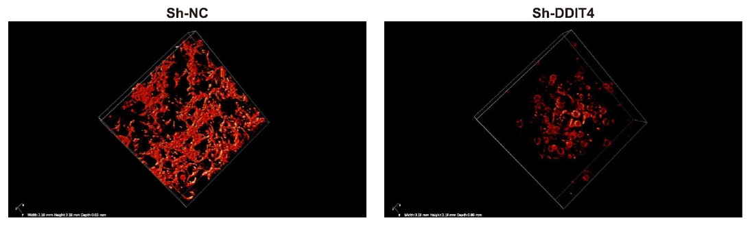
Murine Fibrinogen purchased from MedChemExpress. Usage Cited in: Microvasc Res. 2025 Sep:161:104833. [Abstract]
Fibrinogen (2 mg/mL; DPBS) was used to construct the VMs 3D cell model with or without DDIT4 knockdown.
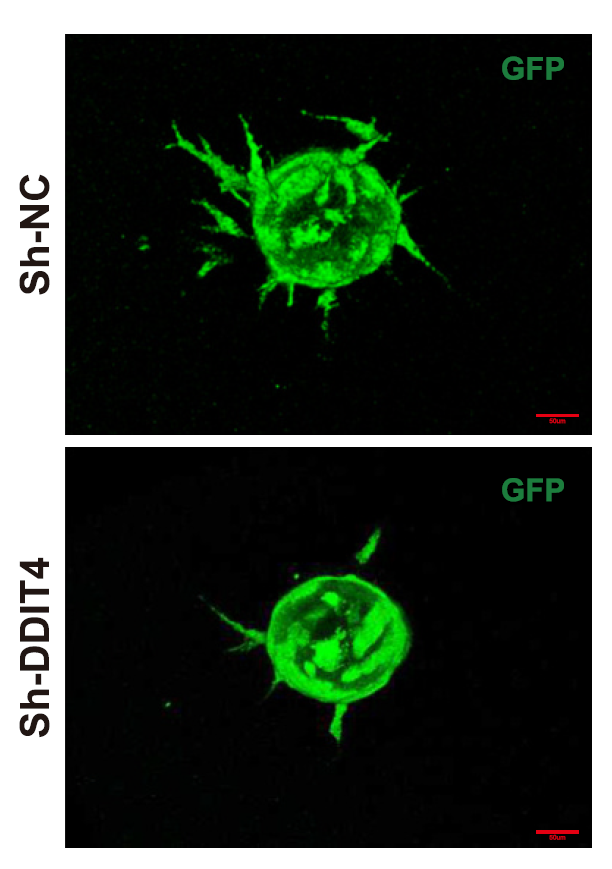
Murine Fibrinogen purchased from MedChemExpress. Usage Cited in: Microvasc Res. 2025 Sep:161:104833. [Abstract]
Fibrinogen (2 mg/mL; DPBS). Vascular sprouting in the VMs 3D cell model after 7 days of culture.
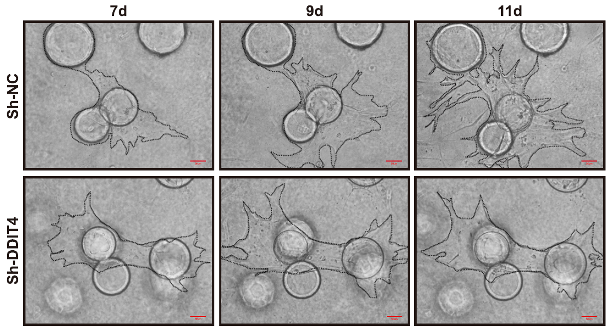
Murine Fibrinogen purchased from MedChemExpress. Usage Cited in: Microvasc Res. 2025 Sep:161:104833. [Abstract]
Fibrinogen (2 mg/mL; DPBS). Time-lapse analysis on days 7, 9, and 11 showed the angiogenesis rate in the VMs 3D cell model.
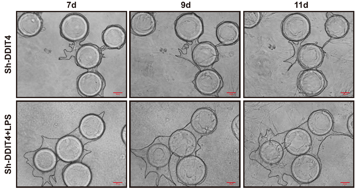
Murine Fibrinogen purchased from MedChemExpress. Usage Cited in: Microvasc Res. 2025 Sep:161:104833. [Abstract]
Fibrinogen (2 mg/mL; DPBS). Time-lapse analysis on days 7, 9, and 11 showed the angiogenesis rate in the DDIT4 knockdown VMs 3D cell model.
Murine Fibrinogen purchased from MedChemExpress. Usage Cited in: Microvasc Res. 2025 Sep:161:104833. [Abstract]
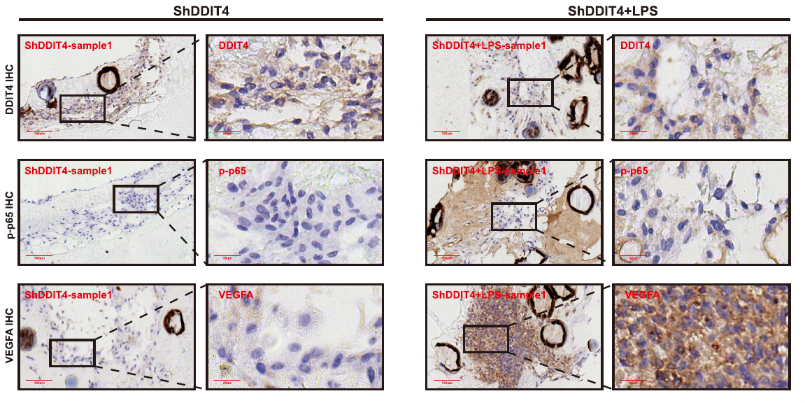
Fibrinogen (2 mg/mL; DPBS). Immunohistochemical (IHC) staining of DDIT4, phospho-p65, and VEGFA in the LPS-stimulated DDIT4 knockdown VMs 3D cell model was performed.

Protocols
-
RNA extraction experimental
By lysing cells, releasing RNA, and removing impurities such as proteins and DNA, high-purity RNA products are finally obtained. The commonly used traditional method is the guanidine isothiocyanate/phenol/chloroform method (Trizol), which is suitable for a variety of animal materials including animal tissues, microorganisms, cultured cells, etc., and most plant materials.
-
Mitochondrial membrane-potential fluorescent assay
Mitochondrial membrane potential fluorescent assays estimate ΔΨm in living cells using lipophilic cationic dyes such as TMRM, TMRE, rhodamine 123, and JC-1, which accumulate in mitochondria according to membrane polarization; loss of signal after FCCP or CCCP treatment is interpreted as mitochondrial depolarization. TMRM/TMRE and rhodamine 123 are commonly used for semi-quantitative live-cell microscopy or flow cytometry, while JC-1 can report a shift from red aggregate fluorescence to green monomer fluorescence during depolarization; interpretation requires controls because dye concentration, quenching mode, cell type, dye efflux, and mitochondrial mass can affect fluorescence independently of ΔΨm.
-
Mitochondrial membrane-potential and mitochondrial mass staining
Mitochondrial membrane potential staining measures the electrochemical polarization across the mitochondrial inner membrane in live cells using lipophilic cationic fluorescent probes; early rhodamine-based work showed that selective mitochondrial dye accumulation is lost when the mitochondrial transmembrane potential is dissipated. JC-1 reports mitochondrial polarization by shifting from green monomer fluorescence to red J-aggregate fluorescence as dye concentration increases within energized mitochondria; therefore, the red/green fluorescence ratio is used as a relative readout of mitochondrial membrane potential. TMRE or TMRM staining provides a single-channel relative readout because these cationic rhodamine esters accumulate in polarized mitochondria, and lower fluorescence indicates reduced mitochondrial polarization when acquisition and dye-loading conditions are controlled. Mitochondrial mass staining is commonly performed with MitoTracker Green FM or related MitoTracker dyes as
-
Transepithelial/transendothelial electrical resistance assay
TEER measures electrical resistance across epithelial or endothelial monolayers cultured on permeable supports, and the readout reflects ionic conductance through the cell barrier, especially the paracellular pathway regulated by junctional integrity. TEER can be measured without destroying the monolayer and is commonly used before or during transport, permeability, barrier-disruption, and barrier-maturation experiments. TEER values are influenced by biological maturation and technical conditions; reported factors include temperature, medium formulation, passage number, electrode geometry, membrane properties, and junctional length during early monolayer maturation. Therefore, TEER should be interpreted with blank-insert subtraction, area normalization, repeated readings, and, when possible, orthogonal barrier readouts such as FITC-dextran flux or tight-junction staining.


Purity & Documentation
-
Data Sheet (268 KB)
-
SDS (251 KB)
- English - EN (251 KB)
- Français - FR (251 KB)
- Deutsch - DE (251 KB)
- Norwegian - NO (251 KB)
- Español - ES (251 KB)
- Swedish - SV (251 KB)
- Italian - IT (251 KB)
- Korean - KR (251 KB)
- Portuguese - PT (251 KB)
-
Handling Instructions (2659 KB)
References

Calculators
Concentration (start) × Volume (start) = Concentration (final) × Volume (final)


Murine Fibrinogen Related Classifications
HY-125864B Related Classifications
 Powered by Bioz