From 11:00 pm to 12:00 pm EST ( 8:00 pm to 9:00 pm PST ) on January 6th, the website will be under maintenance. We are sorry for the inconvenience. Please arrange your schedule properly.
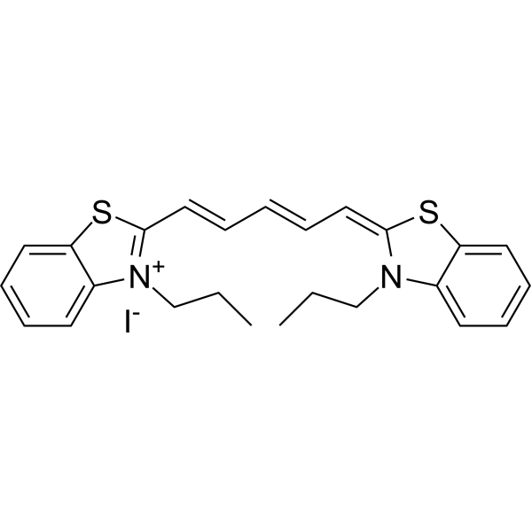
DiSC3(5) is a fluorescent probe commonly used as a tracer dye to evaluate mitochondrial membrane potential. The excitation/emission wavelength of DiSC3(5) is up to 622/670 nm. DiSC3(5) can inhibit the respiratory system associated with mitochondrial NAD, and the IC50 value is 8 μM. DiSC3(5) in the presence of Na +/K +-ATPase inhibitor ouabain 2 can induce membrane hyperpolarization of Ehrlich ascites tumorcells .
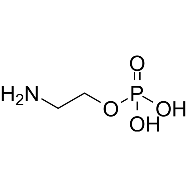
Phosphorylethanolamine (Monoaminoethyl phosphate) is a membrane phospholipid and an important precursor of Phosphatidylcholine (HY-B2233B). It is found in most animal tissues and various human extracranial tumors, playing a critical role in membrane integrity, cell division, mitochondrial respiratory function, and more. Studies have shown that changes in the abundance of Phosphorylethanolamine are associated with Alzheimer's disease and Parkinson's disease. Lowering the ratio of Phosphorylethanolamine to Phosphatidylcholine in the liver can improve insulin signaling. Phosphorylethanolamine holds promise for research in the fields of cancer, neurodegenerative disorders, and metabolic diseases .
Cevostamab (BFCR4350A; RG6160; RO7187797) is a humanized IgG1-based BsAb that targets membrane-proximal extracellular domain of FcRH5 on multiple myeloma (MM) cells as well as CD3 on T cells. Moreover, Cevostamab facilitates efficient synapse formation, improves killing activity of T cells against MM tumorcells .
IR-780 is a near-infrared fluorescent probe for in vivo imaging of tumorcells. IR-780 is transported into tumorcells via OATPs and ABCB10, with uptake dependent on glycolytic activity and plasma membrane potential. IR-780 preferentially accumulates in tumorcell mitochondria, including those of drug-resistant cancer cells, without chemical conjugation. IR-780 generates reactive oxygen species (ROS), induces hyperthermia and apoptosis, inhibits tumor growth and recurrence, and modulates HSP70 expression upon ultrasound or 808 nm laser exposure. IR-780 acts as a sonosensitizer, photodynamic and photothermal agent, and drug delivery carrier, with low acute imaging-dose toxicity and rapid vital organ clearance. IR-780 can be used for the research of cancer, such as breast cancer, lung cancer, and non-small cell lung cancer (NSCLC) .
Squalamine (MSI-1256) is an aminosterol compound with broad-spectrum antiviral activity. Squalamine makes cells less conducive to certain viral replication by altering the electrostatic interactions in the inner membrane of host cells. Squalamine also has antibacterial and antitumor activities. Squalamine has broad-spectrum antibacterial activity against Gram-negative and Gram-positive bacteria, fungi and protozoa. Squalamine inhibits tumor-related angiogenesis and the growth of human breast cancer cells. Squalamine restores the function of enteric nervous system in Parkinson ,s disease mouse models .
N-Acetyl-L-tyrosine is an orally active endogenous mitochondrial stress response regulator that can permeate the cellmembrane by passive diffusion. N-Acetyl-L-tyrosine induces low-level reactive oxygen species (ROS) generation by transiently perturbing mitochondrial membrane potential, triggering reverse signaling to activate FoxO and Keap1 pathways. As a result, N-Acetyl-L-tyrosine enhances the expression of antioxidant enzyme genes, exerting anti-stress and cytoprotective effects. N-Acetyl-L-tyrosine can improve heat stress tolerance, inhibit tumor growth, and regulate energy metabolism. N-Acetyl-L-tyrosine can be used in the research of aging, metabolic diseases (such as diabetes), and cancer .
Tri-GalNAc(OAc)3 TFA is a trivalent N-acetylgalactosamine (GalNAc) derivative that can be used to synthesize GalNAc-LYTAC. Tri-GalNAc is a specific ligand targeting the asialoglycoprotein receptor (ASGPR), mediating the endocytosis and transport of cell surface proteins (such as EGFR, HER2) to lysosomes for degradation by lysosomal targeting chimeras (LYTACs). Tri-GalNAc significantly reduces the level of target proteins and inhibits downstream signaling pathways (such as EGFR-mediated Akt and MAPK signals). Tri-GalNAc(OAc)3 TFA can be used for hepatocyte targeting studies, and can degrade carcinogenic membrane proteins and inhibit tumorcell proliferation in liver cancer cell models .
DSPE-PEG2000-Cy5 is a Cy5 (HY-D0821) fluorophore-labeled conjugate of distearoylphosphatidylethanolamine and polyethylene glycol, as well as a liposome component. The Cy5 fluorophore is commonly used for labeling proteins and nucleic acids in imaging, flow cytometry and genomic applications. DSPE-PEG2000-Cy5 supports cellmembrane modification, in vivo tumor targeting research and long-term in vivo circulation of its liposomal formulations (Ex/Em=633/670 nm) .
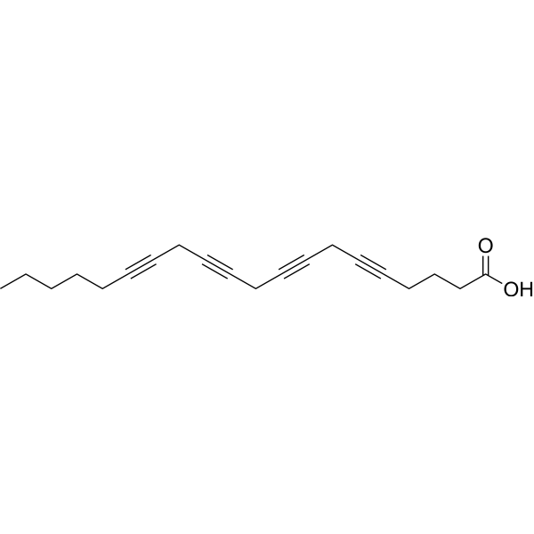
Eicosatetraynoic acid (ETYA) is a non-metabolizable analog of Arachidonic acid (HY-109590) and also an inhibitor of the lipoxygenase (LOX)/cyclooxygenase (COX) pathway (ID50= 8 μM and 4 μM). Eicosatetraynoic acid acts as a suicide substrate to inhibit the production of inflammatory mediators such as leukotrienes and prostaglandins. Eicosatetraynoic acid acts directly on cellmembranes and membrane proteins to exert a wide range of effects, including blocking potassium channels, increasing cellmembrane fluidity, elevating intracellular calcium levels, inhibiting DNA synthesis in tumorcells, inducing differentiation of certain cells, and specifically inhibiting the assembly and replication of orthopoxviruses. Eicosatetraynoic acid alleviates acute lung injury induced by chemicals such as phosgene .
Lactonic sophorolipid is an apoptosis inducer and antimicrobial surfactant with antitumor activity. Lactonic sophorolipid regulates Bax/Bcl-gene expression through caspase-3/9 and induces apoptosis in tumorcells. Lactonic sophorolipid can disrupt cellmembrane permeability and exert antibacterial effects (MIC for oral pathogens is 100-400 μg/mL). Lactonic sophorolipid promotes mitochondrial membrane potential depolarization, activates the intrinsic apoptotic pathway, and can synergize with antibiotics to enhance the antibacterial effect. Lactonic sophorolipid can be used in liver cancer research and the development of oral hygiene antibacterial agents[1][2][3].
Antimycin A2 is a selective inhibitor of the cytochrome b-c1 complex in the mitochondrial electron transport chain. Antimycin A2 disrupts mitochondrial membrane potential and produces reactive oxygen species (ROS) by inhibiting electron transfer between cytochrome b and c. Antimycin A2 has bactericidal and piscicidal activity, as well as tumorcell growth inhibitory effects, and can induce S-phase cell cycle arrest and apoptosis in HeLa cells. Antimycin A2 is suitable for research of cervical cancer and fisheries management. Antimycin A2 can be naturally isolated from the fermentation products of Streptomyces sp. strains .
PARG-IN-4 (Formula (A)) is an orally available PARG inhibitor (EC50=1.9 nM) with cellmembrane permeability. PARG-IN-4 can effectively inhibit tumor growth in mouse models. PARG-IN-4 can be used in cancer research .
BMS-214662 is a farnesyl transferase inhibitor. BMS-214662 can effectively block the localization and function of Ras protein in the cellmembrane by inhibiting the pro-group modification of Ras protein, thereby exerting anti-tumor activity. BMS-214662 has an IC50 value of 1.3 nM for H-Ras and 8.4 nM for K-Ras. BMS-214662 can be used to study Ras-related tumor diseases .
2-Amino-2-deoxyglucose hydrochloride (D-Glucosamine Hydrochloride) is a glucose analog that is specifically recognized and transported by the cellmembraneGLUT1. 2-Amino-2-deoxyglucose hydrochloride acts as a tumor-targeting ligand and a guiding molecule for the synthesis of prodrug conjugates, thus delivering drugs precisely to tumorcells. 2-Amino-2-deoxyglucose hydrochloride is applicable to diagnostic imaging and therapeutic efficacy monitoring of solid tumors and various cancers (e.g., breast cancer, glioblastoma). 2-Amino-2-deoxyglucose hydrochloride also helps bacteria resist lysozyme digestion by integrating into the non-N-acetylated residues of Streptococcus pneumoniae peptidoglycan. 2-Amino-2-deoxyglucose hydrochloride is used in studies on tumor metabolism and the exploration of bacterial drug resistance mechanisms .
IGN523 is an anti-CD98 antibody (hCD98, KD = 0.55 nM). IGN523 induces antibody-dependent cell-mediated cytotoxicity (ADCC) activity, lysosomal membrane permeabilization, and inhibition of essential amino acid transport, ultimately leading to caspase-3 and caspase-7-mediated apoptosis of tumorcells. IGN523 inhibits tumor growth in multiple tumor xenograft models. IGN523 is useful in the research of non-small cell lung cancer (NSCLC), acute myeloid leukemia (AML), and other cancers. .
Cysmethynil is an Icmt inhibitor(IC50 = 2.4 μM). Cysmethynil inhibites RAS membrane binding and EGF signal transduction. Cysmethynil prevents the cells in the G1 phase and induces autophagy. Cysmethynil inhibits PC3 cells proliferation, has synergistic effect with Paclitaxel (HY-B0015) and Doxorubicin (HY-15142A). Cysmethynil has anti-tumor effects and can be used for solid tumor (such as prostate cancer et al.) research .
Chlorophosphonazo III is a cellmembrane-permeable contrast agent and spectrophotometric reagent. Chlorophosphonazo III forms stable 1:1 complexes with intracellular calcium, magnesium, uranium, titanium, zirconium, thorium, scandium, equivalent actinides and protactinium. Chlorophosphonazo III generates photoacoustic signals correlated with Ca 2+ concentrations, which is applicable for imaging 3D tumorcell cultures and tumor spheroids. Chlorophosphonazo III exhibits no cytotoxicity, can be used for spectrophotometric detection of target ions, and the absorbance of its complexes remains stable for up to one week within the pH range of 2.2 to 7.0 .
API-1 is a potent selective Akt/PKB inhibitor that reduces the level of phosphorylated Akt (IC50 = 0.8 μM). API-1 binds to the PH domain and inhibits Akt membrane translocation. API-1 induces c-FLIP degradation. API-1 reduces cell proliferation and induces apoptosis. API-1 decreases tumor growth in mouse xenograft model .
MANS peptide is an inhibitor for myristoylated alanine-rich C kinase substrate (MARCKS), which competes with MARCKS in cells for membrane binding, and thus inhibits the stimulation of mucin secretion and tumor metastasis .
Biotin-NH-PSMA-617 is a biotin-tagged PSMA-617. PSMA-617 is a small molecule targeting the prostate-specific membrane antigen (PSMA), which is directly expressed by the tumorcells .
IMAB027 (ASP1650) is a specific anti-CLDN6 mAb, while CLDN6 (Claudin 6) is a tight junction membrane protein, aberrantly expressed in various human cancer types, ovarian cancers particularly. IMAB 027 shows anti-tumor activity, and induces apoptosis in CLDN6 + ovarian and testicular cancer cell lines .
DiSC3 (5) (solution) is a fluorescent probe commonly used as a tracer dye to evaluate mitochondrial membrane potential. The excitation/emission wavelength of DiSC3 (5) is up to 622/670 nm. DiSC3 (5) can inhibit the respiratory system associated with mitochondrial NAD, and the IC50 value is 8 μM. DiSC3 (5) in the presence of Na +/K +-ATPase inhibitor ouabain 2 can induce membrane hyperpolarization of Ehrlich ascites tumorcells . Solvent and concentration: DMSO: 1 mM The 1 mL volume is defined as the base specification. All larger sizes correspond to incremental volumes of this base.
Tachyplesin I is a β-hairpin antimicrobial peptide that contains 17 amino acid residues. Tachyplesin I exhibits cytotoxic properties against various human tumorcell lines acting primarily by impairing the integrity of the outer cellmembrane .
MANS peptide TFA is the TFA salt form of MANS peptide (HY-P10218). MANS peptide TFA is an inhibitor for myristoylated alanine-rich C kinase substrate (MARCKS), which competes with MARCKS in cells for membrane binding, and thus inhibits the stimulation of mucin secretion and tumor metastasis .
HYNIC-PSMA is a ligand for molecular imaging of tumors. Hynic-psma consists of two components: HYNIC (6-hydrazinonicotinamide) and PSMA (Prostate-Specific Membrane Antigen). HYNIC is a compound used to attach radioactive isotopes to targeted molecules, such as 188Re-HYNIC-PSMA. PSMA is a membrane antigen that is specifically expressed on the surface of prostate cancer cells. HYNIC-PSMA can be used in prostate cancer research . HYNIC-PSMA can be used for the synthesis/research of Radionuclide-Drug Conjugates (RDCs).
Lewis X trisaccharide (Lewis X, Le x) is a potent TH2 regulator, antagonizes LPS-induced IL-12 immune expression. Lewis X trisaccharide is a human histo-blood group antigen, plays an key role in cell-cell adhesion, and servers as a tumor marker. Lewis X trisaccharide is highly expressed in the outer membrane of the parasite, can be used for the immunology research of schistosomiasis .
IPH5201 is a selective CD39 inhibitor and a humanized IgG1 monoclonal antibody. IPH5201 selectively binds to and inhibits the enzymatic activity of both membrane-bound and soluble CD39, blocking ATP hydrolysis. IPH5201 enhances the phenotypic maturation and activation of dendritic cells and macrophages. IPH5201 potentiates the anti-tumor effect of Oxaliplatin (HY-17371). IPH5201 shows preliminary evidence of disease stabilization in advanced solid tumor models when used as a single agent or in combination with Durvalumab (HY-P9919). IPH5201 can be used for the research of advanced solid tumors .
EC1169 is a cytotoxic maytansinoid conjugate that specifically binds to prostate-specific membrane antigen (PSMA). EC1169 precisely delivers the maytansinoid B hydrazide payload to PSMA-positive cells to exert antitumor activity. EC1169 only inhibits the growth of PSMA-positive cells but has no effect on PSMA-negative cells, and enables complete recovery in mice bearing PSMA-positive tumors. EC1169 exhibits safety with no body weight loss or major organ damage induced. EC1169 is used in studies of prostate cancer and other PSMA-expressing malignancies .
GR 159897 is a highly potent, selective, competitive, brain-penetrated non-peptide neurokinin 2 (NK2) receptor antagonist. GR 159897 has little or no affinity for NK1 and NK3 receptors. GR 159897 inhibits binding of [ 3H]GR100679 to human NK2 (hNK2)-CHO cells and rat colon membranes with pKis of 9.51 and 10, respectively. Antagonizes bronchoconstriction. Anxiolytic-like and anti-tumor effects .
PENAO TFA is the trifluoroacetic acid of PENAO (HY-16386). PENAO is a potent tumour cell mitochondrial toxin. PENAO inactivates tumorcell mitochondria by targeting inner-membrane adenine nucleotide transferase .
Glycerophosphoglycerol is a precursor for phospholipid biosynthesis. Glycerophosphoglycerol supports tumorcellmembrane reconstruction and proliferation by promoting phospholipid synthesis. Glycerophosphoglycerol is promising for research of breast cancers .
NADH oxidase is a cyanide-resistant oxidase located on the plasma membrane of animals and plants, which is regulated by growth factors and hormones. NADH oxidase catalyzes the electron transfer from NADH to oxygen, and its activity is closely related to cell growth. The hormone response of NADH oxidase is attenuated in tumor-transformed cells, and it can serve as an anti-tumor target .
RS17 is an anti-tumor peptide designed to bind specifically to the CD47 molecule and block the interaction between CD47 and its ligand, SIRPα, on the surface membrane of macrophages. The main regulatory mechanism of RS17 is to prevent CD47 from transmitting selective phagocytosis signals to SIRPα by binding to CD47, so that macrophages do not recognize tumorcells as their own tissue, but phagocytose them as foreign substances, thereby inhibiting immune escape of tumorcells. RS17 can be used to study the mechanism of anti-tumor response and immune escape .
Defensin HNP-3 human is an α-defensin stored in the azurophilic granules of human neutrophils. Defensin HNP-3 human exerts broad-spectrum bactericidal, antifungal and antiviral activities mainly by forming bacterial membrane pores, and acts as a chemoattractant for monocytes and T cells. Defensin HNP-3 human maintains epithelial integrity to support periodontal tissue homeostasis, and exerts concentration-dependent effects on epithelial cell proliferation, adhesion and bacterial adhesion. Defensin HNP-3 human targets solid tumors and leukemia by inducing single-strand DNA breaks and membrane permeabilization in tumorcells via electrostatic binding and pore formation. Defensin HNP-3 human is abundant in human tongue squamous cell carcinoma and neutrophils infiltrating oral squamous cell carcinoma. Defensin HNP-3 human can be applied to research related to periodontitis and human tongue squamous cell carcinoma .
Phosphorylethanolamine (Standard) is the analytical standard of Phosphorylethanolamine. This product is intended for research and analytical applications. Phosphorylethanolamine (Monoaminoethyl phosphate) is present in most animal tissues and is also present in various human extracranial tumors. Phosphorylethanolamine is considered as the intermediate product of phospholipid metabolism. Phosphorylethanolamine is essential for the formation and maintenance of the cellmembrane .
BMS-214662 hydrochloride is an inhibitor of farnesyltransferase (Farnesyl Transferase). BMS-214662 hydrochloride can effectively block the localization and function of Ras proteins on the cellmembrane by inhibiting the prenylation modification of Ras proteins, thereby exerting anti-tumor activity. The IC50 value of BMS-214662 hydrochloride for H-Ras is 1.3 nM, and for K-Ras it is 8.4 nM. BMS-214662 hydrochloride can be used in the research of tumor diseases related to Ras .
GL0388 is a Bax activator that results in Bax insertion into mitochondrial membrane. GL0388 shows antiproliferative activities against various cancer cells, with IC50s of 0.299-1.57 μM. GL0388 activates Bax and induce Bax-mediated apoptosis. GL0388 suppresses breast cancer xenograft tumor growth in vivo .
d-(KLAKLAK)2, as an antibacterial and anti-tumor polypeptide, is a representative of the antimicrobial peptide group, and also has good anticancer properties. d-(KLAKLAK)2 is able to kill bacteria by damaging their cellmembranes, causing cell contents to leak out. d-(KLAKLAK)2 can also inhibit tumorcell proliferation by causing mitochondrial swelling and mitochondrial membrane destruction, triggering apoptosis (programmed cell death) .
CyPep-1 is a novel cationic lytic peptide with antitumor activity. CyPep-1 is highly cytotoxic by interacting with the negatively charged cellmembrane of cancer cells. CyPep-1 has strong cytolytic activity on cancer cells both in vivo and in vitro, and can be used in the study of solid tumors .
HK2-IN-2 (Compound 26) is a Hexokinase 2 inhibitor that demonstrates significant anti-tumor activity by targeting microtubules and Hexokinase 2, with an IC50 value of 0.764 μM against MD-MBA-231 cells. HK2-IN-2 effectively inhibits the activity of Hexokinase 2, leading to the accumulation of Reactive Oxygen Species and dysfunction of the mitochondrial membrane potential (MMP), thereby promoting apoptosis and blocking the cell cycle .
IPH5301 is a human IgG1 monoclonal antibody (mAb) targeting NT5E/CD73. IPH5301 has a functionally silent Fc domain, specifically inhibits soluble and membrane CD73 enzyme activity, and restores the proliferation of immune T cells. IPH5301 can be used in anti-tumor immunity research. Recommended isotype control: Human IgG1 kappa, Isotype Control (HY-P99001) .
HDAC-IN-92 is a pan-HDAC inhibitor with an IC50 of 12.58 µM in A2780 cells. HDAC-IN-92 demonstrates broad-spectrum, notable cytotoxic activity against a range of human cancer cell lines, including ovarian, liver, and breast carcinomas. HDAC-IN-92 causes apoptosis and demonstrates a notable decrease in tumorcell colony formation. HDAC-IN-92 inhibits the formation of blood vessels in the chick chorioallantoic membrane (CAM). HDAC-IN-92 exhibits anti-tumor effect in a 4T1 tumor-bearing mouse model. HDAC-IN-92 can be used for research targeting solid tumor .
PKMYT1-IN-1 (Compound 1) is an inhibitor for membrane-associated tyrosine and threonine kinase (PKMYT1), with an IC50 of 8.8 nM. PKMYT1-IN-1 inhibits proliferation of tumorcell HCC1569 with IC50 of 42 nM .
EAPB0503 is a quinoline compound with anti-tumor activity, showing strong cytotoxicity against melanoma cells in vitro (IC50=200 nM). EAPB0503 can induce specific cell cycle arrest in mitosis of CML cells and directly activate apoptosis, leading to an increase in the G0 cell population, disruption of mitochondrial membrane potential, PARP cleavage, and DNA fragmentation. EAPB0503 also reduces the levels of BCR-ABL protein .
HsClpP activator-2 is an orally active HsClpP agonist with a KD of 40 nM. HsClpP activator-2 potently inhibits SCLC cells including H69 (IC50 = 0.17 μM) and H82 (IC50 = 0.19 μM). HsClpP activator-2 disrupts mitochondrial membrane potential (MMP), as well as induces apoptosis and ROS in H82 cells. HsClpP activator-2 significantly inhibits tumor growth in non-SMC xenograft models with a tumor growth inhibition. HsClpP activator-2 can be used for the study of small cell lung carcinoma (SCLC) .
γ-Eudesmol ((+)-γ-Eudesmol) is a mitochondrial-mediated apoptosis inducer. γ-Eudesmol binds mitochondrial membrane proteins, triggering depolarization of mitochondrial membrane potential and activating caspase cascades. γ-Eudesmol demonstrates cytotoxicity against multiple tumorcell lines (e.g., HepG2, B16-F10) with IC50 values ranging from 8.86-15.15 μg/mL. γ-Eudesmol is promising for research of cancers, such as hepatocellular carcinoma and melanoma .
Photosensitizer-7 is a endoplasmic reticulum (ER)-targeted photosensitizer (PS) (λab = 610 nm, λem = 622 nm). Photosensitizer-7shows an IC50 of 4.006 μM in HeLa cells and 3.28 μM in MCF-7 cells under light irradiation. Photosensitizer-7 exhibits dose-dependent cellular uptake and predominant colocalization with ER. Photosensitizer-7 induces dose-dependent intracellular ROS generation, reduces mitochondrial membrane potential, and increases apoptosis upon light irradiation in cells. Photosensitizer-7 significantly inhibits tumor growth in MCF-7 tumor-bearing mice. Photosensitizer-7 can be used for the study of photodynamic anticancer applications .
SV119 hydrochloride is a sigma-2 ligand with a Ki value of 5.2 nM. SV119 hydrochloride enhances the efficient transport of drugs across the plasma membrane of cancer cells. The conjugation of dm-Erastin with SV119 hydrochloride successfully overcomes the internalization barrier observed in pancreatic cancer, while preserving the inherent anti-tumor activity of Erastin (HY-15763). Additionally, SV119 hydrochloride demonstrates cytotoxic effects on triple-negative breast cancer (TNBC) cells and induces apoptosis .
BQ7876 is a probe targeting prostate-specific membrane antigen (PSMA) that contains a DOTA chelator. BQ7876, after being radiolabeled with radionuclide (177Lu), functions in both radionuclide imaging and tumorcell destruction by specifically binding to PSMA. BQ7876 shows potential for research in the field of metastatic castration-resistant prostate cancer (mCRPC) . BQ7876 can be used for the synthesis/research of Radionuclide-Drug Conjugates (RDCs).
CP-46665 dihydrochloride is an anticancer agent that inhibits the incorporation of tritiated thymidine into leukemia cells and human solid tumorcells in vitro. It can lead to the loss of surface characteristics in tumorcells and disrupt cellmembranes, inhibiting cell proliferation .
Tolnapersine exhibits multidrug resistance (MDR) reversal activity, which reverses the resistance of tumorcells to multiple chemotherapy drugs by affecting P-glycoprotein (Pgp) on the cellmembrane .
CHD-1 is a a hypoxia-activated antitumor prodrug. CHD-1 impairs mitochondrial morphology and membrane potential in hypoxic tumorcells, further triggering excessive mitophagy and inducing apoptosis. CHD-1 inhibits the growth of hypoxic tumorcells in vitro and the growth of HeLa xenograft in vivo .
IPH10 is an anti-cancer agent that exhibits a strong anti-tumor effect in vivo without hepatic and renal toxicity. IPH10 can significantly increase the content of ROS, decrease the mitochondrial membrane potential, and induce apoptosis in tumorcells .
MTIC-d3 is deuterium labeled MTIC. MTIC is the active metabolite of Temozolomide (TMZ). MITC has lower bioavailability in the brain compared with TMZ, because the agent’s permeability through biological barriers and tumorcellmembranes affects bioavailability. MITC exhibits low affinity to biological membrane .
Tachyplesin I TFA is a β-hairpin antimicrobial peptide that contains 17 amino acid residues. Tachyplesin I TFA exhibits cytotoxic properties against various human tumorcell lines acting primarily by impairing the integrity of the outer cellmembrane .
BTCy is a near-infrared (NIR) fluorescence probe with polarity-responsive and cell plasma membrane-targeting properties. BTCy can be used for in vivo imaging of tumor tissue (λex = 561 nm, λem = 600-700 nm) .
IALYLQQNW is a specific nonapeptide sequence derived from the tumor-associated antigen latent membrane protein 1 (LMP1) encoded by Epstein-Barr virus (EBV). As a latent T-cell epitope, IALYLQQNW is able to activate EBV-specific cytotoxic T lymphocytes (CTLs), which are able to recognize and kill EBV-infected cells expressing LMP1. IALYLQQNW plays an important role in the immune response against EBV-associated tumors and can be used in the study of Hodgkin's disease and nasopharyngeal carcinoma .
FS102 is a selective Fc fragment with antigen binding (Fcab) that targets HER2 with a KD value of 0.8 nM. FS102 induces the degradation of HER2, activates Caspase 3/7 and disrupts the integrity of the cellmembrane, triggering apoptosis of tumorcells. FS102 is promising for research of cancers such as breast cancer, gastric cancer, and colorectal cancer .
HJ-4 is a Piperine (HY-N0144) derivative. HJ-4 potently inhibits the proliferation of CRC cells by dose-dependently reducing colony formation and DNA synthesis. HJ-4 markedly suppresses the adhesion, migration, invasion and induces apoptosis of CRC cells. HJ-4 demonstrates anti-tumor efficacy in chicken embryo chorioallantoic membrane (CAM) model implanted with HCT116/SW480 tumor spheroids. HJ-4 can be used for the study of colorectal cancer (CRC) .
Anticancer agent 59 (compound 11) has inhibitory activity against kinds of cancer cell lines, especially in A549 with IC50 of 0.2 μM. Anticancer agent 59 induces apoptosis and an increase of Ca 2+ and ROS in cancer cells. Anticancer agent 59 significantly decreases mitochondrial membrane potential. Anticancer agent 59 can suppress tumor growth in A549 mouse xenograft model .
Antitumor agent-115 (SS-12) is an effective anti-tumor compound with an IC50 value of 0.34 μM-24.14 μM for cell line 4T1. Antitumor agent-115 can block the cell cycle of mouse breast cancer cell line 4T1, reduce the mitochondrial membrane potential, and induce apoptosis, and the IC50 value is 8-25 μmol/L for cell viability. Antitumor agent-115 can be used for breast cancer research .
Apoptosis inducer 30 (Compound 15a) is an anticancer agent. Apoptosis inducer 30 induces MCF-7 cellsapoptosis through mitochondrial pathway. Apoptosis inducer 30 induces intracellular reactive oxygen species levels and decreases mitochondrial membrane potential, and blocks the cell cycle in the G0/G1 phase. Apoptosis inducer 30 inhibits cell growth, with an IC50 value of 0.32 μM against MCF-7 cells, and inhibits tumor growth in a mouse model of breast cancer .
TAT-327 is cell-penetrating peptide. TAT-327 selectively inserts into cancer cellmembranes and shows potent antitumor activity. TAT-327 effectively inhibits cancer cells proliferation, induces apoptosis and disrupts EGFR signal pathway by inhibiting downstream signals (such as IL-2, TNF-α and IFN-γ) expression and the Eps8/EGFR interaction. TAT-327 significantly inhibits tumor growth in HT-29 xenograft mcie models .
Apoptosis inducer 32 (Compound 7g) is an apoptosis inducer with a KD of 42 μM, showing anti-tumor activity. Apoptosis inducer 32 caused significant cellular morphological changes in MDA-MB-231 cells, including membrane bubbling, nuclear fragmentation, and apoptotic body formation. The IC50 of Apoptosis inducer 32 in MCF-7, MDA-MB-231, and HEK cells is 4.77, 6.56 and 337.8 μM respectively .
Lysosomal P-gp targeted agent 1 (Compound 14) is an anti-tumor agent targeting lysosomal P-glycoprotein (Pgp). Lysosomal P-gp targeted agent 1 is selectively transported into lysosomes by overexpressed Pgp, release nitric oxide (NO) to generate reactive oxygen species (ROS), resulting in lysosomal membrane permeabilization (LMP) and inducing apoptosis. Lysosomal P-gp targeted agent 1 can overcome P-glycoprotein-mediated drug resistance and lead to cell cycle arrest, but relatively low toxicity to normal cells. Lysosomal P-gp targeted agent 1 has antitumor activity, significantly inhibits tumor volume .
DW10075 is a highly selective and orally active VEGFR inhibitor targeting the VEGF/VEGFR pathway. DW10075 selectively inhibits VEGFR-1, VEGFR-2, and VEGFR-3, but has no effect on FGFR and PDGFR. DW10075 inhibits VEGF-induced HUVEC proliferation, migration, and tube formation. And DW10075 inhibits angiogenesis in both the rat aortic ring model and the chick chorionic membrane model. DW10075 also exhibits antiproliferative activity against human cancer cell lines, with IC50s of 2.2 μM and 22.2 μM against U87-MG human glioblastoma cells and A375 melanoma cells, respectively. In the nude mouse U87-MG xenograft tumor model, DW10075 (po) significantly inhibits tumor growth and reduces the expression of CD31 and Ki67 in tumor tissues.
1-Stearoyl-sn-glycerol 3-phosphate sodium is a bioactive phospholipid that plays a crucial role in modulating cellular processes such as motility, proliferation, invasion, survival, and growth factor production, primarily through its interaction with G protein-coupled receptors (GPCRs). Typically found at low concentrations in plasma (~100nM), this compound is synthesized during the formation of membrane phospholipids and is derived from various cell types, including activated platelets, epithelial cells, leukocytes, neuronal cells, and tumorcells. Its unique structure includes stearic acid at the sn-1 position alongside a hydroxyl group at the sn-2 position.
MTIC (Standard) is the analytical standard of MTIC. This product is intended for research and analytical applications. MTIC, the active metabolite of Temozolomide (TMZ), is a DNA alkylating agent. MTIC has antitumor activity
[1][2][3][4].
p38-α MAPK-IN-8 (Compound 13) is a lipophilic cationic derivative. p38-α MAPK-IN-8 is cytotoxic to various tumorcells, and can induce cell cycle arrest, apoptosis, increase reactive oxygen species (ROS) production, and induce mitochondrial membrane potential depolarization. The antitumor activity of p38-α MAPK-IN-8 may be related to p38α MAPK pathway, which can be used in the study of cancer .
Anticancer agent 58 (compound 16) has inhibitory activity against kinds of cancer cell lines, especially in A549 and T24 with IC50s of 0.6 μM and 0.7 μM, respectively. Anticancer agent 58 induces apoptosis by activating caspase 3/8/9 activity, and induces an increase of Ca 2+ and ROS in cancer cells. Anticancer agent 58 significantly decreases mitochondrial membrane potential. Anticancer agent 58 can suppress tumor growth in T24 mouse xenograft model .
Mol4 (AK-918/41759663) is a highly selective BCL-2 protein inhibitor (IC50=153.3 μM). Mol4 induces mitochondrial outer membrane permeabilization (MOMP) and cytochrome c release, showing significant antiproliferative activity against glioblastoma (U87-MG) cell lines. Mol4 is promising for research of BCL-2-dependent tumors (e.g., chronic lymphocytic leukemia) .
1D09C3 is a fully human anti-HLA-DR monoclonal antibody. 1D09C3 induces apoptosis and cell death involving a cascade of events, including ROS generation, JNK activation, mitochondrial membrane depolarization, and AIF release from mitochondria. 1D09C3 shows potent anti-tumor activity and increases overall survival and median survival in JVM-2 cells and GRANTA-519 cells xenograft mice models. 1D09C3 can be used for the researches of cancer, such as chronic lymphocytic leukemia (CLL) .
Azacyclonol (γ-pipradol) hydrochloride is a compound with promising anticancer activity, showing effectiveness in inhibiting NOX-derived ROS in A549 human lung cancer cells. Azacyclonol hydrochloride exhibits enhanced proliferation inhibition against androgen-refractory cancer cell lines, specifically DU145 and PC-3. Azacyclonol hydrochloride demonstrates antitumor activity in DU145-xenografted chorioallantoic membranetumor models. Azacyclonol hydrochloride also acts as a ligand for the M3 muscarinic acetylcholine receptor, which is overexpressed in ARPC. Azacyclonol hydrochloride effectively blocks carbachol-induced proliferation and NOX activity in DU145 cells. Azacyclonol hydrochloride can also be utilized for the treatment of chronic schizophrenia.
VEGFR-2-IN-51 (compound 19) is an orally active dual-target inhibitor of VEGFR-2 (IC50=15.33 μM) and tubulin (IC50=0.76 μM) with anti-tumor activity. VEGFR-2-IN-51 induces tumorcellapoptosis by reducing mitochondrial membrane potential and increasing reactive oxygen species (ROS) levels. VEGFR-2-IN-51 exerts anti-angiogenic effects by blocking the VEGFR-2/PI3K/AKT signaling pathway. In addition, VEGFR-2-IN-51 has significant anti-proliferative activity against the gastric cancer cell line MGC-803 (IC50=0.005 μM) .
huAA98 is a humanized monoclonal antibody and also a CD146 inhibitor. huAA98 binds to human CD146, regulates its activity and inhibits cancer-related angiogenesis, as well as tube formation, motility, proliferation and migration of endothelial cells. huAA98 inhibits angiogenesis in the chick chorioallantoic membrane assay and reduces microvessel density in human tumor xenograft models. huAA98 shows immunoreactivity only to neovascular vessels within tumors and inhibits the growth of tumor xenograft models. huAA98 can be used in studies related to cancer-related angiogenesis, liver cancer, leiomyosarcoma and pancreatic cancer .
GT103 is a human-derived monoclonal antibody targeting complement factor H (CFH). GT103 binds to a conformationally distinct epitope of CFH on tumorcells. GT103 activates the classical complement pathway, induces complement-dependent cytotoxicity, and triggers antibody-dependent cellular phagocytosis (ADCP) of tumorcells. GT103 increases calreticulin translocation to tumorcell plasma membranes. GT103 mediates B-cell activation via Syk kinase phosphorylation. GT103 inhibits tumor growth and metastasis in animal models. GT103 can be used for the research of non-small cell lung cancer .
WX-132-18B is a tubulin inhibitor with an IC50 of 0.45-0.99 nM. WX-132-18B selectively binds to the colchicine-binding site on tubulin, reduces microtubule content via depolymerization, and inhibits tubulin polymerization. WX-132-18B induces tumorcell cycle arrest, apoptosis and changes in nuclear membrane permeability, and decreases mitochondrial membrane potential. WX-132-18B exhibits antiproliferative activity against endothelial cells and human tumorcells, and inhibits the proliferation and growth of xenograft tumors in mice. WX-132-18B can be used in research related to sarcoma, non-small cell lung cancer, gastric cancer and breast cancer .
PSMA ligand 2 is a prostate specific membrane antigen (PSMA) ligand with a glutamate-urea-lysine (GUL) moiety. PSMA ligand 2 can be efficiently labeled with radioactive nuclides such as 68Ga and 177Lu and specifically binds to PSMA-positive tumorcells. PSMA ligand 2 can be used for imaging of PSMA-expressing tumors or cells via PET or SPECT imaging. PSMA ligand 2 can be used for the research of prostate cancer .
PDI2 (PSMA-DOTA-PEI2) is a prostate-specific membrane antigen (PSMA) ligand that acts as a tumor retention agent, renal uptake reducer, imaging agent and antitumor agent, applicable in SPECT diagnostic imaging and radiotheranostics. PDI2 specifically binds to PSMA on prostate cancer cells, enters cells via clathrin-dependent endocytosis, and exhibits higher tumor retention rate and lower renal uptake level. PDI2 is applicable in research related to prostate cancer and castration-resistant metastatic prostate cancer .
W-GTF01 is a MGAT1 inhibitor. W-GTF01 blocks the MGAT1-catalyzed glycosylation of CD73, and inhibits CD73 dimerization, membrane translocation and adenosine production. W-GTF01 restores the function of IFNγ-producing CD8 + T cells, enhances tumor infiltration and activation of CD8 + T cells, and suppresses tumor growth. W-GTF01 can be used in research related to triple-negative breast cancer .
Anticancer agent 292 (Compound P4) is a photosensitizer for anti-melanoma. Anticancer agent 292 shows good biocompatibility under dark conditions and, upon illumination, induces apoptosis and partial necrosis in B16-F10 cells by disrupting the lysosomal membrane. Anticancer agent 292 not only directly kills tumorcells through the generation of reactive oxygen species (ROS), but also induces immune-prone cell death (ICD), thereby generating anti-tumor immune effects. Anticancer agent 292 can be used in the research of photodynamic therapy for melanoma .
Myr-transportan-Cys is a derivative of the cell-penetrating peptide Transportan (HY-P1732), and its conjugated myristoyl group (Myr) enhances the interaction between the peptide and cellmembranes. Myr-transportan-Cys integrates three key delivery functions: nucleic acid condensation, cell penetration, and endosomal escape. Myr-transportan-Cys can form immunostimulatory tandem peptide nanocomplexes (iTPNCs) for encapsulating and delivering immunostimulatory oligonucleotide cargos to tumors .
Microtubule-IN-14 (Compound 10u) is a microtubule inhibitor. Microtubule-IN-14 inhibits the polymerization of tubulin, thereby preventing the formation of the spindle apparatus in cell mitosis and blocking the cell cycle at the G2/M phase. Microtubule-IN-14 induces a decrease in mitochondrial membrane potential and a burst of reactive oxygen species (ROS), promoting tumorcellapoptosis. Microtubule-IN-14 can be used for the study of non-small cell lung cancer and liver cancer .
Microtubule-IN-15 (Compound 10v) is a microtubule inhibitor. Microtubule-IN-15 inhibits the polymerization of tubulin, thereby preventing the formation of the spindle apparatus in cell mitosis and blocking the cell cycle at the G2/M phase. Microtubule-IN-15 induces a decrease in mitochondrial membrane potential and a burst of reactive oxygen species (ROS), promoting tumorcellapoptosis. Microtubule-IN-15 can be used for the study of non-small cell lung cancer and liver cancer .
PDL1 degrader-3 (comppund e24) is a PD-L1 degrader. PDL1 degrader-3 inhibits CSN5 enzymatic activity, increases PD-L1 ubiquitination, and induces PD-L1 degradation via the ubiquitin-proteasome pathway, reducing PD-L1 expression on tumorcellmembranes. PDL1 degrader-3 blocks PD-1/PD-L1 interaction, activates the tumor immune microenvironment, enhances tumor-infiltrating T-cell immunity, and inhibits activation of immunosuppressive MDSCs and Tregs. PDL1 degrader-3 exerts antitumor effects in mouse tumor models. PDL1 degrader-3 can be used for the research of colorectal cancer, lung cancer .
Ferroptosis inducer-15 is a ferroptosis inducer. Ferroptosis inducer-15 downregulates GPX4 expression, triggers lipid peroxidation via ROS accumulation, and disrupts mitochondrial membrane potential to drive ferroptosis. Ferroptosis inducer-15 increases splenic CD4 + T cell proportion, promotes CD8 + cytotoxic T celltumor infiltration, and activates antitumor immune responses. Ferroptosis inducer-15 exerts antiproliferative activity against colorectal cancer cells and inhibits tumor growth in xenograft mice models without significant body weight loss. Ferroptosis inducer-15 can be used for the research of cancer, such as colorectal cancer .
MTHFD2-IN-8 is a selective MTHFD2 inhibitor with an IC50 of 0.066 μM. MTHFD2-IN-8 directly binds to intracellular mitochondrially localized protein MTHFD2 and accumulates selectively in tumor mitochondria. MTHFD2-IN-8 increases intracellular ROS levels, induces mitochondrial membrane potential depolarization, arrests cell cycle at G0/G1 phase, promotes apoptosis in cancer cells. MTHFD2-IN-8 inhibits tumor growth in a mouse colon cancer graft model .
VEGFR-2-IN-71 is a dual VEGFR2/tubulin inhibitor. VEGFR-2-IN-71 inhibits tumorcell proliferation and induces apoptosis and cell cycle arrest. VEGFR-2-IN-71 inhibits angiogenesis in the chick chorioallantoic membrane (CAM) model. VEGFR-2-IN-71 inhibits tumor growth in the HGC-27 xenograft model by inhibiting VEGFR2 and tubulin. VEGFR-2-IN-71 has low oral bioavailability in rats. VEGFR-2-IN-71 can be used in cancer research .
NIR-ASM is a near-infrared fluorescent probe that can cross cellmembranes and be activated by NQO1. NIR-ASM can distinguish NQO1-expressing cancer cells from normal cells via fluorescence microscopy and flow cytometry. NIR-ASM generates near-infrared fluorescence with a high signal-to-noise ratio in tumor models with NQO1 activity, enabling the detection of endogenous NQO1 activity in vivo. NIR-ASM is applicable to the research of lung cancer and breast cancer .
Clausenidin is a selective inhibitor targeting apoptosis-related pathways, including the mitochondrial pathway and death receptor pathway, and vascular endothelial growth factor (VEGF). Clausenidin induces mitochondrial membrane depolarization by activating caspase-3, caspase-8 and caspase-9, upregulating the pro-apoptotic protein Bax and downregulating the anti-apoptotic protein Bcl-2. Clausenidin also inhibits VEGF expression and blocks angiogenesis, exerting anti-tumor activity. Clausenidin has inhibitory effects against Mycobacterium tuberculosis (MIC=200 μg/mL). Clausenidin can induce apoptosis in liver cancer cells, arrest the cell cycle in the G2/M phase, and inhibit tumor angiogenesis. Clausenidin can be used in the research of malignant tumors such as liver cancer .
P-LPK is a dodecapeptide that specifically targets SLC1A5, which is highly expressed on the membrane of colorectal cancer cells, with a Kd value of 1.19 μM. P-LPK has no intrinsic cell proliferation regulatory activity. Gallium-68-labeled P-LPK selectively accumulates at colorectal cancer tumor sites in xenograft mouse models. P-LPK can serve as a targeted carrier to deliver Camptothecin (HY-16560) to colorectal cancer cells, forming the conjugate P-LPK-CPT. P-LPK can be used in the research of colorectal cancer .
Cajanol is an isoflavanone that can be isolated from the roots of Cajanus cajan (L.) Millsp. . Cajanol inhibits cancer cell proliferation and induces cancer cellapoptosis. Cajanol promotes the expression of Bax, inhibits the expression of Bcl-2, activates caspase-9 and caspase-3, induces PARP cleavage, arrests the cell cycle at the G2/M phase, generates ROS, disrupts mitochondrial membrane potential and triggers cytochrome c release. Cajanol induces bacterial DNA damage, disrupts bacterial cellmembranes, and exerts antibacterial activity in vitro. Cajanol reduces the expression of PI3K, inhibits the phosphorylation of Akt and NF-κB, downregulates the expression and transport function of P-gp, restores the sensitivity of drug-resistant cancer cells to Paclitaxel, and inhibits the growth of Paclitaxel-resistant metastatic ovarian tumors. Cajanol is applicable to research related to breast cancer, ovarian cancer and bacterial infections .
Mcl-1-IN-21 is a selective Mcl-1 protein inhibitor. Mcl-1-IN-21 can induce apoptosis, elevate intracellular ROS, reduce mitochondrial membrane potential, exert cytotoxicity against human cervical cancer cells, and inhibit tumor growth in a human cervical cancer xenograft mouse model. Mcl-1-IN-21 can be used for the research of cervical cancer .
PKCδ is a PKC isoform. PKCδ is expressed ubiquitously among cells and tissues. It is activated by diacylglycerol produced by receptor-mediated hydrolysis of membrane inositol phospholipids as well as by tumor-promoting phorbol ester through the binding of these compounds to the C1 region in its regulatory domain. PKCδ Recombinant Human Active Protein Kinase is a recombinant PKCδ protein that can be used to study PKCδ-related functions .
SKF-83566-PEG1-pomalidomide is an anti-tumor agent and is formed by the covalent connection of SKF-83566 (HY-103430A) (dopamine D1 receptor antagonist) and Pomalidomide (HY-10984) (immunomodulatory agent). SKF-83566-PEG1-pomalidomide can inhibit cancer cells invasion, migration and colony formation. SKF-83566-PEG1-pomalidomide can inhibit tumor growth and angiogenesis in MDA-MB-231 cell chicken embryo chorioallantoic membrane (CAM) xenograft model. SKF-83566-PEG1-pomalidomide can be used for research of breast cancer .
Difopein is a 14-3-3 protein inhibitor. Difopein acts as an apoptosis inducer, regulates apoptosis-related proteins, downregulates Bcl-2, upregulates Bax, activates caspase-9 and caspase-3, and induces nuclear fragmentation, membrane-enclosed apoptotic bodies and DNA ladder formation. Difopein serves as a tumor growth inhibitor, which inhibits the proliferation of glioma cells and induces their apoptosis in in vivo nude mouse models. Difopein is applicable to glioma-related research .
AHL-7160 is a covalent DGKα inhibitor that possesses dual functions of enzyme activity inhibition (IC₅₀ ≈ 12 nM) and molecular gel-mediated degradation. AHL-7160 can rapidly recruit endogenous DGKα to the cellmembrane (EC₅₀ = 39 nM), and this effect has isozyme selectivity. AHL-7160 can stereospecifically block phosphatidylcholine acid (PA) production mediated by DGKα (IC₅₀ = 340 nM). AHL-7160 enhances T cell activation and promotes anti-tumor immune responses. AHL-7160 can be used for research on immunotherapy .
Difopein TFA is a 14-3-3 protein inhibitor. Difopein TFA acts as an apoptosis inducer, regulates apoptosis-related proteins, downregulates Bcl-2, upregulates Bax, activates caspase-9 and caspase-3, and induces nuclear fragmentation, membrane-enclosed apoptotic bodies and DNA ladder formation. Difopein TFA serves as a tumor growth inhibitor, which inhibits the proliferation of glioma cells and induces their apoptosis in in vivo nude mouse models. Difopein TFA is applicable to glioma-related research .
Necroptosis inducer 2 is a necroptosis inducer and copper chelator. Necroptosis inducer 2 chelates intracellular free copper ions, disrupts redox homeostasis, elevates ROS levels, disrupts mitochondrial membrane potential, and induces cancer cell necroptosis. Necroptosis inducer 2 upregulates the necroptosis markers p-MLKL and p-RIP3 expression. Necroptosis inducer 2 exhibits anti-tumor activity in mice. Necroptosis inducer 2 can be used for the research of cancer, such as triple-negative breast cancer .
Edelfosine (ET-18-OCH3) is an orally active lipid raft modulator and apoptosis inducer that alters membrane fluidity and preferentially inserts into tumorcellmembranes. Edelfosine recruits death receptor ligands (FasL/CD95L, TRAIL) and Bid to lipid rafts to form death-inducing signaling complexes, thereby initiating mitochondria-dependent apoptosis and inducing cytochrome c release. Edelfosine also exerts anti-inflammatory effects, promotes L-Selectin shedding, and causes no gastrointestinal or organ toxicity. In addition, Edelfosine inhibits nucleic acid and protein synthesis in Leishmania donovani and exhibits antiproliferative activity. Edelfosine can be used in research on multiple myeloma, inflammatory bowel diseases (such as ulcerative colitis and Crohn's disease), and visceral leishmaniasis .
ATI-1 is an autophagy initiation inhibitor. ATI-1 targets valosin-containing protein (VCP/p97, disrupts its interaction with UFL1, impairs UFMylation homeostasis associated with VCP, promotes polyubiquitination and degradation of Beclin1, and blocks the formation of early autophagosomes. ATI-1 induces synergistic death of autophagy-dependent malignant tumorcells under nutrient deprivation conditions, accompanied by decreased mitochondrial membrane potential, reduced ROS levels and lysosomal stress. ATI-1 exhibits anti-tumor efficacy in a pancreatic adenocarcinoma xenograft mouse model. ATI-1 can be used for the research of pancreatic adenocarcinoma and lung cancer .
Anti-MRSA agent 38 is a broad-spectrum antibacterial agent (MIC = 0.0625-2 µg/mL). Anti-MRSA agent 38 can inhibit ribosomal protein synthesis. Anti-MRSA agent 38 exerts multiple bactericidal effects by disrupting bacterial membrane structure and inducing ROS accumulation. Anti-MRSA agent 38 can selectively kill tumorcells, such as HGC-27 (IC50 = 0.86 µM), MRC-5 (IC50 = 5.52 µM), and RPC (IC50 = 6.09 µM) cells. Anti-MRSA agent 38 can be used to study infectious diseases such as bacterial infection .
Hellebrigenin is an inhibitor that selectively targets the MAPK signaling pathway (ERK, p38, JNK) and XIAP, and can inhibit Akt expression and phosphorylation. Hellebrigenin can activate endogenous apoptosis pathways (such as mitochondrial membrane potential disruption, Caspase family activation, PARP cleavage), downregulate anti-apoptotic proteins (Bcl-2, Bcl-xL) and upregulate pro-apoptotic proteins (Bax, Bak). Hellebrigenin can also induce DNA double-strand breaks to activate the ATM pathway. Hellebrigenin can inhibit tumorcell proliferation and clone formation, and is mainly used in the study of oral squamous cell carcinoma, liver cancer and other cancers .
Collagenase, Type VI (EC 3.4.24.3) is a collagenase that can degrade type VI collagen. Type VI collagen is a component of cellmembranes in various tissues (such as skin, heart, blood vessels, cartilage, and synovial fluid). Excessive collagenase can cause extracellular matrix lesions. Collagenase is also a biomarker for tumor invasion and metastasis. Collagenase, Type VI can specifically act on the peptide bond between proline and glycine. This feature can be used to quickly and sensitively detect its concentration level in experiments using corresponding modified electrodes .
Hyaluronic acid is a biopolymer composed of repeating units of disaccharides with various applications. Hyaluronic acid is a major component of the extracellular matrix (ECM). Hyaluronic acid is synthesized at the plasma membrane. Increased hyaluronic acid levels are associated with tumorcell growth, adhesion, migration, invasion and angiogenesis in digestive cancers. Hyaluronic acid participates in tissue remodeling and rapid cell proliferation in some physiological processes including embryonic morphogenesis and wound-healing. Hyaluronic acid activates the PI3K-Akt signaling. Hyaluronic acid acts as a regulator of cancer-associated lymphangiogenesis. Hyaluronic acid also enhances cell invasion and angiogenesis by promoting proteolytic MMP-9 binding to cell surface or stimulating MMP-9 binding to cell surface. Hyaluronic acid can be used as drug delivery for sodium butyrate to improve the anti-proliferative activity on breast cancer cell line. Hyaluronic acid can be studied in joint diseases, wound healing and cancer .
Hyaluronic acid sodium (Sodium hyaluronate) is a biopolymer composed of repeating units of disaccharides with various applications. Hyaluronic acid sodium is a major component of the extracellular matrix (ECM). Hyaluronic acid sodium is synthesized at the plasma membrane. Increased hyaluronic acid sodium levels are associated with tumorcell growth, adhesion, migration, invasion and angiogenesis in digestive cancers. Hyaluronic acid sodium participates in tissue remodeling and rapid cell proliferation in some physiological processes including embryonic morphogenesis and wound-healing. Hyaluronic acid sodium activates the PI3K-Akt signaling. Hyaluronic acid sodium acts as a regulator of cancer-associated lymphangiogenesis. Hyaluronic acid sodium also enhances cell invasion and angiogenesis by promoting proteolytic MMP-9 binding to cell surface or stimulating MMP-9 binding to cell surface. Hyaluronic acid sodium can be used as drug delivery for sodium butyrate to improve the anti-proliferative activity on breast cancer cell line. Hyaluronic acid sodium can be studied in joint diseases, wound healing and cancer .
anti-TNBC agent-8 (Compound TP2) is a photodynamic therapeutic agent targeting mitochondrial DNA G4 (mtG4). Under white light irradiation, its IC50 against 4T1 cells is 0.42 μM. anti-TNBC agent-8 binds tightly to mtG4 and generates a large amount of reactive oxygen species (ROS) under white light irradiation, leading to the loss of mitochondrial membrane potential (MMP), a decrease in ATP production, and an increase in the ROS level. This, in turn, induces significant apoptosis in triple-negative breast cancer (TNBC) cells, exerting the activity of inhibiting tumorcell growth. anti-TNBC agent-8 can be used in the research of triple-negative breast cancer.
PI3Kα-IN-14 (compound F8) is a selective PI3Kα inhibitor with an IC50 of 0.14 nM. PI3Kα-IN-14 induces a great decrease in mitochondrial membrane which caused cell cycle arrest at G1 phase and apoptosis in U87-MG cells. PI3Kα-IN-14 shows significant anti-proliferative activities against three tumor-derived cell lines (PC-3: IC50 of 0.28 μM; HCT-116: IC50 of 0.57 μM; and U87-MG: IC50 of 1.37 μM) .
N-Acetyl-L-tyrosine-d3 is the deuterated form of N-Acetyl-L-tyrosine (HY-W012382). N-Acetyl-L-tyrosine is an orally active endogenous mitochondrial stress response regulator that can permeate the cellmembrane by passive diffusion. N-Acetyl-L-tyrosine induces low-level reactive oxygen species (ROS) generation by transiently perturbing mitochondrial membrane potential, triggering reverse signaling to activate FoxO and Keap1 pathways. As a result, N-Acetyl-L-tyrosine enhances the expression of antioxidant enzyme genes, exerting anti-stress and cytoprotective effects. N-Acetyl-L-tyrosine can improve heat stress tolerance, inhibit tumor growth, and regulate energy metabolism. N-Acetyl-L-tyrosine can be used in the research of aging, metabolic diseases (such as diabetes), and cancer .
N-Acetyl-L-tyrosine (Standard) is the analytical standard of N-Acetyl-L-tyrosine (HY-W012382). This product is intended for research and analytical applications. N-Acetyl-L-tyrosine is an orally active endogenous mitochondrial stress response regulator that can permeate the cellmembrane by passive diffusion. N-Acetyl-L-tyrosine induces low-level reactive oxygen species (ROS) generation by transiently perturbing mitochondrial membrane potential, triggering reverse signaling to activate FoxO and Keap1 pathways. As a result, N-Acetyl-L-tyrosine enhances the expression of antioxidant enzyme genes, exerting anti-stress and cytoprotective effects. N-Acetyl-L-tyrosine can improve heat stress tolerance, inhibit tumor growth, and regulate energy metabolism. N-Acetyl-L-tyrosine can be used in the research of aging, metabolic diseases (such as diabetes), and cancer .
SPT-IN-2 is an orally active serine palmitoyltransferase (SPT) inhibitor (with an IC50 of 0.71 nM against human SPT2). SPT-IN-2 inhibits ceramide synthesis, suppresses cancer cell growth, and exhibits in vivo anti-tumor activity, favorable metabolic stability and cellmembrane permeability in xenograft mouse models. SPT-IN-2 blocks the de novo sphingolipid synthesis pathway, significantly reducing intracellular ceramide levels and the levels of 3-ketodihydrosphingosine (3-KDS), the immediate downstream product of SPT. SPT-IN-2 can be used in research related to lung adenocarcinoma, acute promyelocytic leukemia and breast cancer .
Hyaluronic acid sodium (MW 800kDa) is a biopolymer composed of repeating units of disaccharides with various applications. Hyaluronic acid sodium is a major component of the extracellular matrix (ECM). Hyaluronic acid sodium is synthesized at the plasma membrane. Increased hyaluronic acid sodium levels are associated with tumorcell growth, adhesion, migration, invasion and angiogenesis in digestive cancers. Hyaluronic acid sodium participates in tissue remodeling and rapid cell proliferation in some physiological processes including embryonic morphogenesis and wound-healing. Hyaluronic acid sodium activates the PI3K-Akt signaling. Hyaluronic acid sodium acts as a regulator of cancer-associated lymphangiogenesis. Hyaluronic acid sodium also enhances cell invasion and angiogenesis by promoting proteolytic MMP-9 binding to cell surface or stimulating MMP-9 binding to cell surface. Hyaluronic acid sodium can be used as drug delivery for sodium butyrate to improve the anti-proliferative activity on breast cancer cell line. Hyaluronic acid sodium can be studied in joint diseases, wound healing and cancer .
IKKβ-IN-7 is an IKKβ inhibitor with an IC50 value of 9.44 μM. IKKβ-IN-7 induces DNA damage, S-phase cell cycle arrest, ROS accumulation, mitochondrial membrane potential loss, and apoptosis. IKKβ-IN-7 inhibits phosphorylation of p65 and IκBα, suppresses p65 nuclear translocation, and regulates NF-κB-controlled genes. IKKβ-IN-7 suppresses tumor growth in xenograft models and shows activity against colorectal cancer with low normal cell cytotoxicity. IKKβ-IN-7 can be used for the research of colorectal cancer .
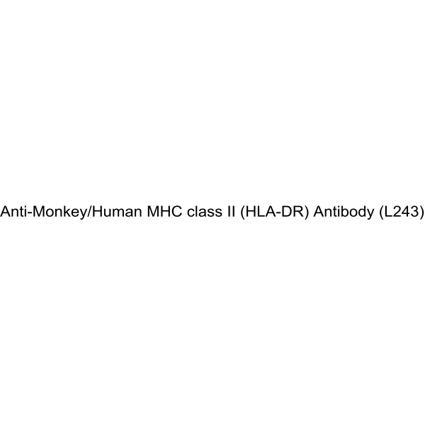
Anti-Monkey/Human MHC class II (HLA-DR) Antibody (L243) is a mouse-derived IgG2a κ type antibody inhibitor, targeting to monkey/human MHC class II. Anti-Monkey/Human MHC class II (HLA-DR) Antibody (L243) can inhibits tumorcells proliferation and induce apoptosis. Anti-Monkey/Human MHC class II (HLA-DR) Antibody (L243) increases cellular reactive oxygen species (ROS) and loss of mitochondrial membrane potential in human endothelial cells. Anti-Monkey/Human MHC class II (HLA-DR) Antibody (L243) can be used for the researches of cancer and infection, such as lymphoma .
ALK/EGFR-IN-1-d5 (Compound (-)-9a) is a deuterated dual-target inhibitor of EGFR and ALK, with an IC50 of 1.08 nM for EGFR and an IC50 of 2.395 nM for ALK. ALK/EGFR-IN-1-d5 inhibits the phosphorylated proteins in the EGFR, ALK, and BRK signaling pathways, blocking the cell cycle, leading to a reduction in mitochondrial membrane potential and cell apoptosis (Apoptosis). ALK/EGFR-IN-1-d5 also significantly inhibits tumor growth in animal models and demonstrates good safety. ALK/EGFR-IN-1-d5 holds promise for research in the field of cancer treatment
α-Casein (90-95) TFA is a partial agonist of opioid receptors and a copper ion ligand, with opioid activity. α-Casein (90-95) TFA inhibits the secretion of β-hexosaminidase by rat peritoneal mast cells (PMC) with IC50= 0.1 μM. α-Casein (90-95) TFA inhibits the proliferation of prostate cancer cells LNCaP, DU145, and PC3 with IC50 of 0.94 nM, 137 nM, and 6.92 nM, respectively. α-Casein (90-95) TFA activates Gi-like proteins through a membrane-assisted, receptor-independent pathway, or reversibly binds to opioid receptors, inducing intracellular calcium release and conformational changes, and exerts the activity of promoting mast cell secretion and inhibiting tumorcell proliferation. α-Casein (90-95) TFA can be used in the study of the mechanisms of allergic diseases and prostate cancer .
α-Casein (90-95) is a partial agonist of opioid receptors and a copper ion ligand, with opioid activity. α-Casein (90-95) inhibits the secretion of β-hexosaminidase by rat peritoneal mast cells (PMC) with IC50= 0.1 μM. α-Casein (90-95) inhibits the proliferation of prostate cancer cells LNCaP, DU145, and PC3 with IC50 of 0.94 nM, 137 nM, and 6.92 nM, respectively. α-Casein (90-95) activates Gi-like proteins through a membrane-assisted, receptor-independent pathway, or reversibly binds to opioid receptors, inducing intracellular calcium release and conformational changes, and exerts the activity of promoting mast cell secretion and inhibiting tumorcell proliferation. α-Casein (90-95) can be used in the study of the mechanisms of allergic diseases and prostate cancer .
Lipid C2 is an ionizable cationic lipid that has been used in the formation of lipid nanoparticles (LNP) for mRNA delivery in vivo. LNPs containing Lipid C2 and encapsulating an mRNA reporter selectively accumulate in the liver and spleen but not the heart, lungs, or kidneys in mice. LNP containing Lipid C2 and encapsulating mRNA encoding the Epstein-Barr virus (EBV) protein latent membrane protein 2 (LMP-2), in combination with an anti-programmed cell death protein 1 (PD-1) antibody, decrease tumor volume and reverse T cell exhaustion, as well as increase the percentage of CD3 +CD8 + central and CD3 +CD8 + effector memory T cells and decrease the percentage of CD3 + T cells expressing Pd-1, in the spleen in a CT26 murine EBV-infected colon cancer model .
MK-1088 is an orally active A2A/A2B adenosine receptor antagonist with human A2AKi of 0.31 nM, human A2BKi 5.3 nM. MK-1088 blocks receptor downstream signaling, inhibits CREB phosphorylation and reverses adenosine-mediated immunosuppression. MK-1088 restores TNF−α release and enhances tumor immune surveillance. MK-1088 can be used for the research of cancer[1].
Fendiline, a diphenylalkylamine type of antianginal agent, is an L-type calcium channel blocker (IC50 of 17 µM). Fendiline is also a selective K-Ras inhibitor, and has no effect on H-Ras and N-Ras. Fendiline inhibits K-Ras plasma membrane localization (IC50 of 9.64 μM), inhibits K-Ras signal output and blocks the proliferation of pancreatic, colon, lung, and endometrial cancer cell lines expressing oncogenic mutant K-Ras. Fendiline is a STING agonist and is able to inhibit the growth of multiple refractory cold tumors (MC38, CT26 and B16F10) .
Fendiline hydrochloride, a diphenylalkylamine type of antianginal agent, is an L-type calcium channel blocker (IC50 of 17 µM). Fendiline hydrochloride is also a selective K-Ras inhibitor, and has no effect on H-Ras and N-Ras. Fendiline hydrochloride inhibits K-Ras plasma membrane localization (IC50 of 9.64 μM), inhibits K-Ras signal output and blocks the proliferation of pancreatic, colon, lung, and endometrial cancer cell lines expressing oncogenic mutant K-Ras. Fendiline hydrochloride is a STING agonist and is able to inhibit the growth of multiple refractory cold tumors (MC38, CT26 and B16F10) .
Meso-tetraphenylchlorin (TPCS2a) is a photosensitizer with poor water solubility, which limits its use in the blood circulation. However, TPCS2a@NPs nanoparticles can be prepared based on polylactic-co-polyethylene glycol acid (PLGA) polymer core loaded with TPCS2. Such nanoparticles can be coated with mesenchymal stem cell-derived plasma membranes (mMSCs) to form mMSC-TPCS2a@NPs, which prolongs blood circulation time and improves tumor targeting ability. Compared with uncoated TPCS2a@NPs, mMSC-TPCS2a@NPs can reduce macrophage uptake by 54% to 70% under different conditions. Both nanoparticle forms are effectively accumulated in MCF7 and MDA-MB-231 breast cancer cells, while uptake in normal breast epithelial cells MCF10A is significantly lower .
Tubulin/VEGFR-2-IN-2 is an orally active tubulin and VEGFR-2 inhibitor with IC50s of 3.27 and 0.09 μM, respectively. Tubulin/VEGFR-2-IN-2 exerts the antitumor effects through multifaceted pathways, including enhancing reactive oxygen species (ROS) generation, disrupting mitochondrial membrane potential, inducing apoptosis, and arresting the cell cycle. Tubulin/VEGFR-2-IN-2 demonstrates anti-angiogenic properties by significantly impairing endothelial cell migration, invasion, and tube formation in vitro. Tubulin/VEGFR-2-IN-2 suppresses angiogenesis, tumor growth, and metastasis in vivo. Tubulin/VEGFR-2-IN-2 can be used for non-small lung cancer, breast cancer, gastric cancer and lymphoma .
β,β-Dimethylacrylalkannin (Arnebin 1) is an orally active FGFR1 inhibitor (IC50=2.5 μM) and the main active component of Lithospermum erythrorhizon. β,β-Dimethylacrylalkannin blocks downstream signaling by binding to the ATP pocket of FGFR1, and regulates the CDK1/Cdc25C pathway and ROS-JNK axis, thereby inducing G2/M phase arrest, necrosis and apoptosis in cancer cells, and inhibiting tumor proliferation. β,β-Dimethylacrylalkannin also acts as a colistin adjuvant to disrupt the cellmembrane of Gram-negative bacteria. β,β-Dimethylacrylalkannin exhibits significant tumor-inhibitory effects with no obvious toxicity in PDX models, but chronic exposure to high doses may alter the relative lung/liver weights of rats, while acute exposure to high doses causes responses such as reduced motor activity. β,β-Dimethylacrylalkannin finds wide application in studies related to hepatocellular carcinoma, colorectal cancer, colistin-resistant bacterial infections, hepatitis and psoriasis .
Low molecular weight protamine acetate (LMWP acetate;TDSP5 acetate) is a truncated arginine-rich protamine peptide, which also acts as an antidote for heparin/low molecular weight heparin and a cell-penetrating delivery vector. Low molecular weight protamine acetate neutralizes heparin-induced anticoagulant activities, including aPTT, anti-Xa and anti-IIa activities. Low molecular weight protamine acetate translocates across mammalian cellmembranes, delivers conjugated impermeable molecules through tumor tissues, enhances the skin permeability of conjugated epidermal growth factor, and accelerates wound healing when conjugated with epidermal growth factor. Low molecular weight protamine acetate retains the in vitro cell proliferation activity of conjugated EGF, and also enables site-specific conjugation with peptides or proteins via genetic recombination. Low molecular weight protamine acetate can be used in studies related to colon cancer, skin wounds and diabetic skin wounds .
Fendiline (hydrochloride) (Standard) is the analytical standard of Fendiline (hydrochloride). This product is intended for research and analytical applications. Fendiline hydrochloride, a diphenylalkylamine type of antianginal agent, is an L-type calcium channel blocker (IC50 of 17 µM). Fendiline hydrochloride is also a selective K-Ras inhibitor, and has no effect on H-Ras and N-Ras. Fendiline hydrochloride inhibits K-Ras plasma membrane localization (IC50 of 9.64 μM), inhibits K-Ras signal output and blocks the proliferation of pancreatic, colon, lung, and endometrial cancer cell lines expressing oncogenic mutant K-Ras. Fendiline hydrochloride is a STING agonist and is able to inhibit the growth of multiple refractory cold tumors (MC38, CT26 and B16F10) .
CBP-1018 is a PDC (peptide-drug conjugate) formed by conjugating Monomethyl auristatin E (HY-15162) to a dual-targeting ligand of FLOR1/PSMA (prostate-specific membrane antigen) via a linker (HY-78738). CBP-1018 binds to FLOR1 and prostate-specific membrane antigen (PSMA). CBP-1018 is applicable to the research of solid tumors and metastatic castration-resistant prostate cancer .
LMP2A (426-434) is a HLA-A2-restricted cytotoxic T lymphocyte (CTL) epitope of Epstein-Barr virus (EBV) latent membrane protein 2A (LMP2A). LMP2A (426-434) can trigger an immune response in individuals expressing different HLA-A*02 subtypes (A*02:01, A*02:03, A*02:06 and A*02:07). LMP2A (426-434) can induce a strong IFN-γ secretion response, stimulating the production of a high proportion of CD8 + IFN-γ + T cells. LMP2A (426-434) induces specific CTLs to effectively kill target cells expressing LMP2A. LMP2A (426-434) can be used to study EBV-related malignant tumors (such as Hodgkin's disease and nasopharyngeal carcinoma) .
PARP1-IN-44, an Olaparib (HY-10162) derivative, is an orally active PARP1 inhibitor (IC50 = 0.6 nM), and also inhibits PARP2 (IC50 = 1.0 nM) and PARP7 (IC50 = 7.5 nM). PARP1-IN-44 has selective antiproliferative activity against BRCA-deficient cancer cells with minimal toxicity to normal cells. PARP1-IN-44 induces G2/M phase arrest, promotes apoptosis, elevates ROS levels, disrupts mitochondrial membrane potential. PARP1-IN-44 suppresses PARylation while increasing γH2AX accumulation. PARP1-IN-44 activates the cGAS-STING pathway, upregulating IFN-β and CXCL10 expression. PARP1-IN-44 enhancing CD8+ T cell infiltration in a CT26 tumor mouse model, demonstrating robust in vivo antitumor efficacy .
Low molecular weight protamine (LMWP;TDSP5) is a truncated arginine-rich protamine peptide, as well as a heparin/low-molecular-weight heparin antidote and a cell-penetrating delivery carrier. Low molecular weight protamine neutralizes heparin-induced anticoagulant activities, including aPTT, anti-Xa and anti-IIa activities, and also neutralizes anti-Xa activity of commercially available low-molecular-weight heparin preparations. Low molecular weight protamine translocates across mammalian cellmembranes, delivers conjugated impermeable molecules across tumor tissues, enhances skin permeability of conjugated epidermal growth factor, and accelerates wound healing when conjugated with epidermal growth factor. Low molecular weight protamine retains the in vitro cell proliferation activity of conjugated EGF, and also enables site-specific conjugation with peptides or proteins via genetic recombination. Low molecular weight protamine can be used in studies related to colon adenocarcinoma, skin wounds and diabetic skin wounds.
Hyaluronic acid sodium (MW 200-1560) is a biopolymer composed of repeating disaccharide units, with a molecular weight of 200-1560. Hyaluronic acid sodium is a major component of the extracellular matrix (ECM). It is synthesized on the plasma membrane. Hyaluronic acid sodium exerts its effects by binding to receptors CD44 and RHAMM. Hyaluronic acid sodium activates PI3K-Akt signaling. Hyaluronic acid sodium also enhances cell invasion and angiogenesis by promoting or stimulating the binding of proteolytic MMP-9 to the cell surface. Elevated hyaluronic acid levels are associated with tumorcell growth, adhesion, migration, invasion, and angiogenesis in digestive system cancers. Hyaluronic acid sodium is involved in tissue remodeling and rapid cell proliferation in several physiological processes, including embryonic morphogenesis and wound healing. Hyaluronic acid sodium can be used as a regulator of cancer-associated lymphangiogenesis. Hyaluronic acid sodium can be used as a drug delivery carrier for sodium butyrate, enhancing its anti-proliferative activity against breast cancer cell lines. Hyaluronic acid sodium can lubricate the corneal endothelium. Hyaluronic acid sodium can improve tissue hydration and enhance the resistance of cells to mechanical damage. Hyaluronic acid sodium has been conjugated with antibodies to ensure that the active compound continues to exert its effects at the site of inflammation. Hyaluronic acid sodium can be used in research in the fields of osteoarthritis, ophthalmology, cosmetic dermatology, oncology, and liver diseases .
Hyaluronic acid, low endotoxin (Hyaluronan, low endotoxin) is a biopolymer composed of repeating disaccharide units containing low levels of endotoxin. Hyaluronic acid is a major component of the extracellular matrix (ECM). It is synthesized on the plasma membrane. Hyaluronic acid exerts its effects by binding to receptors CD44 and RHAMM. Hyaluronic acid activates PI3K-Akt signaling. Hyaluronic acid also enhances cell invasion and angiogenesis by promoting or stimulating the binding of proteolytic MMP-9 to the cell surface. Elevated hyaluronic acid levels are associated with tumorcell growth, adhesion, migration, invasion, and angiogenesis in digestive system cancers. Hyaluronic acid is involved in tissue remodeling and rapid cell proliferation in several physiological processes, including embryonic morphogenesis and wound healing. Hyaluronic acid can be used as a regulator of cancer-associated lymphangiogenesis. Hyaluronic acid can be used as a drug delivery carrier for sodium butyrate, enhancing its anti-proliferative activity against breast cancer cell lines. Hyaluronic acid can lubricate the corneal endothelium. Hyaluronic acid can improve tissue hydration and enhance the resistance of cells to mechanical damage. Hyaluronic acid has been conjugated with antibodies to ensure that the active compound continues to exert its effects at the site of inflammation. Hyaluronic acid can be used in research in the fields of osteoarthritis, ophthalmology, cosmetic dermatology, oncology, and liver diseases .
Tauro-β-muricholic acid (TβMCA) is an orally active trihydroxylated bile acid and a competitive, reversible FXR antagonist (IC50=40 μM). Tauro-β-muricholic acid inhibits bile acid-induced hepatocyte apoptosis by maintaining mitochondrial membrane potential, while simultaneously inhibiting intestinal FXR signaling, affecting bile acid synthesis, hepatic lipid metabolism, and insulin sensitivity. Accumulation of tauro-β-muricholic acid disrupts metabolic homeostasis, promoting cancer stem cell proliferation and tumor progression. The mechanisms of tauro-β-muricholic acid involve two aspects: first, inhibiting the translocation of the pro-apoptotic protein Bax to mitochondria and maintaining mitochondrial membrane potential (MMP); and second, blocking the FXR signaling pathway to regulate bile acid metabolism, reduce serum ceramide production, and downregulate the hepatic SREBP1C/CIDEA pathway. Tauro-β-muricholic acid possesses anti-hepatocyte apoptosis, bile acid homeostasis regulation, and liver fat accumulation reduction properties, and also functions as a biomarker, making it useful in the study of diseases such as bile acid metabolism disorders, non-alcoholic fatty liver disease, colorectal cancer, and liver fibrosis .
Hsp70TAC PD-1 Degrader-2 is a PD-L1 Hsp70TAC (Hsp70-targeting Chimeras) degrader with Kd values of 0.36 μM. Hsp70TAC PD-1 Degrader-2 forms a ternary complex with Hsp70 and PD-L1 to drive PD-L1 degradation. Hsp70TAC PD-1 Degrader-2 induces degradation of mature membrane-bound PD-L1 in an Hsp70-dependent manner and via caveolin-mediated endocytosis and lysosomal trafficking. Hsp70TAC PD-1 Degrader-2 accumulates preferentially in tumorcells with elevated Hsp70 expression for tumor-selective PD-L1 degradation. Hsp70TAC PD-1 Degrader-2 can be used for the research of cancer, such as breast invasive carcinoma, glioblastoma multiforme, diffuse large b-cell lymphoma . (Pink: PD-1/PD-L1 ligand (HY-19745A); Blue: Hsp70 ligand (HY-182979); Black: linker (HY-182982)).
KRAS G12D-IN-37 is a KRAS G12D inhibitor. KRAS G12D-IN-37 shows antiproliferative activity against KRAS G12D mutant tumorcells and minimal cytotoxicity toward normal cells. KRAS G12D-IN-37 binds stably to KRAS G12D via hydrogen bond interactions with residues His 95, Arg 68, and Asp 12, and inhibits downstream ERK/AKT signaling pathways. KRAS G12D-IN-37 elevates ROS levels, induces apoptosis, disrupts mitochondrial membrane potential. KRAS G12D-IN-37 downregulates the level of anti-apoptotic protein Bcl-2, and upregulates the levels of pro-apoptotic proteins Bax and caspase 3. KRAS G12D-IN-37 can be used for the research of cancer, such as gastric adenocarcinoma and colorectal cancer .
Tabersonine is a selective, orally active NLRP3 inhibitor. Tabersonine directly binds to the NACHT domain of NLRP3, inhibiting its ATPase activity and oligomerization, thereby blocking ASC spot formation and caspase-1 activation, and reducing the release of pro-inflammatory cytokines such as IL-1β. Tabersonine also inhibits K63-linked ubiquitination of TRAF6, blocking NF-κB, PI3K/Akt, and p38 MAPK signaling pathways. Tabersonine can inhibit inflammatory responses, induce apoptosis of liver cancer cells through mitochondrial pathways and death receptor pathways, reduce mitochondrial membrane potential, promote cytochrome c release, and activate caspase proteins. Tabersonine is mainly used in the study of NLRP3-driven inflammatory diseases (such as acute lung injury, sepsis, peritonitis) and tumors such as liver cancer .
Tabersonine hydrochloride is a selective, orally active NLRP3 inhibitor. Tabersonine hydrochloride directly binds to the NACHT domain of NLRP3, inhibiting its ATPase activity and oligomerization, thereby blocking ASC spot formation and caspase-1 activation, and reducing the release of pro-inflammatory cytokines such as IL-1β. Tabersonine hydrochloride also inhibits K63-linked ubiquitination of TRAF6, blocking NF-κB, PI3K/Akt, and p38 MAPK signaling pathways. Tabersonine hydrochloride can inhibit inflammatory responses, induce apoptosis of liver cancer cells through mitochondrial pathways and death receptor pathways, reduce mitochondrial membrane potential, promote cytochrome c release, and activate caspase proteins. Tabersonine hydrochloride is mainly used in the study of NLRP3-driven inflammatory diseases (such as acute lung injury, sepsis, peritonitis) and tumors such as liver cancer .
DUPA-FITC is a fluorescent reagent targeting PSMA, which specifically binds to prostate cancer cells expressing PSMA without non-specific binding to normal blood cells. DUPA-FITC can label PSMA-expressing prostate cancer cells in whole blood, followed by internalization and trafficking to acidic intracellular endosomes, during which the fluorescence is quenched. When combined with flow cytometry and density gradient centrifugation enrichment, DUPA-FITC enables quantitative analysis of circulating tumor cells in peripheral blood samples from prostate cancer patients .
TREM-1 inhibitory peptide GF9 (Human TREM-1 (213-221)) is a TREM-1 inhibitor. TREM-1 inhibitory peptide GF9 blocks the TREM-1 signaling pathway via a ligand-independent mechanism, spontaneously inserts into the cellmembrane to dissociate TREM-1 from DAP-12, and functions through the Signaling Chain Homooligomerization (SCHOOL) model. TREM-1 inhibitory peptide GF9 reduces the levels of TNFα, IL-1β, IL-6, and M-CSF. TREM-1 inhibitory peptide GF9 inhibits tumor growth, prolongs the survival of mice with pancreatic cancer models, ameliorates collagen-induced arthritis, and exerts protective effects on bone and cartilage simultaneously. TREM-1 inhibitory peptide GF9 can be used in research related to arthritis, pancreatic cancer, retinopathy, alcoholic liver disease, and liver cancer .
Mutations in oncogenes and tumor suppressor genes can modify multiple signaling pathways and in turn cell metabolism, which facilitates tumorigenesis. The paramount hallmark of tumor metabolism is “aerobic glycolysis” or the Warburg effect, coined by Otto Warburg in 1926, in which cancer cells produce most of energy from glycolysis pathway regardless of whether in aerobic or anaerobic condition. Usually, cancer cells are highly glycolytic (glucose addiction) and take up more glucose than do normal cells from outside. The increased uptake of glucose is facilitated by the overexpression of several isoforms of membrane glucose transporters (GLUTs). Likewise, the metabolic pathways of glutamine, amino acid and fat metabolism are also altered. Recent trends in anti-cancer drug discovery suggests that targeting the altered metabolic pathways of cancer cells result in energy crisis inside the cancer cells and can selectively inhibit cancer cell proliferation by delaying or suppressing tumor growth.
MCE provides a unique collection of 3,552 compounds which cover various tumor metabolism-related signaling pathways. These compounds can be used for anti-cancer metabolism targets identification, validation as well anti-cancer drug discovery.
DiSC3(5) is a fluorescent probe commonly used as a tracer dye to evaluate mitochondrial membrane potential. The excitation/emission wavelength of DiSC3(5) is up to 622/670 nm. DiSC3(5) can inhibit the respiratory system associated with mitochondrial NAD, and the IC50 value is 8 μM. DiSC3(5) in the presence of Na +/K +-ATPase inhibitor ouabain 2 can induce membrane hyperpolarization of Ehrlich ascites tumorcells .
IR-780 is a near-infrared fluorescent probe for in vivo imaging of tumorcells. IR-780 is transported into tumorcells via OATPs and ABCB10, with uptake dependent on glycolytic activity and plasma membrane potential. IR-780 preferentially accumulates in tumorcell mitochondria, including those of drug-resistant cancer cells, without chemical conjugation. IR-780 generates reactive oxygen species (ROS), induces hyperthermia and apoptosis, inhibits tumor growth and recurrence, and modulates HSP70 expression upon ultrasound or 808 nm laser exposure. IR-780 acts as a sonosensitizer, photodynamic and photothermal agent, and drug delivery carrier, with low acute imaging-dose toxicity and rapid vital organ clearance. IR-780 can be used for the research of cancer, such as breast cancer, lung cancer, and non-small cell lung cancer (NSCLC) .
DiSC3 (5) (solution) is a fluorescent probe commonly used as a tracer dye to evaluate mitochondrial membrane potential. The excitation/emission wavelength of DiSC3 (5) is up to 622/670 nm. DiSC3 (5) can inhibit the respiratory system associated with mitochondrial NAD, and the IC50 value is 8 μM. DiSC3 (5) in the presence of Na +/K +-ATPase inhibitor ouabain 2 can induce membrane hyperpolarization of Ehrlich ascites tumorcells . Solvent and concentration: DMSO: 1 mM The 1 mL volume is defined as the base specification. All larger sizes correspond to incremental volumes of this base.
NIR-ASM is a near-infrared fluorescent probe that can cross cellmembranes and be activated by NQO1. NIR-ASM can distinguish NQO1-expressing cancer cells from normal cells via fluorescence microscopy and flow cytometry. NIR-ASM generates near-infrared fluorescence with a high signal-to-noise ratio in tumor models with NQO1 activity, enabling the detection of endogenous NQO1 activity in vivo. NIR-ASM is applicable to the research of lung cancer and breast cancer .
DUPA-FITC is a fluorescent reagent targeting PSMA, which specifically binds to prostate cancer cells expressing PSMA without non-specific binding to normal blood cells. DUPA-FITC can label PSMA-expressing prostate cancer cells in whole blood, followed by internalization and trafficking to acidic intracellular endosomes, during which the fluorescence is quenched. When combined with flow cytometry and density gradient centrifugation enrichment, DUPA-FITC enables quantitative analysis of circulating tumor cells in peripheral blood samples from prostate cancer patients .
Hyaluronic acid is a biopolymer composed of repeating units of disaccharides with various applications. Hyaluronic acid is a major component of the extracellular matrix (ECM). Hyaluronic acid is synthesized at the plasma membrane. Increased hyaluronic acid levels are associated with tumorcell growth, adhesion, migration, invasion and angiogenesis in digestive cancers. Hyaluronic acid participates in tissue remodeling and rapid cell proliferation in some physiological processes including embryonic morphogenesis and wound-healing. Hyaluronic acid activates the PI3K-Akt signaling. Hyaluronic acid acts as a regulator of cancer-associated lymphangiogenesis. Hyaluronic acid also enhances cell invasion and angiogenesis by promoting proteolytic MMP-9 binding to cell surface or stimulating MMP-9 binding to cell surface. Hyaluronic acid can be used as drug delivery for sodium butyrate to improve the anti-proliferative activity on breast cancer cell line. Hyaluronic acid can be studied in joint diseases, wound healing and cancer .
DSPE-PEG2000-Cy5 is a Cy5 (HY-D0821) fluorophore-labeled conjugate of distearoylphosphatidylethanolamine and polyethylene glycol, as well as a liposome component. The Cy5 fluorophore is commonly used for labeling proteins and nucleic acids in imaging, flow cytometry and genomic applications. DSPE-PEG2000-Cy5 supports cellmembrane modification, in vivo tumor targeting research and long-term in vivo circulation of its liposomal formulations (Ex/Em=633/670 nm) .
Hyaluronic acid, low endotoxin (Hyaluronan, low endotoxin) is a biopolymer composed of repeating disaccharide units containing low levels of endotoxin. Hyaluronic acid is a major component of the extracellular matrix (ECM). It is synthesized on the plasma membrane. Hyaluronic acid exerts its effects by binding to receptors CD44 and RHAMM. Hyaluronic acid activates PI3K-Akt signaling. Hyaluronic acid also enhances cell invasion and angiogenesis by promoting or stimulating the binding of proteolytic MMP-9 to the cell surface. Elevated hyaluronic acid levels are associated with tumorcell growth, adhesion, migration, invasion, and angiogenesis in digestive system cancers. Hyaluronic acid is involved in tissue remodeling and rapid cell proliferation in several physiological processes, including embryonic morphogenesis and wound healing. Hyaluronic acid can be used as a regulator of cancer-associated lymphangiogenesis. Hyaluronic acid can be used as a drug delivery carrier for sodium butyrate, enhancing its anti-proliferative activity against breast cancer cell lines. Hyaluronic acid can lubricate the corneal endothelium. Hyaluronic acid can improve tissue hydration and enhance the resistance of cells to mechanical damage. Hyaluronic acid has been conjugated with antibodies to ensure that the active compound continues to exert its effects at the site of inflammation. Hyaluronic acid can be used in research in the fields of osteoarthritis, ophthalmology, cosmetic dermatology, oncology, and liver diseases .
Chlorophosphonazo III is a cellmembrane-permeable contrast agent and spectrophotometric reagent. Chlorophosphonazo III forms stable 1:1 complexes with intracellular calcium, magnesium, uranium, titanium, zirconium, thorium, scandium, equivalent actinides and protactinium. Chlorophosphonazo III generates photoacoustic signals correlated with Ca 2+ concentrations, which is applicable for imaging 3D tumorcell cultures and tumor spheroids. Chlorophosphonazo III exhibits no cytotoxicity, can be used for spectrophotometric detection of target ions, and the absorbance of its complexes remains stable for up to one week within the pH range of 2.2 to 7.0 .
Laminin peptide CDPGYIGSR (Laminin (925-933)) TFA is a 67 kDa laminin receptor ligand and selective cell adhesion inducer. Laminin peptide CDPGYIGSR TFA not only promotes cell adhesion and mediates directed neurite outgrowth via matrix coating or covalent immobilization, but also inhibits neural crest cell migration under specific conditions. Laminin peptide CDPGYIGSR TFA inhibits lung colonization of melanoma cells, and suppresses the growth of Sarcoma 180 solid tumors and Lewis lung carcinoma (3LL) in mice. Laminin peptide CDPGYIGSR TFA also exerts significant anti-angiogenic effects by inhibiting embryonic angiogenesis in the chick chorioallantoic membrane and vascular endothelial cell migration induced by tumor-conditioned medium. Laminin peptide CDPGYIGSR TFA can be widely used in studies related to melanoma, Sarcoma 180, Lewis lung carcinoma (3LL), and other relevant areas .
pH-Low Insertion Peptide (pHLIP) is a short, pH-responsive peptide capable of inserting across a cellmembrane to form a transmembrane helix at acidic pH. pH-Low Insertion Peptide targets the acidic tumor microenvironment for tumors at early and metastatic stages with high specificity, used as a specific ligand. pH-Low Insertion Peptide successfully modify polylysine polymers to have the pH-responsive capability. pH-Low Insertion Peptide-based targeting of cancer presents an opportunity to monitor metabolic changes, and to selectively deliver imaging and therapeutic agents to tumors .
MANS peptide is an inhibitor for myristoylated alanine-rich C kinase substrate (MARCKS), which competes with MARCKS in cells for membrane binding, and thus inhibits the stimulation of mucin secretion and tumor metastasis .
Difopein TFA is a 14-3-3 protein inhibitor. Difopein TFA acts as an apoptosis inducer, regulates apoptosis-related proteins, downregulates Bcl-2, upregulates Bax, activates caspase-9 and caspase-3, and induces nuclear fragmentation, membrane-enclosed apoptotic bodies and DNA ladder formation. Difopein TFA serves as a tumor growth inhibitor, which inhibits the proliferation of glioma cells and induces their apoptosis in in vivo nude mouse models. Difopein TFA is applicable to glioma-related research .
TREM-1 inhibitory peptide GF9 (Human TREM-1 (213-221)) is a TREM-1 inhibitor. TREM-1 inhibitory peptide GF9 blocks the TREM-1 signaling pathway via a ligand-independent mechanism, spontaneously inserts into the cellmembrane to dissociate TREM-1 from DAP-12, and functions through the Signaling Chain Homooligomerization (SCHOOL) model. TREM-1 inhibitory peptide GF9 reduces the levels of TNFα, IL-1β, IL-6, and M-CSF. TREM-1 inhibitory peptide GF9 inhibits tumor growth, prolongs the survival of mice with pancreatic cancer models, ameliorates collagen-induced arthritis, and exerts protective effects on bone and cartilage simultaneously. TREM-1 inhibitory peptide GF9 can be used in research related to arthritis, pancreatic cancer, retinopathy, alcoholic liver disease, and liver cancer .
Tachyplesin I is a β-hairpin antimicrobial peptide that contains 17 amino acid residues. Tachyplesin I exhibits cytotoxic properties against various human tumorcell lines acting primarily by impairing the integrity of the outer cellmembrane .
MANS peptide TFA is the TFA salt form of MANS peptide (HY-P10218). MANS peptide TFA is an inhibitor for myristoylated alanine-rich C kinase substrate (MARCKS), which competes with MARCKS in cells for membrane binding, and thus inhibits the stimulation of mucin secretion and tumor metastasis .
HYNIC-PSMA is a ligand for molecular imaging of tumors. Hynic-psma consists of two components: HYNIC (6-hydrazinonicotinamide) and PSMA (Prostate-Specific Membrane Antigen). HYNIC is a compound used to attach radioactive isotopes to targeted molecules, such as 188Re-HYNIC-PSMA. PSMA is a membrane antigen that is specifically expressed on the surface of prostate cancer cells. HYNIC-PSMA can be used in prostate cancer research . HYNIC-PSMA can be used for the synthesis/research of Radionuclide-Drug Conjugates (RDCs).
α-Casein (90-95) is a partial agonist of opioid receptors and a copper ion ligand, with opioid activity. α-Casein (90-95) inhibits the secretion of β-hexosaminidase by rat peritoneal mast cells (PMC) with IC50= 0.1 μM. α-Casein (90-95) inhibits the proliferation of prostate cancer cells LNCaP, DU145, and PC3 with IC50 of 0.94 nM, 137 nM, and 6.92 nM, respectively. α-Casein (90-95) activates Gi-like proteins through a membrane-assisted, receptor-independent pathway, or reversibly binds to opioid receptors, inducing intracellular calcium release and conformational changes, and exerts the activity of promoting mast cell secretion and inhibiting tumorcell proliferation. α-Casein (90-95) can be used in the study of the mechanisms of allergic diseases and prostate cancer .
LMP2A (426-434) is a HLA-A2-restricted cytotoxic T lymphocyte (CTL) epitope of Epstein-Barr virus (EBV) latent membrane protein 2A (LMP2A). LMP2A (426-434) can trigger an immune response in individuals expressing different HLA-A*02 subtypes (A*02:01, A*02:03, A*02:06 and A*02:07). LMP2A (426-434) can induce a strong IFN-γ secretion response, stimulating the production of a high proportion of CD8 + IFN-γ + T cells. LMP2A (426-434) induces specific CTLs to effectively kill target cells expressing LMP2A. LMP2A (426-434) can be used to study EBV-related malignant tumors (such as Hodgkin's disease and nasopharyngeal carcinoma) .
RS17 is an anti-tumor peptide designed to bind specifically to the CD47 molecule and block the interaction between CD47 and its ligand, SIRPα, on the surface membrane of macrophages. The main regulatory mechanism of RS17 is to prevent CD47 from transmitting selective phagocytosis signals to SIRPα by binding to CD47, so that macrophages do not recognize tumorcells as their own tissue, but phagocytose them as foreign substances, thereby inhibiting immune escape of tumorcells. RS17 can be used to study the mechanism of anti-tumor response and immune escape .
Defensin HNP-3 human is an α-defensin stored in the azurophilic granules of human neutrophils. Defensin HNP-3 human exerts broad-spectrum bactericidal, antifungal and antiviral activities mainly by forming bacterial membrane pores, and acts as a chemoattractant for monocytes and T cells. Defensin HNP-3 human maintains epithelial integrity to support periodontal tissue homeostasis, and exerts concentration-dependent effects on epithelial cell proliferation, adhesion and bacterial adhesion. Defensin HNP-3 human targets solid tumors and leukemia by inducing single-strand DNA breaks and membrane permeabilization in tumorcells via electrostatic binding and pore formation. Defensin HNP-3 human is abundant in human tongue squamous cell carcinoma and neutrophils infiltrating oral squamous cell carcinoma. Defensin HNP-3 human can be applied to research related to periodontitis and human tongue squamous cell carcinoma .
CBP-1018 is a PDC (peptide-drug conjugate) formed by conjugating Monomethyl auristatin E (HY-15162) to a dual-targeting ligand of FLOR1/PSMA (prostate-specific membrane antigen) via a linker (HY-78738). CBP-1018 binds to FLOR1 and prostate-specific membrane antigen (PSMA). CBP-1018 is applicable to the research of solid tumors and metastatic castration-resistant prostate cancer .
Low molecular weight protamine (LMWP;TDSP5) is a truncated arginine-rich protamine peptide, as well as a heparin/low-molecular-weight heparin antidote and a cell-penetrating delivery carrier. Low molecular weight protamine neutralizes heparin-induced anticoagulant activities, including aPTT, anti-Xa and anti-IIa activities, and also neutralizes anti-Xa activity of commercially available low-molecular-weight heparin preparations. Low molecular weight protamine translocates across mammalian cellmembranes, delivers conjugated impermeable molecules across tumor tissues, enhances skin permeability of conjugated epidermal growth factor, and accelerates wound healing when conjugated with epidermal growth factor. Low molecular weight protamine retains the in vitro cell proliferation activity of conjugated EGF, and also enables site-specific conjugation with peptides or proteins via genetic recombination. Low molecular weight protamine can be used in studies related to colon adenocarcinoma, skin wounds and diabetic skin wounds.
d-(KLAKLAK)2, as an antibacterial and anti-tumor polypeptide, is a representative of the antimicrobial peptide group, and also has good anticancer properties. d-(KLAKLAK)2 is able to kill bacteria by damaging their cellmembranes, causing cell contents to leak out. d-(KLAKLAK)2 can also inhibit tumorcell proliferation by causing mitochondrial swelling and mitochondrial membrane destruction, triggering apoptosis (programmed cell death) .
CyPep-1 is a novel cationic lytic peptide with antitumor activity. CyPep-1 is highly cytotoxic by interacting with the negatively charged cellmembrane of cancer cells. CyPep-1 has strong cytolytic activity on cancer cells both in vivo and in vitro, and can be used in the study of solid tumors .
pH-Low Insertion Peptide TFA (pHLIP TFA) is a short, pH-responsive peptide capable of inserting across a cellmembrane to form a transmembrane helix at acidic pH. pH-Low Insertion Peptide TFA targets the acidic tumor microenvironment for tumors at early and metastatic stages with high specificity, used as a specific ligand. pH-Low Insertion Peptide TFA successfully modifys polylysine polymers to have the pH-responsive capability. pH-Low Insertion Peptide TFA -based targeting of cancer presents an opportunity to monitor metabolic changes and to selectively deliver imaging and therapeutic agents to tumors .
IT9302 is a synthetic IL-10 agonist with the activity of inducing tolerogenic dendritic cells. IT9302 is able to mimic multiple effects of IL-10, including downregulating the antigen presentation machinery and increasing the sensitivity of tumorcells to natural killer cell-mediated lysis. IT9302 can also hinder the response of human monocytes to differentiation factors and reduce the antigen presentation and co-stimulatory capacity of dendritic cells. Dendritic cells treated with IT9302 showed a weakened ability to stimulate T cell proliferation and interferon-γ production. IT9302 exerts its effects through mechanisms that are partially different from IL-10, involving STAT3 inactivation and regulation of the NF-κB intracellular pathway. IT9302-treated dendritic cells showed enhanced expression of membrane-bound TGF-β, associated with the effective induction of foxp3+ regulatory T cells .
Cys-Asp-Pro-Gly-Tyr-Ile-Gly-Ser-Arg-NH2 is a linear peptide from laminin B1 chain that interferes with tumorcell attachment and invasion into basement membrane and has anti-angiogenic effects .
HN-1 is a 12-mer peptide with specific activity to head and neck squamous cell cancer (HNSCC) cells. HN-1, as a tumor-specific peptide, is capable of penetrating tumor tissues. HN-1 is capable of translocating agents across cellmembranes .
BQ7876 is a probe targeting prostate-specific membrane antigen (PSMA) that contains a DOTA chelator. BQ7876, after being radiolabeled with radionuclide (177Lu), functions in both radionuclide imaging and tumorcell destruction by specifically binding to PSMA. BQ7876 shows potential for research in the field of metastatic castration-resistant prostate cancer (mCRPC) . BQ7876 can be used for the synthesis/research of Radionuclide-Drug Conjugates (RDCs).
α-Casein (90-95) TFA is a partial agonist of opioid receptors and a copper ion ligand, with opioid activity. α-Casein (90-95) TFA inhibits the secretion of β-hexosaminidase by rat peritoneal mast cells (PMC) with IC50= 0.1 μM. α-Casein (90-95) TFA inhibits the proliferation of prostate cancer cells LNCaP, DU145, and PC3 with IC50 of 0.94 nM, 137 nM, and 6.92 nM, respectively. α-Casein (90-95) TFA activates Gi-like proteins through a membrane-assisted, receptor-independent pathway, or reversibly binds to opioid receptors, inducing intracellular calcium release and conformational changes, and exerts the activity of promoting mast cell secretion and inhibiting tumorcell proliferation. α-Casein (90-95) TFA can be used in the study of the mechanisms of allergic diseases and prostate cancer .
Low molecular weight protamine acetate (LMWP acetate;TDSP5 acetate) is a truncated arginine-rich protamine peptide, which also acts as an antidote for heparin/low molecular weight heparin and a cell-penetrating delivery vector. Low molecular weight protamine acetate neutralizes heparin-induced anticoagulant activities, including aPTT, anti-Xa and anti-IIa activities. Low molecular weight protamine acetate translocates across mammalian cellmembranes, delivers conjugated impermeable molecules through tumor tissues, enhances the skin permeability of conjugated epidermal growth factor, and accelerates wound healing when conjugated with epidermal growth factor. Low molecular weight protamine acetate retains the in vitro cell proliferation activity of conjugated EGF, and also enables site-specific conjugation with peptides or proteins via genetic recombination. Low molecular weight protamine acetate can be used in studies related to colon cancer, skin wounds and diabetic skin wounds .
Tachyplesin I TFA is a β-hairpin antimicrobial peptide that contains 17 amino acid residues. Tachyplesin I TFA exhibits cytotoxic properties against various human tumorcell lines acting primarily by impairing the integrity of the outer cellmembrane .
IALYLQQNW is a specific nonapeptide sequence derived from the tumor-associated antigen latent membrane protein 1 (LMP1) encoded by Epstein-Barr virus (EBV). As a latent T-cell epitope, IALYLQQNW is able to activate EBV-specific cytotoxic T lymphocytes (CTLs), which are able to recognize and kill EBV-infected cells expressing LMP1. IALYLQQNW plays an important role in the immune response against EBV-associated tumors and can be used in the study of Hodgkin's disease and nasopharyngeal carcinoma .
TAT-327 is cell-penetrating peptide. TAT-327 selectively inserts into cancer cellmembranes and shows potent antitumor activity. TAT-327 effectively inhibits cancer cells proliferation, induces apoptosis and disrupts EGFR signal pathway by inhibiting downstream signals (such as IL-2, TNF-α and IFN-γ) expression and the Eps8/EGFR interaction. TAT-327 significantly inhibits tumor growth in HT-29 xenograft mcie models .
PDI2 (PSMA-DOTA-PEI2) is a prostate-specific membrane antigen (PSMA) ligand that acts as a tumor retention agent, renal uptake reducer, imaging agent and antitumor agent, applicable in SPECT diagnostic imaging and radiotheranostics. PDI2 specifically binds to PSMA on prostate cancer cells, enters cells via clathrin-dependent endocytosis, and exhibits higher tumor retention rate and lower renal uptake level. PDI2 is applicable in research related to prostate cancer and castration-resistant metastatic prostate cancer .
Myr-transportan-Cys is a derivative of the cell-penetrating peptide Transportan (HY-P1732), and its conjugated myristoyl group (Myr) enhances the interaction between the peptide and cellmembranes. Myr-transportan-Cys integrates three key delivery functions: nucleic acid condensation, cell penetration, and endosomal escape. Myr-transportan-Cys can form immunostimulatory tandem peptide nanocomplexes (iTPNCs) for encapsulating and delivering immunostimulatory oligonucleotide cargos to tumors .
P-LPK is a dodecapeptide that specifically targets SLC1A5, which is highly expressed on the membrane of colorectal cancer cells, with a Kd value of 1.19 μM. P-LPK has no intrinsic cell proliferation regulatory activity. Gallium-68-labeled P-LPK selectively accumulates at colorectal cancer tumor sites in xenograft mouse models. P-LPK can serve as a targeted carrier to deliver Camptothecin (HY-16560) to colorectal cancer cells, forming the conjugate P-LPK-CPT. P-LPK can be used in the research of colorectal cancer .
Difopein is a 14-3-3 protein inhibitor. Difopein acts as an apoptosis inducer, regulates apoptosis-related proteins, downregulates Bcl-2, upregulates Bax, activates caspase-9 and caspase-3, and induces nuclear fragmentation, membrane-enclosed apoptotic bodies and DNA ladder formation. Difopein serves as a tumor growth inhibitor, which inhibits the proliferation of glioma cells and induces their apoptosis in in vivo nude mouse models. Difopein is applicable to glioma-related research .
Anticancer peptoid 1 (Peptoid 1) is a peptoid with antitumor activity. Anticancer peptoid 1 exerts cytotoxicity primarily via plasma membrane damage. Anticancer peptoid 1 exerts cytotoxicity against a broad range of cancer cell lines, including those with multidrug resistance. Anticancer peptoid 1 inhibits tumor growth in a human breast cancer xenotransplantation mouse model without noticeable acute adverse effects. Anticancer peptoid 1 can be used for the research of cancer, such as breast cancer, and prostate cancer .
MCE Basement Membrane Matrix IPSC-qualified is primarily composed of natural basement membrane matrix extracted from mouse tumors. This product is mainly used for stem cells culture.
MCE Basement Membrane Matrix GFR&HC (Growth Factor Reduced, High Concentration) is primarily composed of natural basement membrane matrix extracted from mouse tumors, is suitable for cell cultures that need to reduce growth factor induction and studies related to growth factors, signaling pathways, etc.
Cevostamab (BFCR4350A; RG6160; RO7187797) is a humanized IgG1-based BsAb that targets membrane-proximal extracellular domain of FcRH5 on multiple myeloma (MM) cells as well as CD3 on T cells. Moreover, Cevostamab facilitates efficient synapse formation, improves killing activity of T cells against MM tumorcells .
IGN523 is an anti-CD98 antibody (hCD98, KD = 0.55 nM). IGN523 induces antibody-dependent cell-mediated cytotoxicity (ADCC) activity, lysosomal membrane permeabilization, and inhibition of essential amino acid transport, ultimately leading to caspase-3 and caspase-7-mediated apoptosis of tumorcells. IGN523 inhibits tumor growth in multiple tumor xenograft models. IGN523 is useful in the research of non-small cell lung cancer (NSCLC), acute myeloid leukemia (AML), and other cancers. .
IMAB027 (ASP1650) is a specific anti-CLDN6 mAb, while CLDN6 (Claudin 6) is a tight junction membrane protein, aberrantly expressed in various human cancer types, ovarian cancers particularly. IMAB 027 shows anti-tumor activity, and induces apoptosis in CLDN6 + ovarian and testicular cancer cell lines .
IPH5201 is a selective CD39 inhibitor and a humanized IgG1 monoclonal antibody. IPH5201 selectively binds to and inhibits the enzymatic activity of both membrane-bound and soluble CD39, blocking ATP hydrolysis. IPH5201 enhances the phenotypic maturation and activation of dendritic cells and macrophages. IPH5201 potentiates the anti-tumor effect of Oxaliplatin (HY-17371). IPH5201 shows preliminary evidence of disease stabilization in advanced solid tumor models when used as a single agent or in combination with Durvalumab (HY-P9919). IPH5201 can be used for the research of advanced solid tumors .
Anti-Monkey/Human MHC class II (HLA-DR) Antibody (L243) is a mouse-derived IgG2a κ type antibody inhibitor, targeting to monkey/human MHC class II. Anti-Monkey/Human MHC class II (HLA-DR) Antibody (L243) can inhibits tumorcells proliferation and induce apoptosis. Anti-Monkey/Human MHC class II (HLA-DR) Antibody (L243) increases cellular reactive oxygen species (ROS) and loss of mitochondrial membrane potential in human endothelial cells. Anti-Monkey/Human MHC class II (HLA-DR) Antibody (L243) can be used for the researches of cancer and infection, such as lymphoma .
IPH5301 is a human IgG1 monoclonal antibody (mAb) targeting NT5E/CD73. IPH5301 has a functionally silent Fc domain, specifically inhibits soluble and membrane CD73 enzyme activity, and restores the proliferation of immune T cells. IPH5301 can be used in anti-tumor immunity research. Recommended isotype control: Human IgG1 kappa, Isotype Control (HY-P99001) .
FS102 is a selective Fc fragment with antigen binding (Fcab) that targets HER2 with a KD value of 0.8 nM. FS102 induces the degradation of HER2, activates Caspase 3/7 and disrupts the integrity of the cellmembrane, triggering apoptosis of tumorcells. FS102 is promising for research of cancers such as breast cancer, gastric cancer, and colorectal cancer .
1D09C3 is a fully human anti-HLA-DR monoclonal antibody. 1D09C3 induces apoptosis and cell death involving a cascade of events, including ROS generation, JNK activation, mitochondrial membrane depolarization, and AIF release from mitochondria. 1D09C3 shows potent anti-tumor activity and increases overall survival and median survival in JVM-2 cells and GRANTA-519 cells xenograft mice models. 1D09C3 can be used for the researches of cancer, such as chronic lymphocytic leukemia (CLL) .
huAA98 is a humanized monoclonal antibody and also a CD146 inhibitor. huAA98 binds to human CD146, regulates its activity and inhibits cancer-related angiogenesis, as well as tube formation, motility, proliferation and migration of endothelial cells. huAA98 inhibits angiogenesis in the chick chorioallantoic membrane assay and reduces microvessel density in human tumor xenograft models. huAA98 shows immunoreactivity only to neovascular vessels within tumors and inhibits the growth of tumor xenograft models. huAA98 can be used in studies related to cancer-related angiogenesis, liver cancer, leiomyosarcoma and pancreatic cancer .
GT103 is a human-derived monoclonal antibody targeting complement factor H (CFH). GT103 binds to a conformationally distinct epitope of CFH on tumorcells. GT103 activates the classical complement pathway, induces complement-dependent cytotoxicity, and triggers antibody-dependent cellular phagocytosis (ADCP) of tumorcells. GT103 increases calreticulin translocation to tumorcell plasma membranes. GT103 mediates B-cell activation via Syk kinase phosphorylation. GT103 inhibits tumor growth and metastasis in animal models. GT103 can be used for the research of non-small cell lung cancer .
Hyaluronic acid is a biopolymer composed of repeating units of disaccharides with various applications. Hyaluronic acid is a major component of the extracellular matrix (ECM). Hyaluronic acid is synthesized at the plasma membrane. Increased hyaluronic acid levels are associated with tumorcell growth, adhesion, migration, invasion and angiogenesis in digestive cancers. Hyaluronic acid participates in tissue remodeling and rapid cell proliferation in some physiological processes including embryonic morphogenesis and wound-healing. Hyaluronic acid activates the PI3K-Akt signaling. Hyaluronic acid acts as a regulator of cancer-associated lymphangiogenesis. Hyaluronic acid also enhances cell invasion and angiogenesis by promoting proteolytic MMP-9 binding to cell surface or stimulating MMP-9 binding to cell surface. Hyaluronic acid can be used as drug delivery for sodium butyrate to improve the anti-proliferative activity on breast cancer cell line. Hyaluronic acid can be studied in joint diseases, wound healing and cancer .
Hyaluronic acid sodium (Sodium hyaluronate) is a biopolymer composed of repeating units of disaccharides with various applications. Hyaluronic acid sodium is a major component of the extracellular matrix (ECM). Hyaluronic acid sodium is synthesized at the plasma membrane. Increased hyaluronic acid sodium levels are associated with tumorcell growth, adhesion, migration, invasion and angiogenesis in digestive cancers. Hyaluronic acid sodium participates in tissue remodeling and rapid cell proliferation in some physiological processes including embryonic morphogenesis and wound-healing. Hyaluronic acid sodium activates the PI3K-Akt signaling. Hyaluronic acid sodium acts as a regulator of cancer-associated lymphangiogenesis. Hyaluronic acid sodium also enhances cell invasion and angiogenesis by promoting proteolytic MMP-9 binding to cell surface or stimulating MMP-9 binding to cell surface. Hyaluronic acid sodium can be used as drug delivery for sodium butyrate to improve the anti-proliferative activity on breast cancer cell line. Hyaluronic acid sodium can be studied in joint diseases, wound healing and cancer .
Phosphorylethanolamine (Monoaminoethyl phosphate) is a membrane phospholipid and an important precursor of Phosphatidylcholine (HY-B2233B). It is found in most animal tissues and various human extracranial tumors, playing a critical role in membrane integrity, cell division, mitochondrial respiratory function, and more. Studies have shown that changes in the abundance of Phosphorylethanolamine are associated with Alzheimer's disease and Parkinson's disease. Lowering the ratio of Phosphorylethanolamine to Phosphatidylcholine in the liver can improve insulin signaling. Phosphorylethanolamine holds promise for research in the fields of cancer, neurodegenerative disorders, and metabolic diseases .
Squalamine (MSI-1256) is an aminosterol compound with broad-spectrum antiviral activity. Squalamine makes cells less conducive to certain viral replication by altering the electrostatic interactions in the inner membrane of host cells. Squalamine also has antibacterial and antitumor activities. Squalamine has broad-spectrum antibacterial activity against Gram-negative and Gram-positive bacteria, fungi and protozoa. Squalamine inhibits tumor-related angiogenesis and the growth of human breast cancer cells. Squalamine restores the function of enteric nervous system in Parkinson ,s disease mouse models .
Tauro-β-muricholic acid (TβMCA) is an orally active trihydroxylated bile acid and a competitive, reversible FXR antagonist (IC50=40 μM). Tauro-β-muricholic acid inhibits bile acid-induced hepatocyte apoptosis by maintaining mitochondrial membrane potential, while simultaneously inhibiting intestinal FXR signaling, affecting bile acid synthesis, hepatic lipid metabolism, and insulin sensitivity. Accumulation of tauro-β-muricholic acid disrupts metabolic homeostasis, promoting cancer stem cell proliferation and tumor progression. The mechanisms of tauro-β-muricholic acid involve two aspects: first, inhibiting the translocation of the pro-apoptotic protein Bax to mitochondria and maintaining mitochondrial membrane potential (MMP); and second, blocking the FXR signaling pathway to regulate bile acid metabolism, reduce serum ceramide production, and downregulate the hepatic SREBP1C/CIDEA pathway. Tauro-β-muricholic acid possesses anti-hepatocyte apoptosis, bile acid homeostasis regulation, and liver fat accumulation reduction properties, and also functions as a biomarker, making it useful in the study of diseases such as bile acid metabolism disorders, non-alcoholic fatty liver disease, colorectal cancer, and liver fibrosis .
N-Acetyl-L-tyrosine is an orally active endogenous mitochondrial stress response regulator that can permeate the cellmembrane by passive diffusion. N-Acetyl-L-tyrosine induces low-level reactive oxygen species (ROS) generation by transiently perturbing mitochondrial membrane potential, triggering reverse signaling to activate FoxO and Keap1 pathways. As a result, N-Acetyl-L-tyrosine enhances the expression of antioxidant enzyme genes, exerting anti-stress and cytoprotective effects. N-Acetyl-L-tyrosine can improve heat stress tolerance, inhibit tumor growth, and regulate energy metabolism. N-Acetyl-L-tyrosine can be used in the research of aging, metabolic diseases (such as diabetes), and cancer .
Antimycin A2 is a selective inhibitor of the cytochrome b-c1 complex in the mitochondrial electron transport chain. Antimycin A2 disrupts mitochondrial membrane potential and produces reactive oxygen species (ROS) by inhibiting electron transfer between cytochrome b and c. Antimycin A2 has bactericidal and piscicidal activity, as well as tumorcell growth inhibitory effects, and can induce S-phase cell cycle arrest and apoptosis in HeLa cells. Antimycin A2 is suitable for research of cervical cancer and fisheries management. Antimycin A2 can be naturally isolated from the fermentation products of Streptomyces sp. strains .
2-Amino-2-deoxyglucose hydrochloride (D-Glucosamine Hydrochloride) is a glucose analog that is specifically recognized and transported by the cellmembraneGLUT1. 2-Amino-2-deoxyglucose hydrochloride acts as a tumor-targeting ligand and a guiding molecule for the synthesis of prodrug conjugates, thus delivering drugs precisely to tumorcells. 2-Amino-2-deoxyglucose hydrochloride is applicable to diagnostic imaging and therapeutic efficacy monitoring of solid tumors and various cancers (e.g., breast cancer, glioblastoma). 2-Amino-2-deoxyglucose hydrochloride also helps bacteria resist lysozyme digestion by integrating into the non-N-acetylated residues of Streptococcus pneumoniae peptidoglycan. 2-Amino-2-deoxyglucose hydrochloride is used in studies on tumor metabolism and the exploration of bacterial drug resistance mechanisms .
Tabersonine is a selective, orally active NLRP3 inhibitor. Tabersonine directly binds to the NACHT domain of NLRP3, inhibiting its ATPase activity and oligomerization, thereby blocking ASC spot formation and caspase-1 activation, and reducing the release of pro-inflammatory cytokines such as IL-1β. Tabersonine also inhibits K63-linked ubiquitination of TRAF6, blocking NF-κB, PI3K/Akt, and p38 MAPK signaling pathways. Tabersonine can inhibit inflammatory responses, induce apoptosis of liver cancer cells through mitochondrial pathways and death receptor pathways, reduce mitochondrial membrane potential, promote cytochrome c release, and activate caspase proteins. Tabersonine is mainly used in the study of NLRP3-driven inflammatory diseases (such as acute lung injury, sepsis, peritonitis) and tumors such as liver cancer .
β,β-Dimethylacrylalkannin (Arnebin 1) is an orally active FGFR1 inhibitor (IC50=2.5 μM) and the main active component of Lithospermum erythrorhizon. β,β-Dimethylacrylalkannin blocks downstream signaling by binding to the ATP pocket of FGFR1, and regulates the CDK1/Cdc25C pathway and ROS-JNK axis, thereby inducing G2/M phase arrest, necrosis and apoptosis in cancer cells, and inhibiting tumor proliferation. β,β-Dimethylacrylalkannin also acts as a colistin adjuvant to disrupt the cellmembrane of Gram-negative bacteria. β,β-Dimethylacrylalkannin exhibits significant tumor-inhibitory effects with no obvious toxicity in PDX models, but chronic exposure to high doses may alter the relative lung/liver weights of rats, while acute exposure to high doses causes responses such as reduced motor activity. β,β-Dimethylacrylalkannin finds wide application in studies related to hepatocellular carcinoma, colorectal cancer, colistin-resistant bacterial infections, hepatitis and psoriasis .
Lewis X trisaccharide (Lewis X, Le x) is a potent TH2 regulator, antagonizes LPS-induced IL-12 immune expression. Lewis X trisaccharide is a human histo-blood group antigen, plays an key role in cell-cell adhesion, and servers as a tumor marker. Lewis X trisaccharide is highly expressed in the outer membrane of the parasite, can be used for the immunology research of schistosomiasis .
Phosphorylethanolamine (Standard) is the analytical standard of Phosphorylethanolamine. This product is intended for research and analytical applications. Phosphorylethanolamine (Monoaminoethyl phosphate) is present in most animal tissues and is also present in various human extracranial tumors. Phosphorylethanolamine is considered as the intermediate product of phospholipid metabolism. Phosphorylethanolamine is essential for the formation and maintenance of the cellmembrane .
Hellebrigenin is an inhibitor that selectively targets the MAPK signaling pathway (ERK, p38, JNK) and XIAP, and can inhibit Akt expression and phosphorylation. Hellebrigenin can activate endogenous apoptosis pathways (such as mitochondrial membrane potential disruption, Caspase family activation, PARP cleavage), downregulate anti-apoptotic proteins (Bcl-2, Bcl-xL) and upregulate pro-apoptotic proteins (Bax, Bak). Hellebrigenin can also induce DNA double-strand breaks to activate the ATM pathway. Hellebrigenin can inhibit tumorcell proliferation and clone formation, and is mainly used in the study of oral squamous cell carcinoma, liver cancer and other cancers .
Tabersonine hydrochloride is a selective, orally active NLRP3 inhibitor. Tabersonine hydrochloride directly binds to the NACHT domain of NLRP3, inhibiting its ATPase activity and oligomerization, thereby blocking ASC spot formation and caspase-1 activation, and reducing the release of pro-inflammatory cytokines such as IL-1β. Tabersonine hydrochloride also inhibits K63-linked ubiquitination of TRAF6, blocking NF-κB, PI3K/Akt, and p38 MAPK signaling pathways. Tabersonine hydrochloride can inhibit inflammatory responses, induce apoptosis of liver cancer cells through mitochondrial pathways and death receptor pathways, reduce mitochondrial membrane potential, promote cytochrome c release, and activate caspase proteins. Tabersonine hydrochloride is mainly used in the study of NLRP3-driven inflammatory diseases (such as acute lung injury, sepsis, peritonitis) and tumors such as liver cancer .
N-Acetyl-L-tyrosine (Standard) is the analytical standard of N-Acetyl-L-tyrosine (HY-W012382). This product is intended for research and analytical applications. N-Acetyl-L-tyrosine is an orally active endogenous mitochondrial stress response regulator that can permeate the cellmembrane by passive diffusion. N-Acetyl-L-tyrosine induces low-level reactive oxygen species (ROS) generation by transiently perturbing mitochondrial membrane potential, triggering reverse signaling to activate FoxO and Keap1 pathways. As a result, N-Acetyl-L-tyrosine enhances the expression of antioxidant enzyme genes, exerting anti-stress and cytoprotective effects. N-Acetyl-L-tyrosine can improve heat stress tolerance, inhibit tumor growth, and regulate energy metabolism. N-Acetyl-L-tyrosine can be used in the research of aging, metabolic diseases (such as diabetes), and cancer .
γ-Eudesmol ((+)-γ-Eudesmol) is a mitochondrial-mediated apoptosis inducer. γ-Eudesmol binds mitochondrial membrane proteins, triggering depolarization of mitochondrial membrane potential and activating caspase cascades. γ-Eudesmol demonstrates cytotoxicity against multiple tumorcell lines (e.g., HepG2, B16-F10) with IC50 values ranging from 8.86-15.15 μg/mL. γ-Eudesmol is promising for research of cancers, such as hepatocellular carcinoma and melanoma .
Clausenidin is a selective inhibitor targeting apoptosis-related pathways, including the mitochondrial pathway and death receptor pathway, and vascular endothelial growth factor (VEGF). Clausenidin induces mitochondrial membrane depolarization by activating caspase-3, caspase-8 and caspase-9, upregulating the pro-apoptotic protein Bax and downregulating the anti-apoptotic protein Bcl-2. Clausenidin also inhibits VEGF expression and blocks angiogenesis, exerting anti-tumor activity. Clausenidin has inhibitory effects against Mycobacterium tuberculosis (MIC=200 μg/mL). Clausenidin can induce apoptosis in liver cancer cells, arrest the cell cycle in the G2/M phase, and inhibit tumor angiogenesis. Clausenidin can be used in the research of malignant tumors such as liver cancer .
Cajanol is an isoflavanone that can be isolated from the roots of Cajanus cajan (L.) Millsp. . Cajanol inhibits cancer cell proliferation and induces cancer cellapoptosis. Cajanol promotes the expression of Bax, inhibits the expression of Bcl-2, activates caspase-9 and caspase-3, induces PARP cleavage, arrests the cell cycle at the G2/M phase, generates ROS, disrupts mitochondrial membrane potential and triggers cytochrome c release. Cajanol induces bacterial DNA damage, disrupts bacterial cellmembranes, and exerts antibacterial activity in vitro. Cajanol reduces the expression of PI3K, inhibits the phosphorylation of Akt and NF-κB, downregulates the expression and transport function of P-gp, restores the sensitivity of drug-resistant cancer cells to Paclitaxel, and inhibits the growth of Paclitaxel-resistant metastatic ovarian tumors. Cajanol is applicable to research related to breast cancer, ovarian cancer and bacterial infections .
Tumor associated calcium signal transducer 2 (TACSTD2) is a carcinoma-associated antigen which is a cell surface receptor that transduces calcium signals and may also function as a growth factor receptor. TACSTD2 is involved in negative regulation of branching involved in ureteric bud morphogenesis; negative regulation of cellular component organization; negative regulation of substrate adhesion-dependent cell spreading; positive regulation of stem cell differentiation; and regulation of epithelial cell proliferation. TROP-2 Protein, Rhesus macaque (HEK293, His) is the recombinant Rhesus Macaque-derived TROP-2 protein, expressed by HEK293 , with C-6*His labeled tag.
The TROP-2 protein emerged as a potential growth factor receptor, implying involvement in cellular processes related to growth and signaling. As a putative receptor, TROP-2 may play a crucial role in transducing signals that regulate cell growth and proliferation. TROP-2 Protein, Human (HEK293, His) is the recombinant human-derived TROP-2 protein, expressed by HEK293, with C-His labeled tag.
The TROP-2 protein emerged as a potential growth factor receptor, implying involvement in cellular processes related to growth and signaling. As a putative receptor, TROP-2 may play a crucial role in transducing signals that regulate cell growth and proliferation. TROP-2 Protein, Human (HEK293, His-Avi) is the recombinant human-derived TROP-2 protein, expressed by HEK293 , with C-Avi, C-6*His labeled tag.
The TROP-2 protein emerged as a potential growth factor receptor, implying involvement in cellular processes related to growth and signaling. As a putative receptor, TROP-2 may play a crucial role in transducing signals that regulate cell growth and proliferation. TROP-2 Protein, Human (248a.a, HEK293, His) is the recombinant human-derived TROP-2 protein, expressed by HEK293 , with C-6*His labeled tag.
The TROP-2 protein emerged as a potential growth factor receptor, implying involvement in cellular processes related to growth and signaling. As a putative receptor, TROP-2 may play a crucial role in transducing signals that regulate cell growth and proliferation. TROP-2 Protein, Human (187a.a, HEK293, His) is the recombinant human-derived TROP-2 protein, expressed by HEK293 , with C-6*His labeled tag.
The TROP-2 protein emerged as a potential growth factor receptor, implying involvement in cellular processes related to growth and signaling. As a putative receptor, TROP-2 may play a crucial role in transducing signals that regulate cell growth and proliferation. TROP-2 Protein, Human (187a.a, HEK293, Fc) is the recombinant human-derived TROP-2 protein, expressed by HEK293 , with C-hFc labeled tag.
The TROP-2 protein emerged as a potential growth factor receptor, implying involvement in cellular processes related to growth and signaling. As a putative receptor, TROP-2 may play a crucial role in transducing signals that regulate cell growth and proliferation. TROP-2 Protein, Human (HEK293, hFc) is the recombinant human-derived TROP-2 protein, expressed by HEK293 , with C-hFc labeled tag.
The EpCAM/TROP1 protein serves as an important homogeneous interacting molecule that promotes direct contact between intestinal epithelial cells (IEC) and intraepithelial lymphocytes (IEL) in the mucosal epithelium. This feature helps establish an immune barrier against mucosal infections. EpCAM/TROP1 Protein, Human (His-SUMO) is the recombinant human-derived EpCAM/TROP1 protein, expressed by E. coli , with N-6*His, N-SUMO labeled tag.
N-Acetyl-L-tyrosine-d3 is the deuterated form of N-Acetyl-L-tyrosine (HY-W012382). N-Acetyl-L-tyrosine is an orally active endogenous mitochondrial stress response regulator that can permeate the cellmembrane by passive diffusion. N-Acetyl-L-tyrosine induces low-level reactive oxygen species (ROS) generation by transiently perturbing mitochondrial membrane potential, triggering reverse signaling to activate FoxO and Keap1 pathways. As a result, N-Acetyl-L-tyrosine enhances the expression of antioxidant enzyme genes, exerting anti-stress and cytoprotective effects. N-Acetyl-L-tyrosine can improve heat stress tolerance, inhibit tumor growth, and regulate energy metabolism. N-Acetyl-L-tyrosine can be used in the research of aging, metabolic diseases (such as diabetes), and cancer .
MTIC-d3 is deuterium labeled MTIC. MTIC is the active metabolite of Temozolomide (TMZ). MITC has lower bioavailability in the brain compared with TMZ, because the agent’s permeability through biological barriers and tumorcellmembranes affects bioavailability. MITC exhibits low affinity to biological membrane .
ALK/EGFR-IN-1-d5 (Compound (-)-9a) is a deuterated dual-target inhibitor of EGFR and ALK, with an IC50 of 1.08 nM for EGFR and an IC50 of 2.395 nM for ALK. ALK/EGFR-IN-1-d5 inhibits the phosphorylated proteins in the EGFR, ALK, and BRK signaling pathways, blocking the cell cycle, leading to a reduction in mitochondrial membrane potential and cell apoptosis (Apoptosis). ALK/EGFR-IN-1-d5 also significantly inhibits tumor growth in animal models and demonstrates good safety. ALK/EGFR-IN-1-d5 holds promise for research in the field of cancer treatment
Hyaluronic acid is a biopolymer composed of repeating units of disaccharides with various applications. Hyaluronic acid is a major component of the extracellular matrix (ECM). Hyaluronic acid is synthesized at the plasma membrane. Increased hyaluronic acid levels are associated with tumorcell growth, adhesion, migration, invasion and angiogenesis in digestive cancers. Hyaluronic acid participates in tissue remodeling and rapid cell proliferation in some physiological processes including embryonic morphogenesis and wound-healing. Hyaluronic acid activates the PI3K-Akt signaling. Hyaluronic acid acts as a regulator of cancer-associated lymphangiogenesis. Hyaluronic acid also enhances cell invasion and angiogenesis by promoting proteolytic MMP-9 binding to cell surface or stimulating MMP-9 binding to cell surface. Hyaluronic acid can be used as drug delivery for sodium butyrate to improve the anti-proliferative activity on breast cancer cell line. Hyaluronic acid can be studied in joint diseases, wound healing and cancer .
Edelfosine (ET-18-OCH3) is an orally active lipid raft modulator and apoptosis inducer that alters membrane fluidity and preferentially inserts into tumorcellmembranes. Edelfosine recruits death receptor ligands (FasL/CD95L, TRAIL) and Bid to lipid rafts to form death-inducing signaling complexes, thereby initiating mitochondria-dependent apoptosis and inducing cytochrome c release. Edelfosine also exerts anti-inflammatory effects, promotes L-Selectin shedding, and causes no gastrointestinal or organ toxicity. In addition, Edelfosine inhibits nucleic acid and protein synthesis in Leishmania donovani and exhibits antiproliferative activity. Edelfosine can be used in research on multiple myeloma, inflammatory bowel diseases (such as ulcerative colitis and Crohn's disease), and visceral leishmaniasis .
DSPE-PEG2000-Cy5 is a Cy5 (HY-D0821) fluorophore-labeled conjugate of distearoylphosphatidylethanolamine and polyethylene glycol, as well as a liposome component. The Cy5 fluorophore is commonly used for labeling proteins and nucleic acids in imaging, flow cytometry and genomic applications. DSPE-PEG2000-Cy5 supports cellmembrane modification, in vivo tumor targeting research and long-term in vivo circulation of its liposomal formulations (Ex/Em=633/670 nm) .
Glycerophosphoglycerol is a precursor for phospholipid biosynthesis. Glycerophosphoglycerol supports tumorcellmembrane reconstruction and proliferation by promoting phospholipid synthesis. Glycerophosphoglycerol is promising for research of breast cancers .
Lipid C2 is an ionizable cationic lipid that has been used in the formation of lipid nanoparticles (LNP) for mRNA delivery in vivo. LNPs containing Lipid C2 and encapsulating an mRNA reporter selectively accumulate in the liver and spleen but not the heart, lungs, or kidneys in mice. LNP containing Lipid C2 and encapsulating mRNA encoding the Epstein-Barr virus (EBV) protein latent membrane protein 2 (LMP-2), in combination with an anti-programmed cell death protein 1 (PD-1) antibody, decrease tumor volume and reverse T cell exhaustion, as well as increase the percentage of CD3 +CD8 + central and CD3 +CD8 + effector memory T cells and decrease the percentage of CD3 + T cells expressing Pd-1, in the spleen in a CT26 murine EBV-infected colon cancer model .
Inquiry Online
Your information is safe with us. * Required Fields.
Western blot analysis of extracts from THP-1(lane 2(20μg), Jurkat (lane 3(20μg) and NIH3T3(lane 4(20μg) using FOXO1A (HY-P80132) Rabbit mAb. Proteins were transferred
to a PVDF membrane and blocked with 5% non-fat milk in TBST for 2 hour at room temperature. The primary antibody (1/1000) and Loading control antibody (Beta Actin, HY-P80438, 1/10000) was
used in 5% non-fat milk in TBST at 4°C overnight. Goat Anti-Mouse/Rabbit IgG-HRP Secondary Antibody (1/10000) was used for 1 hour at room temperature.
Western blot analysis of extracts from THP-1(lane 2(20μg), Jurkat (lane 3(20μg) and NIH3T3(lane 4(20μg) using FOXO1A (HY-P80132) Rabbit mAb. Proteins were transferred
to a PVDF membrane and blocked with 5% non-fat milk in TBST for 2 hour at room temperature. The primary antibody (1/1000) and Loading control antibody (Beta Actin, HY-P80438, 1/10000) was
MedchemExpress Validation 03
Western blot analysis of extracts from THP-1(lane 2(20μg), Jurkat (lane 3(20μg) and NIH3T3(lane 4(20μg) using FOXO1A (HY-P80132) Rabbit mAb. Proteins were transferred
MedchemExpress Validation 04
Western blot analysis of extracts from THP-1(lane 2(20μg), Jurkat (lane 3(20μg) and NIH3T3(lane 4(20μg) using FOXO1A (HY-P80132) Rabbit mAb. Proteins were transferred
to a PVDF membrane and blocked with 5% non-fat milk in TBST for 2 hour at room temperature. The primary antibody (1/1000) and Loading control antibody (Beta Actin, HY-P80438, 1/10000) was
used in 5% non-fat milk in TBST at 4°C overnight. Goat Anti-Mouse/Rabbit IgG-HRP Secondary Antibody (1/10000) was used for 1 hour at room temperature.
MedchemExpress Validation
Western blot analysis of extracts from THP-1(lane 2(20μg), Jurkat (lane 3(20μg) and NIH3T3(lane 4(20μg) using FOXO1A (HY-P80132) Rabbit mAb. Proteins were transferred
to a PVDF membrane and blocked with 5% non-fat milk in TBST for 2 hour at room temperature. The primary antibody (1/1000) and Loading control antibody (Beta Actin, HY-P80438, 1/10000) was
used in 5% non-fat milk in TBST at 4°C overnight. Goat Anti-Mouse/Rabbit IgG-HRP Secondary Antibody (1/10000) was used for 1 hour at room temperature.
MedchemExpress Validation
Western blot analysis of extracts from THP-1(lane 2(20μg), Jurkat (lane 3(20μg) and NIH3T3(lane 4(20μg) using FOXO1A (HY-P80132) Rabbit mAb. Proteins were transferred
to a PVDF membrane and blocked with 5% non-fat milk in TBST for 2 hour at room temperature. The primary antibody (1/1000) and Loading control antibody (Beta Actin, HY-P80438, 1/10000) was
used in 5% non-fat milk in TBST at 4°C overnight. Goat Anti-Mouse/Rabbit IgG-HRP Secondary Antibody (1/10000) was used for 1 hour at room temperature.
MedChemExpress values your privacy and your trust is important to us. We use cookies to enhance your website experience. Some cookies are necessary to run the website.
Privacy and Cookie Policy