



Phosphorylethanolamine
Based on 5 publication(s) in Google Scholar
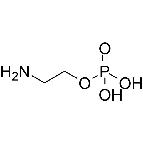
Phosphorylethanolamine (Monoaminoethyl phosphate) is a membrane phospholipid and an important precursor of Phosphatidylcholine (HY-N18891). It is found in most animal tissues and various human extracranial tumors, playing a critical role in membrane integrity, cell division, mitochondrial respiratory function, and more. Studies have shown that changes in the abundance of Phosphorylethanolamine are associated with Alzheimer's disease and Parkinson's disease. Lowering the ratio of Phosphorylethanolamine to Phosphatidylcholine in the liver can improve insulin signaling. Phosphorylethanolamine holds promise for research in the fields of cancer, neurodegenerative disorders, and metabolic diseases.
Nos produits utilisent uniquement pour la recherche. Nous ne vendons pas aux patients.
- Pureté : 99.90%
- CAS No.: 1071-23-4
- Formule: C2H8NO4P
- Masse moléculaire:141.06
-
Stockage:Powder -20°C, 3 years , 4°C, 2 years ; In solvent -80°C, 2 years , -20°C, 1 year

To place orders, for customer services and technical support, please contact: MedChemExpress USA
Tel: 609-228-6898 E-mail: [email protected] [email protected]


-
 Activité biologique
-
Chemical Information
-
Solvant et solubilité
- Protocole
- Pureté et documentation
- Références
-
 Help & FAQs


-
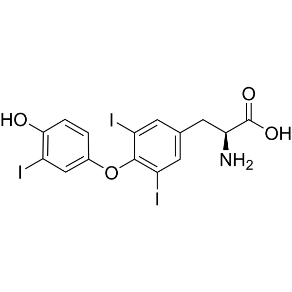
HY-A0070A
LiothyronineTIGIT/PVR Blocker -
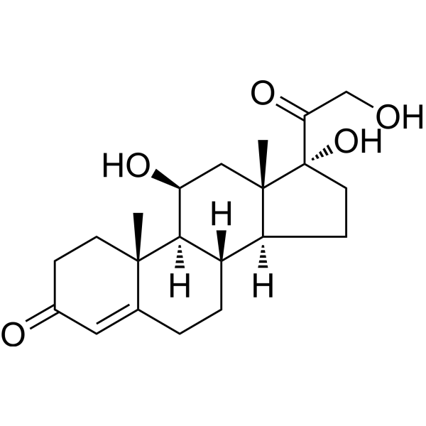
HY-N0583
HydrocortisoneGlucocorticoid Receptor Agonist -
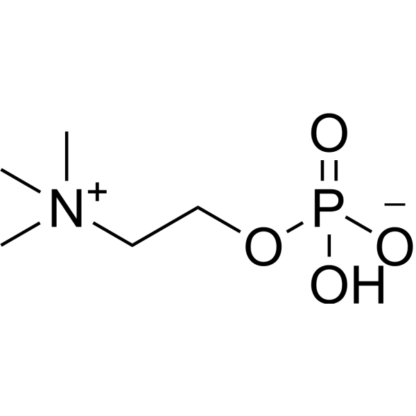
HY-B2233B
PhosphorylcholineEndogenous Metabolite -
HY-N5034R
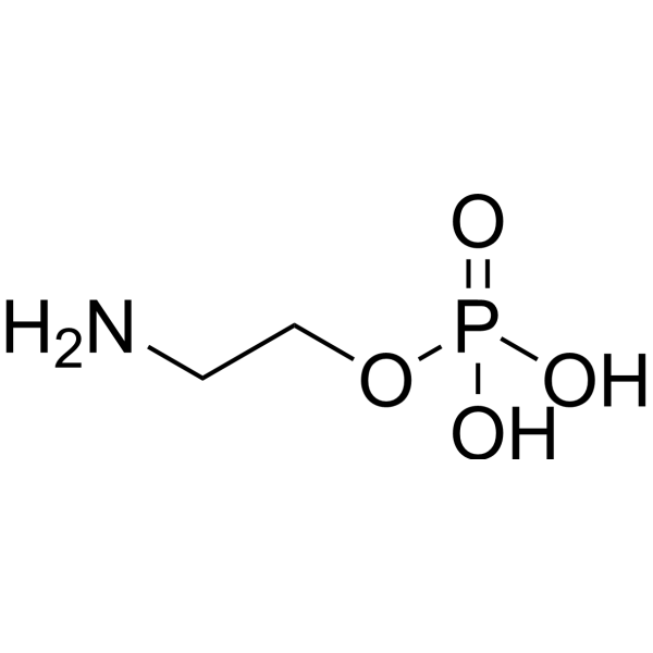
Phosphorylethanolamine (Standard)Endogenous Metabolite -

HY-113498
SphingomyelinEndogenous Metabolite -
HY-N0437
ProgesteroneEndogenous Metabolite -
HY-B2176
ATPEndogenous Metabolite -
HY-B1337
Choline chlorideBronchodilator -
HY-B1654
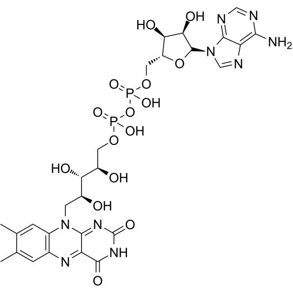
Flavin adenine dinucleotideRedox Cofactor -

HY-125853
Phosphatidylcholines,soyaPhosphatidylcholine -
HY-N0322
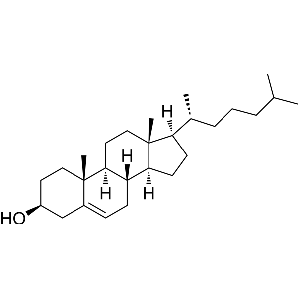
Cholesterol (from animal)ERRα Agonist -

HY-P0035
Insulin (human)Recombinant Human Insulin
Publications Citing Use of MedChemExpress (MCE) Phosphorylethanolamine
MoreVoir tous les produits spécifiques à Isoform Endogenous Metabolite
More

Activité biologique
Description
IC50 & Target
|
Microbial Metabolite |
Human Endogenous Metabolite |
In Vitro
Phosphorylethanolamine (Monoaminoethyl phosphate) arrests A2058 cells at the G2/M phase of the cell cycle and inhibits their proliferation, migration, and adhesion capabilities[2].
MedChemExpress (MCE) has not independently confirmed the accuracy of these methods. They are for reference only.
-
Cell Line:A2058 cells, B16F10 cells
-
Concentration:4.69-300 mM
-
Incubation Time:24 h
-
Result:Not only reduced viability but also blocked the capacity of A2058 cells of forming new clones.


Chemical Information
-
CAS No. 1071-23-4
-
Appearance Solid
-
Masse moléculaire 141.06
-
Formule C2H8NO4P
-
Color White to off-white
-
SMILES
NCCOP(O)(O)=O
-
Synonyms
Monoaminoethyl phosphate; NSC 254167; O-Phosphoethanolamine
-
Structure Classification
-
Initial Source
-
Livraison
Room temperature in continental US; may vary elsewhere.
-
Stockage
Powder -20°C 3 years 4°C 2 years In solvent -80°C 2 years -20°C 1 year


Publications (5)
-
Journal Impact Factor
-
Most Recent
-
Nature
2025 Jul;643(8070):192-200. PMID: 39695227 -
Cell Death Discov
SNORA14A inhibits hepatoblastoma cell proliferation by regulating SDHB-mediated succinate metabolism. [Abstract]2023 Jan 30;9(1):36. PMID: 36717552 -
-
Biochem Biophys Res Commun
CircRNA circBACH1 (hsa_circ_0061395) serves as a miR-656-3p sponge to facilitate hepatocellular carcinoma progression through increasing SERBP1 expression. [Abstract]2021 Jun 4:556:1-8. PMID: 33831787 -


Solvant et solubilité
In Vitro:
H2O : 125 mg/mL (886.15 mM; Need ultrasonic)
Please refer to the solubility information to select the appropriate solvent. Once prepared, please aliquot and store the solution to prevent product inactivation from repeated freeze-thaw cycles.
Storage method and period of stock solution: -80°C, 2 years; -20°C, 1 year. When stored at -80°C, please use it within 2 years. When stored at -20°C, please use it within 1 year.
* Note: If you choose water as the stock solution, please dilute it to the working solution, then filter and sterilize it with a 0.22 μm filter before use.
Please refer to the solubility information to select the appropriate solvent. Once prepared, please aliquot and store the solution to prevent product inactivation from repeated freeze-thaw cycles.
Storage method and period of stock solution: -80°C, 2 years; -20°C, 1 year. When stored at -80°C, please use it within 2 years. When stored at -20°C, please use it within 1 year.
* Note: If you choose water as the stock solution, please dilute it to the working solution, then filter and sterilize it with a 0.22 μm filter before use.
Concentration (start) × Volume (start) = Concentration (final) × Volume (final)
In Vivo:
For the following dissolution methods, please prepare the working solution directly:
It is recommended to prepare fresh solutions and use them promptly within a short period of time.
The percentages shown for the solvents indicate their volumetric ratio in the final prepared solution. If precipitation or phase separation occurs during preparation, heat and/or sonication can be used to aid dissolution.
Add each solvent one by one: PBS
Solubility: 100 mg/mL (708.92 mM); Clear solution; Need ultrasonic


Pureté et documentation
-
Fiche technique (279 KB)
-
SDS (477 KB)
- English - EN (477 KB)
- Français - FR (477 KB)
- Deutsch - DE (477 KB)
- Norwegian - NO (477 KB)
- Español - ES (477 KB)
- Swedish - SV (477 KB)
- Italian - IT (477 KB)
- Korean - KR (477 KB)
- Portuguese - PT (477 KB)
-
Instruction de manipulation (2659 KB)
Références
[1]. Kinoshita Y, et al. Phosphorylethanolamine content of human brain tumors. Neurol Med Chir (Tokyo). 1994 Dec;34(12):803-6. [Content Brief]
[2]. Hui SCN, et al. In vivo spectral editing of phosphorylethanolamine. Magn Reson Med. 2022 Jan;87(1):50-56. [Content Brief]
[3]. Calzada E,et al. Phosphatidylethanolamine Metabolism in Health and Disease. Int Rev Cell Mol Biol. 2016;321:29-88. doi: 10.1016/bs.ircmb.2015.10.001. [Content Brief]
[4]. van der Veen JN, et al. A role for phosphatidylcholine and phosphatidylethanolamine in hepatic insulin signaling. FASEB J. 2019 Apr;33(4):5045-5057. [Content Brief]
[5]. Nesic I, et al. Alterations in phosphatidylethanolamine levels affect the generation of Aβ. Aging Cell. 2012 Feb;11(1):63-72. [Content Brief]


Complete Stock Solution Preparation Table
Please refer to the solubility information to select the appropriate solvent. Once prepared, please aliquot and store the solution to prevent product inactivation from repeated freeze-thaw cycles.
Storage method and period of stock solution: -80°C, 2 years; -20°C, 1 year. When stored at -80°C, please use it within 2 years. When stored at -20°C, please use it within 1 year.
| Optional Solvent | Concentration Solvent Mass | 1 mg | 5 mg | 10 mg | 25 mg |
|---|---|---|---|---|---|
| H2O | 1 mM | 7.0892 mL | 35.4459 mL | 70.8918 mL | 177.2296 mL |
| 5 mM | 1.4178 mL | 7.0892 mL | 14.1784 mL | 35.4459 mL | |
| 10 mM | 0.7089 mL | 3.5446 mL | 7.0892 mL | 17.7230 mL | |
| 15 mM | 0.4726 mL | 2.3631 mL | 4.7261 mL | 11.8153 mL | |
| 20 mM | 0.3545 mL | 1.7723 mL | 3.5446 mL | 8.8615 mL | |
| 25 mM | 0.2836 mL | 1.4178 mL | 2.8357 mL | 7.0892 mL | |
| 30 mM | 0.2363 mL | 1.1815 mL | 2.3631 mL | 5.9077 mL | |
| 40 mM | 0.1772 mL | 0.8861 mL | 1.7723 mL | 4.4307 mL | |
| 50 mM | 0.1418 mL | 0.7089 mL | 1.4178 mL | 3.5446 mL | |
| 60 mM | 0.1182 mL | 0.5908 mL | 1.1815 mL | 2.9538 mL | |
| 80 mM | 0.0886 mL | 0.4431 mL | 0.8861 mL | 2.2154 mL | |
| 100 mM | 0.0709 mL | 0.3545 mL | 0.7089 mL | 1.7723 mL |
* Note: If you choose water as the stock solution, please dilute it to the working solution, then filter and sterilize it with a 0.22 μm filter before use.


Phosphorylethanolamine Related Classifications
HY-N5034 Related Classifications
 Powered by Bioz