



Thiazole Orange (solution)
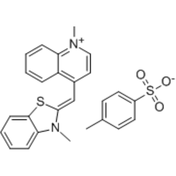
Thiazole Orange (solution) is an asymmetric anthocyanin dye that can be coupled with oligonucleotides (ONs) to prepare fluorescent hybridization probes. Thiazole Orange has been widely used in biomolecular detection and staining of DNA/ RNA in gels and can be used for reticulocyte analysis. Thiazole orange generates a significant fluorescence enhancement and high quantum yield when it binds with nucleic acids, especially RNA. Thiazole orange can permeate living cell membranes. Thiazole orange can use UV light for detection, but can also be detected with blue light. The excitation and emission of Thiazole orange are λex = 510 nm (488 nm and 470 nm also show strong excitation) and λem = 527 nm, respectively.
Solvent and concentration: DMSO: 10 mM
The 1 mL volume is defined as the base specification. All larger sizes correspond to incremental volumes of this base.
For research use only. We do not sell to patients.
- CAS No.: 107091-89-4
- Formula: C26H24N2O3S2
- Molecular Weight:476.61
-
Storage:
Please store the product under the recommended conditions in the Certificate of Analysis.


To place orders, for customer services and technical support, please contact: MedChemExpress USA
Tel: 609-228-6898 E-mail: [email protected] [email protected]


-
 Biological Activity
-
Chemical Information
- Purity & Documentation
- References
-
 Help & FAQs




Biological Activity
Description
Solvent and concentration: DMSO: 10 mM
The 1 mL volume is defined as the base specification. All larger sizes correspond to incremental volumes of this base.
In Vitro
Guide (The following is our recommended protocol. This protocol is only a guide and should be modified according to your specific needs).
Preparation of Thiazole Orange Staining Solution:
1.1 Preparing the working solution.
Dilute the 1 mg/mL Thiazole Orange stock solution in phosphate buffered saline containing 0.002 M EDTA and 0.02% sodium azide at a ratio of 1:10000.
Thiazole Orange staining
1 Reticulocyte analysis[1]
1)Whole blood (5 pL) is added to the diluted dye solution (1 mL) in a 12 x 75 mm culture tube and incubated at room temperature for 1 h. The sample is analyzed on a FACS IV, FACS 440, or FACStar.
2)Data analysis (flow cytometry) : Mature red blood cells between the left and middle marker and reticulocytes between the middle and right marker. The classification of cell type is determined through the analysis of mixed whole blood from human peripheral mononuclear cells.
2 DNA Electrophoresis[3]
1)Mix agarose (~1% w/v, percentage can be varied for particular size separations) in buffer (approximately 70 mL for a mini-gel (8 x 7 cm)). Buffers are commonly TAE (tris-acetate-EDTA, 40 mM Tris, 20 mM acetate, 1 mM EDTA, pH approximately 8.6) or TBE (tris-borate-EDTA, 90 mM Tris, 90 mM borate, 2 mM EDTA, pH approximately 8.3)).
2)Add thiazole orange to a final concentration of 1.3 μg/mL.
NOTE: The gel can also be stained with Thiazole Orange after electrophoresis.
3)Microwave the mixture of agarose, buffer, and thiazole orange to dissolve agarose (approximately 60 s). This step is commonly referred to as “melting”. Swirl (5 s) to aid dissolution if needed. Allow the agarose solution to solidify into a gel.
4)Place the gel in the electrophoresis apparatus. Load DNA samples (commonly 10 μL) using a loading dye. Attach the cover and electrodes. Apply voltage (typically ~100V for a mini-gel) until loading dye has traveled an appropriate distance (approximately 4-7 cm for a mini-gel).
NOTE: Thiazole orange is positively charged, migrating in the opposite direction of electrophoresing DNA.
5)Remove the gel from the electrophoresis apparatus and place on a UV transilluminator and a blue-light transilluminator (~470 nm maximum emission wavelength).
Select appropriate excitation and emission settings in gel-imaging apparatus. The excitation and emission of thiazole orange (λex,max = 510 nm (488 nm and 470 nm also show strong excitation, in addition to strong excitation at UV wavelengths); λem = 527 nm).
MedChemExpress (MCE) has not independently confirmed the accuracy of these methods. They are for reference only.


Chemical Information
-
CAS No. 107091-89-4
-
Molecular Weight 476.61
-
Formula C26H24N2O3S2
-
SMILES
CN1/C(SC2=CC=CC=C12)=C/C3=C4C=CC=CC4=[N+](C)C=C3.O=S(C5=CC=C(C)C=C5)([O-])=O
-
Shipping
Room temperature in continental US; may vary elsewhere.
-
Storage
Please store the product under the recommended conditions in the Certificate of Analysis.


Purity & Documentation
References
[1]. Suss O, et al. Broad Applications of Thiazole Orange in Fluorescent Sensing of Biomolecules and Ions. Molecules. 2021 May 10;26(9):2828. [Content Brief]
[2]. Long W, et al. Molecular Recognition and Imaging of Human Telomeric G-Quadruplex DNA in Live Cells: A Systematic Advancement of Thiazole Orange Scaffold To Enhance Binding Specificity and Inhibition of Gene Expression. J Med Chem. 2021 Feb 25;64(4):2125-2138. [Content Brief]
[3]. Lu YJ, et al. Molecular Engineering of Thiazole Orange Dye: Change of Fluorescent Signaling from Universal to Specific upon Binding with Nucleic Acids in Bioassay. ACS Chem Biol. 2016 Apr 15;11(4):1019-29. [Content Brief]
[4]. O'Neil CS, et al. DNA Electrophoresis Using Thiazole Orange Instead of Ethidium Bromide or Alternative Dyes. J Vis Exp. 2019 Mar 31;(145) [Content Brief]
[5]. Piotr Klimkowski, et al. Design of thiazole orange oligonucleotide probes for detection of DNA and RNA by fluorescence and duplex melting. Org Biomol Chem. 2019 Jun 28; 17(24): 5943–5950. [Content Brief]
[6]. Linda G. Le, et al. Thiazole Orange: A New Dye for Reticulocyte Analysis. cytometry 7:508-517 (1986)

Calculators
Concentration (start) × Volume (start) = Concentration (final) × Volume (final)


Thiazole Orange (solution) Related Classifications
HY-DY1097 Related Classifications
 Powered by Bioz