



High Sensitivity ECL Kit
Based on 24 publication(s) in Google Scholar
High Sensitivity ECL Kit enables low picogram detection of antigen by oxidizing luminol in the presence of HRP and peroxide. This reaction produces a prolonged chemiluminescence which can be visualized on X-ray film or digital imaging systems. The 100 mL is defined as the base specification. All larger sizes correspond to incremental volumes of this base.
-
Storage :
4°C, 1 year.
Protect from light
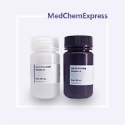
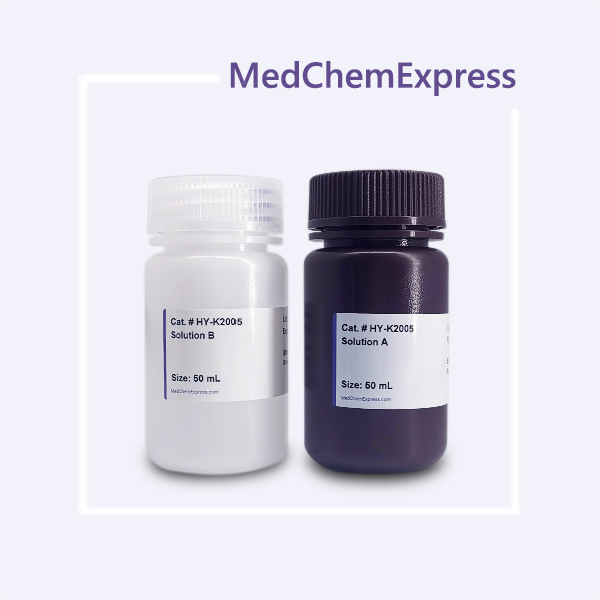





Description & Advantages
MCE High Sensitivity ECL Kit enables low picogram detection of antigen by oxidizing luminol in the presence of HRP and peroxide. This reaction produces a prolonged chemiluminescence which can be visualized on X-ray film or digital imaging systems.
The Kit produces a strong, long-lived signal (6-8 hours), which, combined with very low background levels, allows for long exposure times and enables the detection of low-abundance proteins. In the Western Blot experiment, the storage solution of primary antibody (1 mg/mL) can be diluted at a ratio of 1:1,000-1:50,000. (the final concentration is about 0.2-1 μg/mL). The secondary antibody (1 mg/mL) storage solution can be diluted at a ratio of 1:20,000-1:100,000 (the final concentration is about 10-50 ng/mL).


Protocol
1. Take the High Sensitivity ECL Kit out of the refrigerator and allow it to equilibrate at room temperature for about 20 minutes.
2. Prepare the ECL working solution.
Note: Approximately 0.125 mL of High Sensitivity ECL Reagent is required per cm2 of membrane area.
a). Mix solutions A and B in a 1:1 ratio. Use a in sufficient volume to ensure that the blot is completely wetted with the ECL working solution.
b). It is recommended to prepare the working solution just before use, although the mixed reagent solution is stable for several hours at room temperature.
3. Place the blot, protein-side up, in a clean container, and add ECL working solution.
4. Incubate the blot for 3-5 minutes at room temperature.
5. Drain the excess substrate and place the blot in a plastic membrane protector. Remove all air bubbles between the blot and the surface of the membrane protector.
6. Visualize the blot using X-ray film or a CCD-based imaging system.


Publications
-
Journal Impact Factor
-
Most Recent
-
Adv Sci (Weinh)Alpinetin Nanoparticles Alleviate Optic Nerve Injury Induced by Acute Glaucoma via LRP1-PPARγ Mediated Regulation of Microglial Lipid Metabolism. [Abstract]2025 Nov 12:e13270. PMID: 41220285
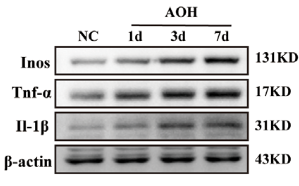
High Sensitivity ECL Kit purchased from MedChemExpress. Usage Cited in: Adv Sci (Weinh). 2025 Nov 12:e13270. [Abstract]
Protein expression levels were assessed by Western blotting.Protein bands were visualized using an enhanced chemiluminescence (ECL) kit.
-
Inflamm Res5-oxoETE promote thrombosis in antiphospholipid syndrome by triggering NETs formation through PLC/PKC/ERK pathway. [Abstract]2024 Dec;73(12):2165-2177. PMID: 39377801
-
BMC CancerTRPM2 channels mediate ROS-induced actin remodeling and cell migration of prostate cancer cells. [Abstract]2025 May 28;25(1):956. PMID: 40437388
Storage
4°C, 1 year.
Protect from light


Components
| Components | HY-K2005-100 mL | HY-K2005-500 mL |
|---|---|---|
| Solution A | 50 mL | 50 mL × 5 |
| Solution B | 50 mL | 50 mL × 5 |


 Please enter the correct Lot Number
