



RhoNox-1 (solution)

Based on 1 Customer Validation
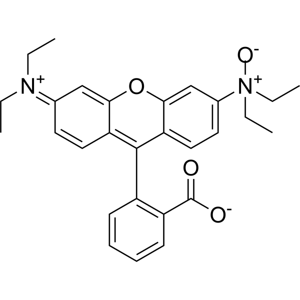
RhoNox-1 (solution) is a fluorescent probe for the specific detection of divalent iron ions, and when RhoNox-1 reacts with Fe2+. RhoNox-1 can generate an irreversible orange (red) fluorescent product (Ex/Em:540/575 nm). FeRhoNox-1 can enter the cell well, suitable for the detection of Fe2+ in living cells, and tends to be localized in the Golgi apparatus.
Solvent and concentration: DMSO: 5 mM
商品は「研究用試薬」です。人や動物の医療用・臨床診断用・食品用の製品ではありません。
研究用途以外に使用した場合、当社は一切の責任を負いかねます。
- 純度 : 94.95%
- CAS 番号: 1447815-38-4
- 分子式: C28H30N2O4
- 分子量:458.56
-
保管条件:
Please store the product under the recommended conditions in the Certificate of Analysis.


To place orders, for customer services and technical support, please contact: MedChemExpress USA
Tel: 609-228-6898 E-mail: [email protected] [email protected]


-
 生物活性
生物活性
-
化学情報
- 純度とドキュメンテーション
- 参考文献
-
 Help & FAQs




生物活性
製品説明
Solvent and concentration: DMSO: 5 mM
体外実験
Guidelines (Following is our recommended protocol. This protocol only provides a guideline, and should be modified according to your specific needs).
1. Preparation of RhoNox-1 working solution
Dilute the stock solution in serum-free cell culture medium or PBS. The corresponding stock solution can be diluted according to the actual situation. Note that if the solvent is DMSO, the cytotoxicity of DMSO must be considered, and a solvent control should be prepared; if the solvent is pure water, the working solution needs to be filtered and sterilized before adding cells.
Note: Please adjust the concentration of RhoNox-1 working solution according to the actual situation.
2. Cell staining (Suspension cells)
2.1 Centrifuge at 1000 g at 4°C for 3-5 minutes and then discard the supernatant. Wash twice with PBS, 5 minutes each time. The cell density is 1×10⁶/mL.
2.2 Add 1 mL of working solution, and then incubate at room temperature for 5-30 minutes.
2.3 Centrifuge at 400 g at 4°C for 3-4 minutes and then discard the supernatant.
2.4 Wash twice with PBS, 5 minutes each time.
2.5 Resuspend cells with serum-free cell culture medium or PBS. Observation by fluorescence microscopy or flow cytometry.
3. Cell staining (Adherent cells)
3.1 Culture adherent cells on sterile coverslips.
3.2 Remove the coverslip from the medium and aspirate excess medium.
3.3 Add 100 μL of working solution, gently shake it to completely cover the cells, and then incubate at room temperature for 5-30 minutes.
3.4 Wash twice with medium, 5 minutes each time. Observation by fluorescence microscopy.
MedChemExpress (MCE) has not independently confirmed the accuracy of these methods. They are for reference only.


化学情報
-
CAS 番号 1447815-38-4
-
性状 Liquid
-
分子量 458.56
-
分子式 C28H30N2O4
-
Color Purple to purplish red
-
SMILES
O=C(C1=C(C2=C3C=C/C(C=C3OC4=CC([N+](CC)(CC)[O-])=CC=C42)=[N+](CC)\CC)C=CC=C1)[O-]
-
輸送条件
Room temperature in continental US; may vary elsewhere.
-
保管条件
Please store the product under the recommended conditions in the Certificate of Analysis.


純度とドキュメンテーション
-
データシート (276 KB)
-
SDS (251 KB)
- English - EN (251 KB)
- Français - FR (251 KB)
- Deutsch - DE (251 KB)
- Norwegian - NO (251 KB)
- Español - ES (251 KB)
- Swedish - SV (251 KB)
- Italian - IT (251 KB)
- Korean - KR (251 KB)
- Portuguese - PT (251 KB)
-
取扱説明書 (2659 KB)
参考文献
[1]. Mukaide T, et al. Histological detection of catalytic ferrous iron with the selective turn-on fluorescent probe RhoNox-1 in a Fenton reaction-based rat renal carcinogenesis model. Free Radic Res. 2014 Sep;48(9):990-5. [Content Brief]
[2]. Jamnongkan W, et al. Upregulation of transferrin receptor-1 induces cholangiocarcinoma progression via induction of labile iron pool. Tumour Biol. 2017 Jul;39(7):1010428317717655. [Content Brief]
[3]. Ito F, et al. Contrasting intra- and extracellular distribution of catalytic ferrous iron in ovalbumin-induced peritonitis. Biochem Biophys Res Commun. 2016 Aug 5;476(4):600-606. [Content Brief]

Calculators
濃度 (開始) × 体積 (開始) = 濃度 (終了) × 体積 (終了)


RhoNox-1 (solution) 関連分類
HY-DY1030 関連分類
 Powered by Bioz