



CREB Antibody (YA491)

Based on 1 Customer Validation
CREB Antibody (YA491) is a Rabbit-derived and non-conjugated IgG monoclonal antibody, targeting to CREB.
-
Host:
Rabbit
-
Isotype:
IgG
-
Application:
WB, IHC-P, ICC/IF, FC, IF-Tissue
-
Reactivity :
Human, Mouse, Rat, Zebrafish
-
Formulation:
Supplied in 1*TBS (pH7.4), 0.05% BSA and 40% Glycerol. Preservative: 0.05% Sodium Azide.
-
Conjugation:
Non-conjugated


To place orders, for customer services and technical support, please contact: MedChemExpress USA
Tel: 609-228-6898 E-mail: [email protected] [email protected]





Applications
| Application |
WB

WB: Western Blot
|
ICC/IF
ICC/IF: Immunocytochemistry/
Immunofluorescence |
IHC-P
IHC-P: Immunohistochemistry-Paraffin
|
FC
FC: Flow Cytometry
|
IF-Tissue
IF-Tissue: Immunofluorescence-Tissue
|
|---|---|---|---|---|---|
| Dilution Ratio | 1:500 | 1:500 | 1:500-1:5000 | 1:1000 | 1:200 |


Product Details
CREB Antibody (YA491) is a Rabbit-derived and non-conjugated IgG monoclonal antibody, targeting to CREB.
-
Host Rabbit

-
Clonality Recombinant,Monoclonal

-
Species ReactivityHuman, Mouse, Rat, Zebrafish
-
Observed Molecular WeightObserved band size: 42 kDa
Note: Due to possible protein modifications or aggregation, the molecular weight should be confirmed by actual measurement, and the predicted value is for reference only.

-
Calculated Molecular Weight Predicted band size: 35 kDa;

Synthetic peptide corresponding to Human CREB.AA range:250-290.
Endogenous
Protein A affinity purified.
Non-conjugated
Unmodified
IgG


Product Properties
-
Appearance
Liquid
-
Formulation
Supplied in 1*TBS (pH7.4), 0.05% BSA and 40% Glycerol. Preservative: 0.05% Sodium Azide.
-
Concentration
Batch-dependent, Please check the COA for the concentration of each lot. Check Lot Concentration
-
Storage & Stability
Stored at -20°C for 1 year. Avoid repeated freeze / thaw cycles.
-
Shipping
Shipping with blue ice.


Verification Images
-
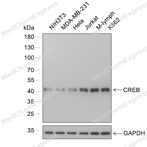 Western blot analysis of extracts from NIH3T3 (lane 1) and MDA-MB-231 (lane 2) and Hela (lane 3) and Jurkat (lane 4) and M-lymph (lane 5) and K562 (lane 6) using CREB antibody. Proteins were transferred to a PVDF membrane and blocked with 5% nonfat powdered milk in PBST for 2 hour at room temperature. The primary antibody (1/1000) and Loading control antibody (GAPDH, 1/3000) was diluted with 5% nonfat powdered milk in PBST at 4°C overnight. Goat Anti-Rabbit IgG-HRP Secondary Antibody (1/8,000) was incubated for 45min at room temperature.
 -
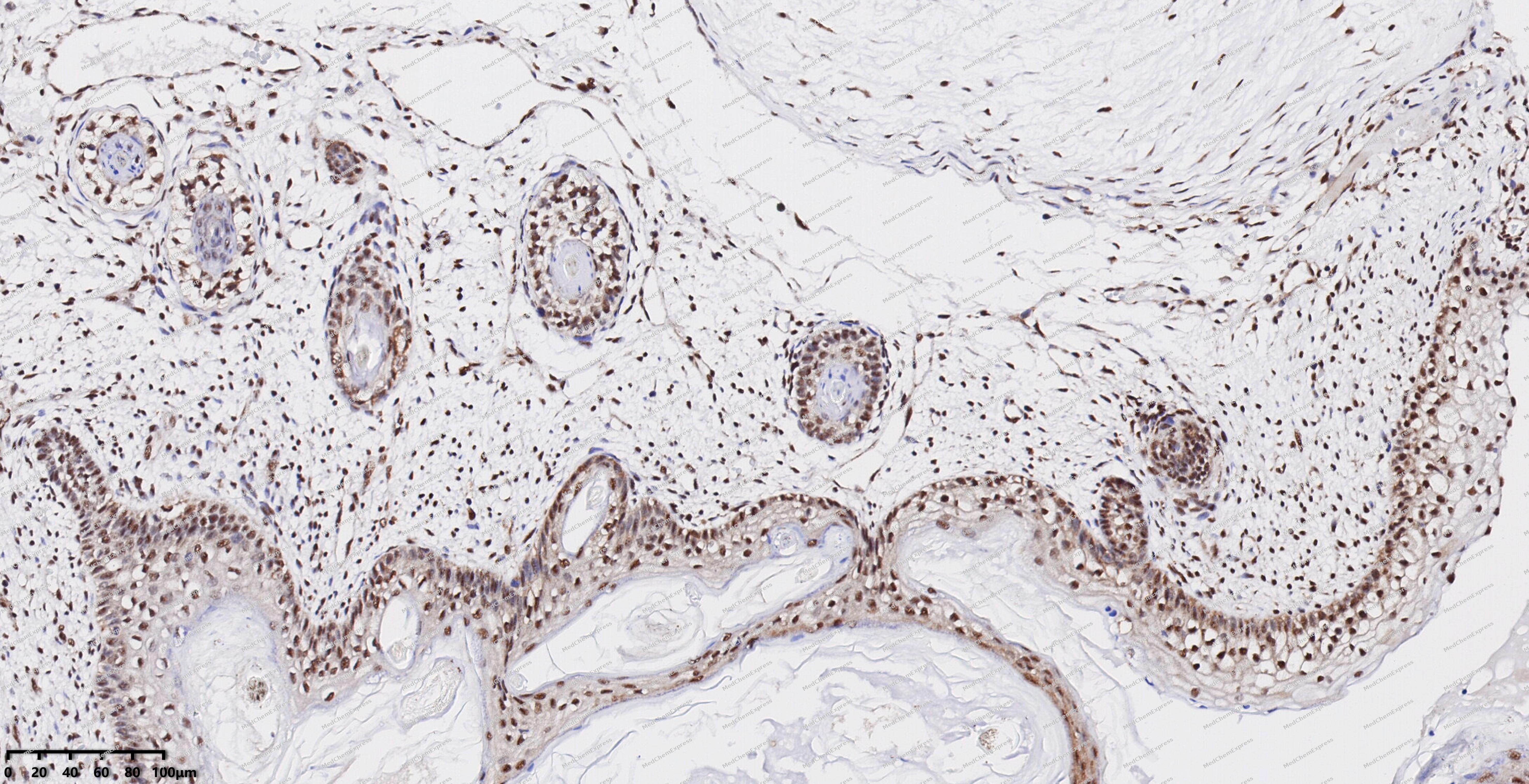 Immunohistochemical analysis of paraffin-embedded human ovarian cancer using CREB antibody. The section was pre-treated using heat mediated antigen retrieval with EDTA (pH 9.0) for 14 minutes. The tissues were blocked in 5% BSA for 30 minutes at room temperature, washed with TBST, and then probed with the primary antibody (HY-P80092, 1/1000) for 30 minutes at room temperature. The detection was performed using Polymer HRP-conjugated Goat Anti-Mouse/Rabbit lgG(H&L) secondary antibody (HY-P83652). DAB was used as the chromogen. Tissues were counterstained with hematoxylin and mounted with DPX.
 -
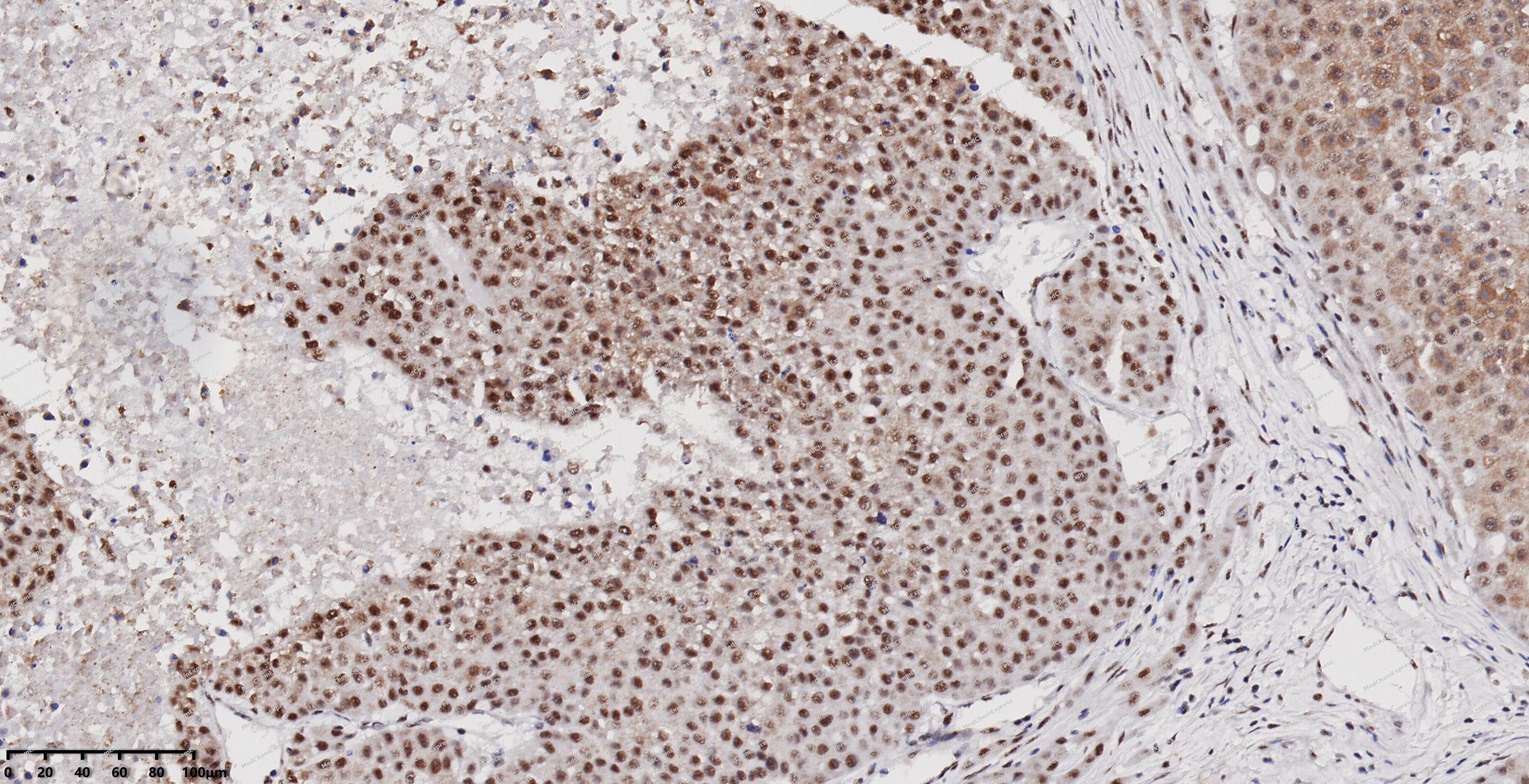 Immunohistochemical analysis of paraffin-embedded human liver cancer using CREB antibody. The section was pre-treated using heat mediated antigen retrieval with EDTA (pH 9.0) for 14 minutes. The tissues were blocked in 5% BSA for 30 minutes at room temperature, washed with TBST, and then probed with the primary antibody (HY-P80092, 1/1000) for 30 minutes at room temperature. The detection was performed using Polymer HRP-conjugated Goat Anti-Mouse/Rabbit lgG(H&L) secondary antibody (HY-P83652). DAB was used as the chromogen. Tissues were counterstained with hematoxylin and mounted with DPX.
 -
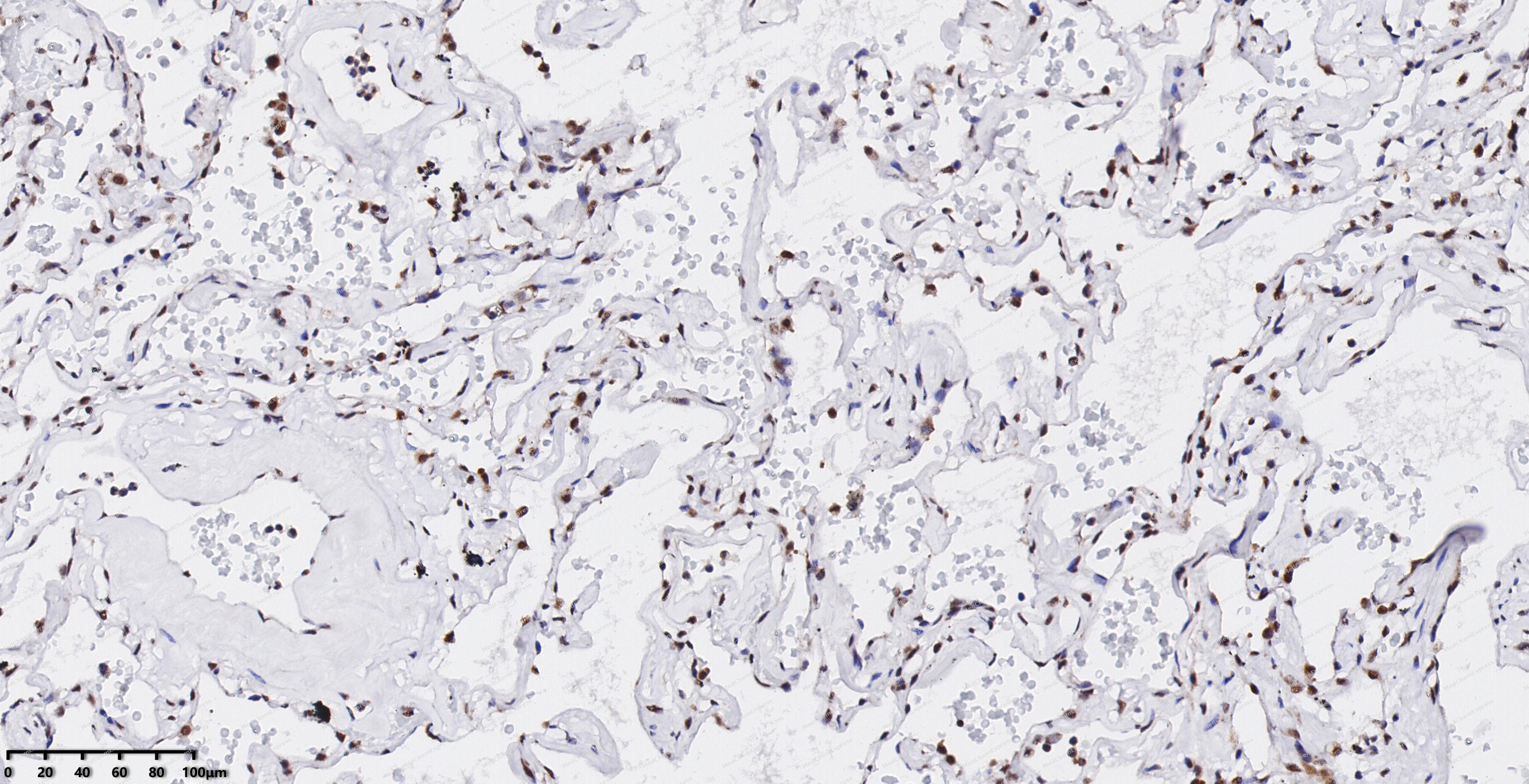 Immunohistochemical analysis of paraffin-embedded human lung cancer using CREB antibody. The section was pre-treated using heat mediated antigen retrieval with EDTA (pH 9.0) for 14 minutes. The tissues were blocked in 5% BSA for 30 minutes at room temperature, washed with TBST, and then probed with the primary antibody (HY-P80092, 1/1000) for 30 minutes at room temperature. The detection was performed using Polymer HRP-conjugated Goat Anti-Mouse/Rabbit lgG(H&L) secondary antibody (HY-P83652). DAB was used as the chromogen. Tissues were counterstained with hematoxylin and mounted with DPX.
 -
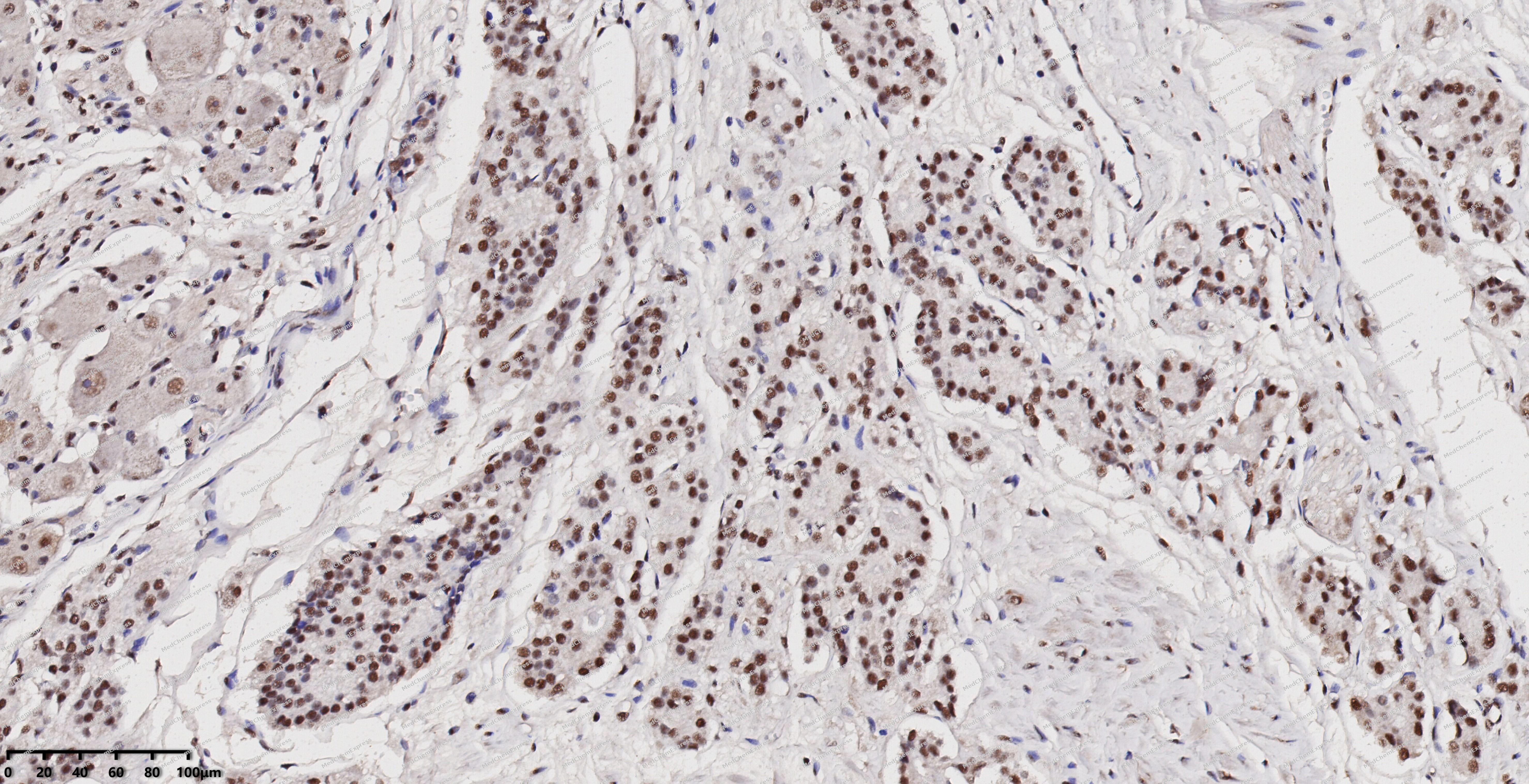 Immunohistochemical analysis of paraffin-embedded human prostate cancer using CREB antibody. The section was pre-treated using heat mediated antigen retrieval with EDTA (pH 9.0) for 14 minutes. The tissues were blocked in 5% BSA for 30 minutes at room temperature, washed with TBST, and then probed with the primary antibody (HY-P80092, 1/1000) for 30 minutes at room temperature. The detection was performed using Polymer HRP-conjugated Goat Anti-Mouse/Rabbit lgG(H&L) secondary antibody (HY-P83652). DAB was used as the chromogen. Tissues were counterstained with hematoxylin and mounted with DPX.
 -
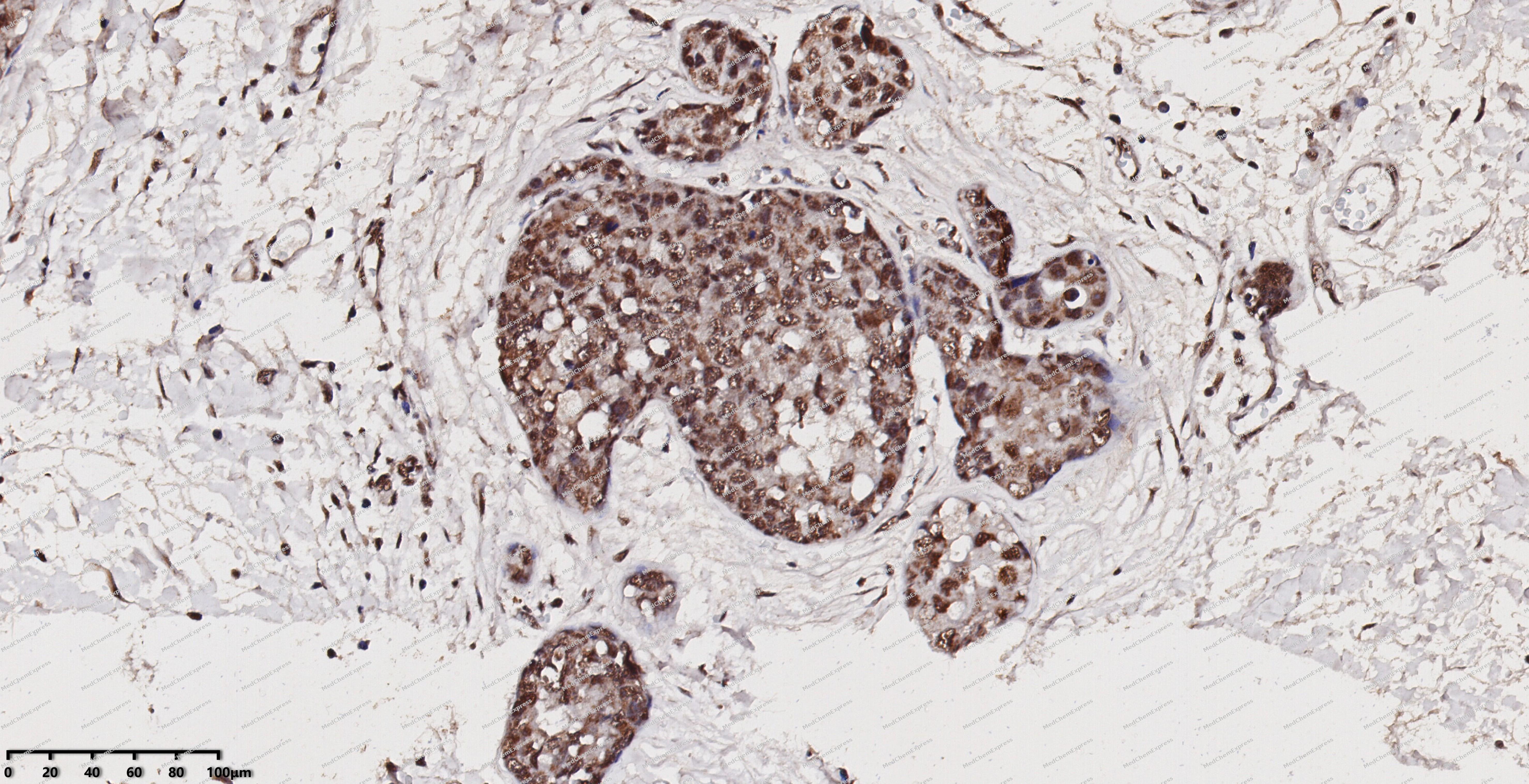 Immunohistochemical analysis of paraffin-embedded human breast cancer using CREB antibody. The section was pre-treated using heat mediated antigen retrieval with EDTA (pH 9.0) for 14 minutes. The tissues were blocked in 5% BSA for 30 minutes at room temperature, washed with TBST, and then probed with the primary antibody (HY-P80092, 1/1000) for 30 minutes at room temperature. The detection was performed using Polymer HRP-conjugated Goat Anti-Mouse/Rabbit lgG(H&L) secondary antibody (HY-P83652). DAB was used as the chromogen. Tissues were counterstained with hematoxylin and mounted with DPX.
 -
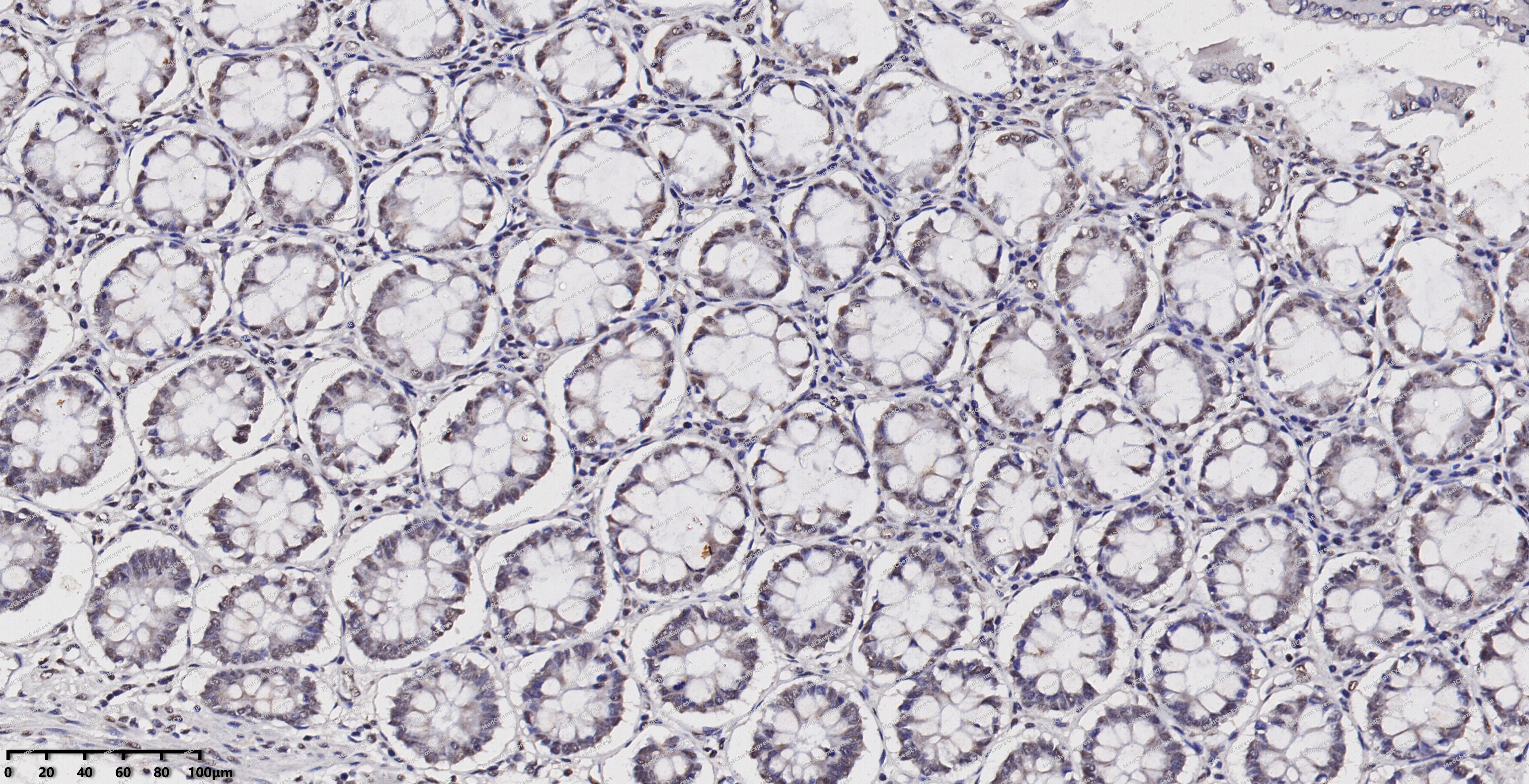 Immunohistochemical analysis of paraffin-embedded human colon using CREB antibody. The section was pre-treated using heat mediated antigen retrieval with EDTA (pH 9.0) for 14 minutes. The tissues were blocked in 5% BSA for 30 minutes at room temperature, washed with TBST, and then probed with the primary antibody (HY-P80092, 1/1000) for 30 minutes at room temperature. The detection was performed using Polymer HRP-conjugated Goat Anti-Mouse/Rabbit lgG(H&L) secondary antibody (HY-P83652). DAB was used as the chromogen. Tissues were counterstained with hematoxylin and mounted with DPX.
 -
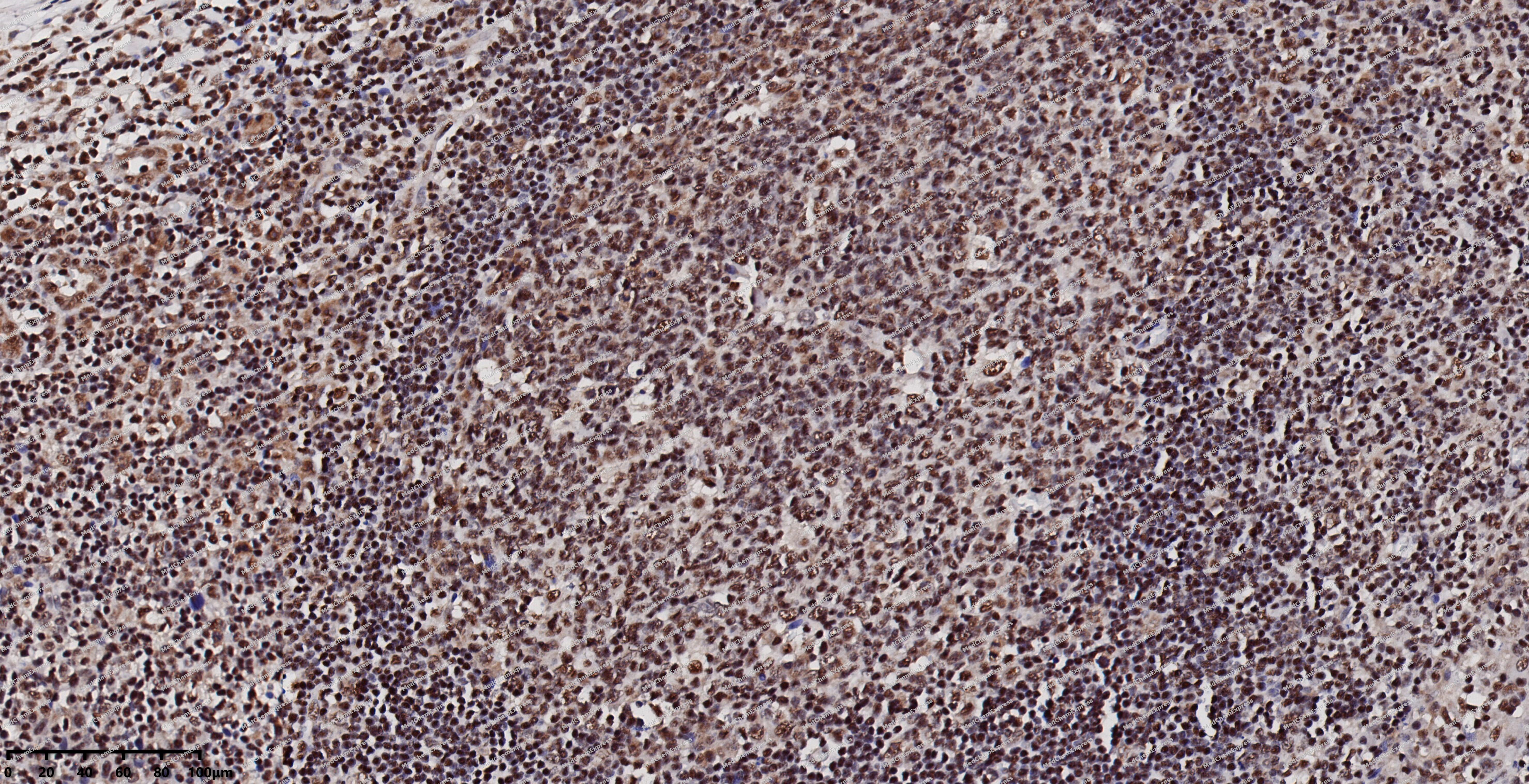 Immunohistochemical analysis of paraffin-embedded human tonsil using CREB antibody. The section was pre-treated using heat mediated antigen retrieval with EDTA (pH 9.0) for 14 minutes. The tissues were blocked in 5% BSA for 30 minutes at room temperature, washed with TBST, and then probed with the primary antibody (HY-P80092, 1/1000) for 30 minutes at room temperature. The detection was performed using Polymer HRP-conjugated Goat Anti-Mouse/Rabbit lgG(H&L) secondary antibody (HY-P83652). DAB was used as the chromogen. Tissues were counterstained with hematoxylin and mounted with DPX.
 -
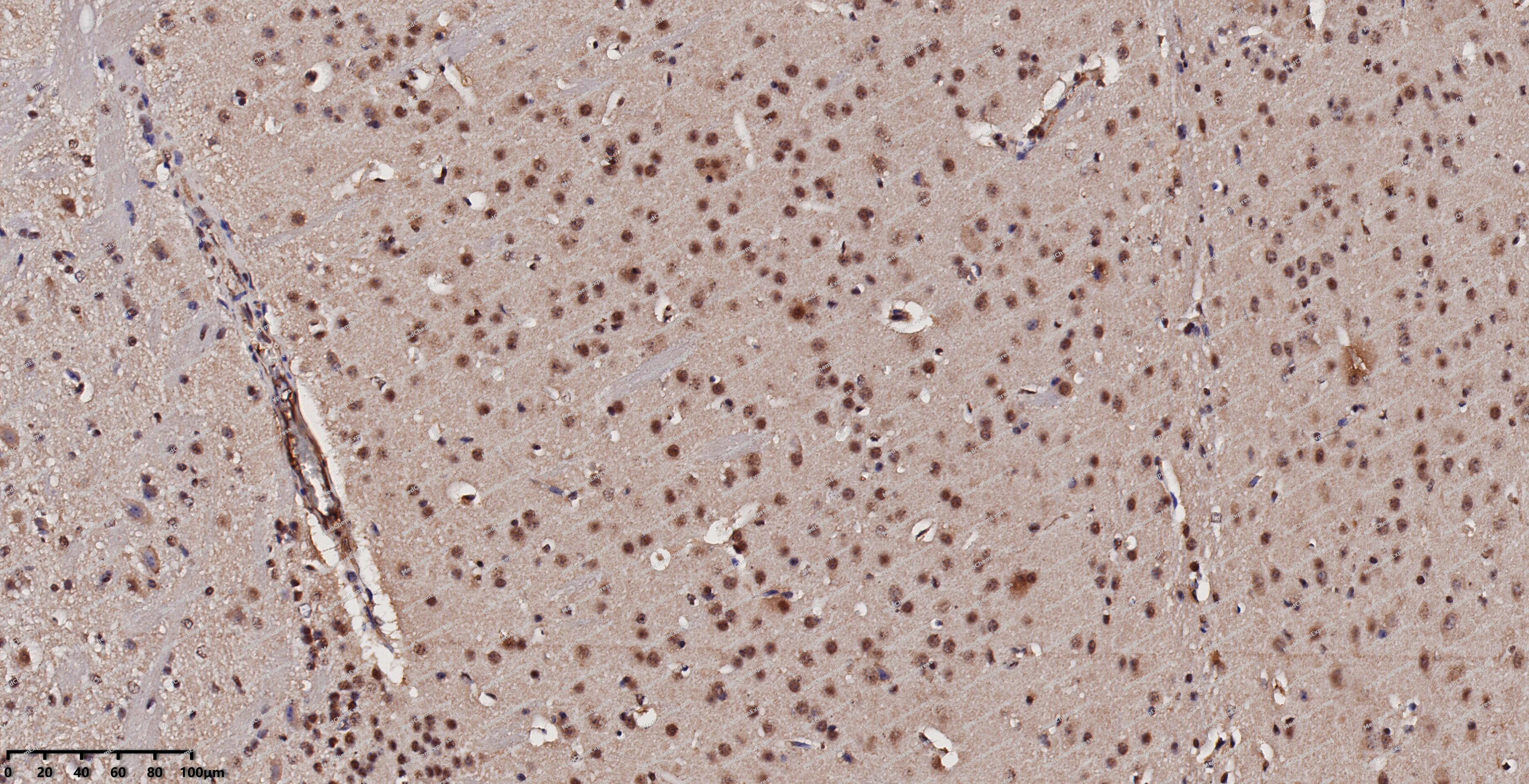 Immunohistochemical analysis of paraffin-embedded mouse brain using CREB antibody. The section was pre-treated using heat mediated antigen retrieval with EDTA (pH 9.0) for 14 minutes. The tissues were blocked in 5% BSA for 30 minutes at room temperature, washed with TBST, and then probed with the primary antibody (HY-P80092, 1/1000) for 30 minutes at room temperature. The detection was performed using Polymer HRP-conjugated Goat Anti-Mouse/Rabbit lgG(H&L) secondary antibody (HY-P83652). DAB was used as the chromogen. Tissues were counterstained with hematoxylin and mounted with DPX.
 -
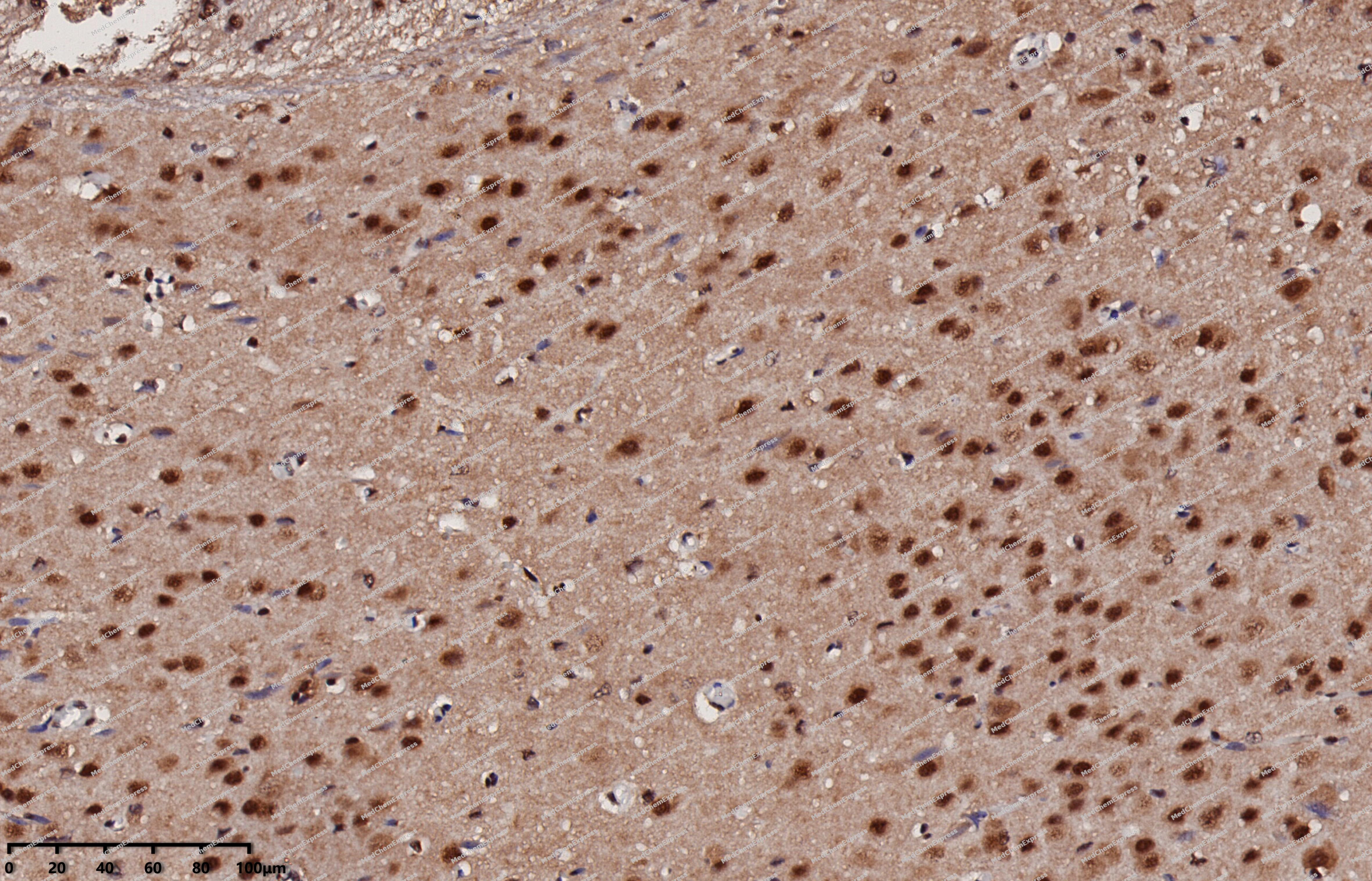 Immunohistochemical analysis of paraffin-embedded rat brain using CREB antibody. The section was pre-treated using heat mediated antigen retrieval with EDTA (pH 9.0) for 14 minutes. The tissues were blocked in 5% BSA for 30 minutes at room temperature, washed with TBST, and then probed with the primary antibody (HY-P80092, 1/1000) for 30 minutes at room temperature. The detection was performed using Polymer HRP-conjugated Goat Anti-Mouse/Rabbit lgG(H&L) secondary antibody (HY-P83652). DAB was used as the chromogen. Tissues were counterstained with hematoxylin and mounted with DPX.
 -
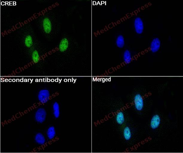 Immunocytochemistry analysis of Hela cells labeling CREB with CREB Antibody (HY-P80092) at 1:50 dilution. Cells were fixed in 4% paraformaldehyde for 15 minutes at room temperature, permeabilized with 0.1% Triton X-100 in PBS for 15 minutes at room temperature, then blocked with quick block buffer for 10 minutes at room temperature. Cells were then incubated with CREB Antibody (HY-P80092) at 1:50 dilution in quick block buffer overnight at 4 ℃. AF488-conjugated Goat Anti-Rabbit IgG H&L(HY-P8002, Green) was used as the secondary antibody at 1/1,000 dilution. PBS instead of the primary antibody was used as the secondary antibody only control. The Nuclear counterstain was DAPI (Blue).
 -
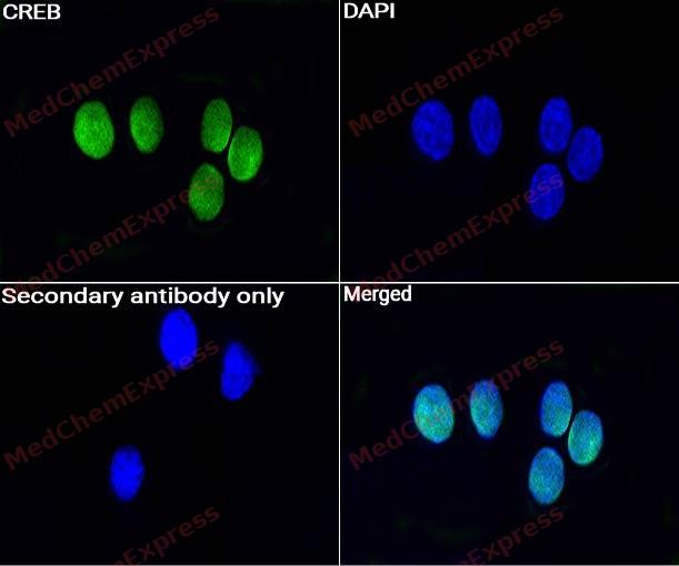 Immunocytochemistry analysis of Neuro-2a cells labeling CREB with CREB Antibody (HY-P80092) at 1:50 dilution. Cells were fixed in 4% paraformaldehyde for 15 minutes at room temperature, permeabilized with 0.1% Triton X-100 in PBS for 15 minutes at room temperature, then blocked with quick block buffer for 10 minutes at room temperature. Cells were then incubated with CREB Antibody (HY-P80092) at 1:50 dilution in quick block buffer overnight at 4 ℃. AF488-conjugated Goat Anti-Rabbit IgG H&L(HY-P8002, Green) was used as the secondary antibody at 1/1,000 dilution. PBS instead of the primary antibody was used as the secondary antibody only control. The Nuclear counterstain was DAPI (Blue).
 -
 Tyramide signaling amplification based immunofluorescence was performed on paraffin-embedded human bladder cancer using CREB antibody. The section was pre-treated using heat mediated antigen retrieval with Sodium citrate (pH 6.0) for 14 minutes. The tissues were blocked in 5% BSA for 10 minutes at room temperature and then probed with the primary antibody (HY-P80092, 1/1000) for 2 hour at room temperature. The detection was performed using Polymer HRP-conjugated Goat Anti-Mouse/Rabbit lgG(H&L) secondary antibody (HY-P83652). Fluorescence was then developed with Vari Fluor 488 TSA(200×) (HY-D1837). The tissues were counterstained with DAPI and mounted with Anti-fade mounting medium (HY-K1047).
 -
 Tyramide signaling amplification based immunofluorescence was performed on paraffin-embedded human lung cancer using CREB antibody. The section was pre-treated using heat mediated antigen retrieval with Sodium citrate (pH 6.0) for 14 minutes. The tissues were blocked in 5% BSA for 10 minutes at room temperature and then probed with the primary antibody (HY-P80092, 1/1000) for 2 hour at room temperature. The detection was performed using Polymer HRP-conjugated Goat Anti-Mouse/Rabbit lgG(H&L) secondary antibody (HY-P83652). Fluorescence was then developed with Vari Fluor 488 TSA(200×) (HY-D1837). The tissues were counterstained with DAPI and mounted with Anti-fade mounting medium (HY-K1047).
 -
 Tyramide signaling amplification based immunofluorescence was performed on paraffin-embedded human colon cancer using CREB antibody. The section was pre-treated using heat mediated antigen retrieval with Sodium citrate (pH 6.0) for 14 minutes. The tissues were blocked in 5% BSA for 10 minutes at room temperature and then probed with the primary antibody (HY-P80092, 1/1000) for 2 hour at room temperature. The detection was performed using Polymer HRP-conjugated Goat Anti-Mouse/Rabbit lgG(H&L) secondary antibody (HY-P83652). Fluorescence was then developed with Vari Fluor 488 TSA(200×) (HY-D1837). The tissues were counterstained with DAPI and mounted with Anti-fade mounting medium (HY-K1047).
 -
 Tyramide signaling amplification based immunofluorescence was performed on paraffin-embedded human ovarian cancer using CREB antibody. The section was pre-treated using heat mediated antigen retrieval with Sodium citrate (pH 6.0) for 14 minutes. The tissues were blocked in 5% BSA for 10 minutes at room temperature and then probed with the primary antibody (HY-P80092, 1/1000) for 2 hour at room temperature. The detection was performed using Polymer HRP-conjugated Goat Anti-Mouse/Rabbit lgG(H&L) secondary antibody (HY-P83652). Fluorescence was then developed with Vari Fluor 488 TSA(200×) (HY-D1837). The tissues were counterstained with DAPI and mounted with Anti-fade mounting medium (HY-K1047).
 -
 Tyramide signaling amplification based immunofluorescence was performed on paraffin-embedded human prostate using CREB antibody. The section was pre-treated using heat mediated antigen retrieval with Sodium citrate (pH 6.0) for 14 minutes. The tissues were blocked in 5% BSA for 10 minutes at room temperature and then probed with the primary antibody (HY-P80092, 1/1000) for 2 hour at room temperature. The detection was performed using Polymer HRP-conjugated Goat Anti-Mouse/Rabbit lgG(H&L) secondary antibody (HY-P83652). Fluorescence was then developed with Vari Fluor 488 TSA(200×) (HY-D1837). The tissues were counterstained with DAPI and mounted with Anti-fade mounting medium (HY-K1047).
 -
 Tyramide signaling amplification based immunofluorescence was performed on paraffin-embedded human prostate cancer using CREB antibody. The section was pre-treated using heat mediated antigen retrieval with Sodium citrate (pH 6.0) for 14 minutes. The tissues were blocked in 5% BSA for 10 minutes at room temperature and then probed with the primary antibody (HY-P80092, 1/1000) for 2 hour at room temperature. The detection was performed using Polymer HRP-conjugated Goat Anti-Mouse/Rabbit lgG(H&L) secondary antibody (HY-P83652). Fluorescence was then developed with Vari Fluor 488 TSA(200×) (HY-D1837). The tissues were counterstained with DAPI and mounted with Anti-fade mounting medium (HY-K1047).
 -
 Tyramide signaling amplification based immunofluorescence was performed on paraffin-embedded human stomach cancer using CREB antibody. The section was pre-treated using heat mediated antigen retrieval with Sodium citrate (pH 6.0) for 14 minutes. The tissues were blocked in 5% BSA for 10 minutes at room temperature and then probed with the primary antibody (HY-P80092, 1/1000) for 2 hour at room temperature. The detection was performed using Polymer HRP-conjugated Goat Anti-Mouse/Rabbit lgG(H&L) secondary antibody (HY-P83652). Fluorescence was then developed with Vari Fluor 488 TSA(200×) (HY-D1837). The tissues were counterstained with DAPI and mounted with Anti-fade mounting medium (HY-K1047).
 -
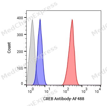 Flow cytometric analysis of 1X10⁶ SH-SY5Y cells labeling CREB Antibody (HY-P80092, red). Cells were fixed with 4% paraformaldehyde and permeabilised with 90% methanol. Then stained with the primary antibody at 1/1000 dilution for an hour at 4℃. AF488-conjugated Goat Anti-Rabbit IgG H&L (HY-P8002) was used as the secondary antibody at 1/1,000 dilution for 30 minutes at 4℃. Rabbit IgG Isotype Control (HY-P80879, blue) was used as the isotype control, cells without incubation with primary antibody were used as the unlabeled control (black).


Background
-
Function
CREB is a Phosphorylation-dependent transcription factor that stimulates transcription upon binding to the DNA cAMP response element (CRE), a sequence present in many viral and cellular promoters. Transcription activation is enhanced by the TORC coactivators which act independently of Ser-119 phosphorylation. Involved in different cellular processes including the synchronization of circadian rhythmicity and the differentiation of adipose cells. Regulates the expression of apoptotic and inflammatory response factors in cardiomyocytes in response to ERFE-mediated activation of AKT signaling[1].
-
Subcellular Localization
Nucleus
-
Subunit
Interacts with PPRC1. Binds DNA as a dimer. This dimer is stabilized by magnesium ions. Interacts, through the bZIP domain, with the coactivators CRTC1/TORC1, CRTC2/TORC2 and CRTC3/TORC3. When phosphorylated on Ser-119, binds CREBBP (By similarity). Interacts with CREBL2; regulates CREB1 phosphorylation, stability and transcriptional activity (By similarity). Interacts (phosphorylated form) with TOX3. Interacts with ARRB1. Binds to HIPK2. Interacts with SGK1. Interacts with TSSK4; this interaction facilitates phosphorylation on Ser-119 (PubMed:15964553). Forms a complex with KMT2A and CREBBP (By similarity) (PubMed:14506290, PubMed:14536081, PubMed:15454081, PubMed:15733869, PubMed:15964553, PubMed:16325578, PubMed:16908542, PubMed:20573984, PubMed:21172805, PubMed:23651431). Interacts with TOX4; CREB1 is required for full induction of TOX4-dependent activity and the interaction is increased by cAMP and inhibited by insulin (By similarity)
-
SwissProt ID
-
Research Field
Epigenetics and Nuclear Signaling


Documentation
-
Data Sheet (235 KB)
-
SDS (313 KB)
- English - EN (313 KB)
- Français - FR (313 KB)
- Deutsch - DE (313 KB)
- Norwegian - NO (313 KB)
- Español - ES (313 KB)
- Swedish - SV (313 KB)
- Italian - IT (313 KB)
- Korean - KR (313 KB)
- Portuguese - PT (313 KB)
-
User Guide for Antibodies (1077 KB)

 Powered by Bioz
