



Cusatuzumab
Based on 1 publication(s) in Google Scholar
Cusatuzumab (ARGX-110) is a selective competitive blocker targeting CD70 (with an equilibrium dissociation constant of 17 pM for binding to human CD70). Cusatuzumab also possesses enhanced antibody-dependent cell-mediated cytotoxicity (ADCC) activity. It is a humanized IgG1 monoclonal antibody, artificially synthesized through humanization and genetic engineering modifications (CH2 region mutation to enhance effector function). Cusatuzumab has a dual mechanism of action: firstly, it competitively blocks the interaction between CD70 and CD27, inhibiting the CD27-NF-κB signaling pathway, reducing regulatory T cell (Treg) activation and tumor cell proliferation; secondly, by enhancing binding to FcγRIIIa, it mediates ADCC and antibody-dependent cellular phagocytosis (ADCP), directly lysing CD70-positive tumor cells. Cusatuzumab can efficiently eliminate leukemia stem cells (LSCs), induce tumor cell differentiation and apoptosis, restore immune surveillance, and target CD70-positive tumors. Cusatuzumab is used in the study of acute myeloid leukemia (AML).
商品は「研究用試薬」です。人や動物の医療用・臨床診断用・食品用の製品ではありません。
研究用途以外に使用した場合、当社は一切の責任を負いかねます。
- 純度 : 98.87%
- CAS 番号: 1864871-20-4
- 分子量:144.36 kDa
-
保管条件:
Please store the product under the recommended conditions in the Certificate of Analysis.


To place orders, for customer services and technical support, please contact: MedChemExpress USA
Tel: 609-228-6898 E-mail: [email protected] [email protected]


-
 生物活性
生物活性
-
化学情報
- プロトコル
- 純度とドキュメンテーション
- 参考文献
-
 Help & FAQs







MedChemExpress(MCE)の使用を引用している文献 Cusatuzumab
More

生物活性
製品説明
Isotype
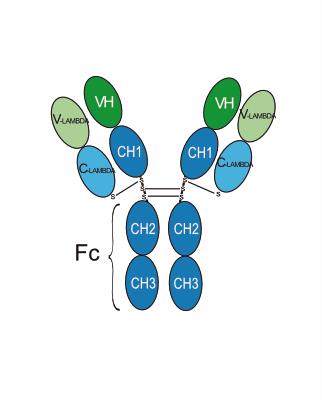
Human IgG1 lambda2
Recommend Isotype Controls
Species Reactivity
Human
IC50 & Target
TNFSF7/CD27L/CD70
体外実験
Cusatuzumab (3-100 nM) specifically binds to human CD70 with an equilibrium dissociation constant of 17 pM, and does not bind to CD70-negative cells (SUP-T1, untransfected HEK293). Its binding affinity to FcγRIIIa is 70 times higher than the fucosylated version[1].
Cusatuzumab (0.01-1 μg/mL; 2 h) induces antibody-dependent cell-mediated cytotoxicity (ADCC), with a 20-fold higher lysis efficiency against 786-O cells compared to the fucosylated version[1].
Cusatuzumab (0.001-10 μg/mL; 2 h) does not affect complement-dependent cytotoxicity (CDC), and its CDC activity against U266 cells is comparable to the fucosylated version[1].
Cusatuzumab retains antibody-dependent cellular phagocytosis (ADCP) activity and can mediate the phagocytosis of 786-O cells by monocyte-derived macrophages[1].
Cusatuzumab (5 μg/mL; 2 days) blocks the proliferation of regulatory T cells (Tregs) induced by CD70-positive tumor cells (Raji, SU-DHL-6, U266) and reduces the level of soluble CD27 (sCD27) in the supernatant[1].
Cusatuzumab (10 μg/mL; 72 h) in combination with NK cells significantly reduces acute myeloid leukemia stem cell (LSC) colony formation, induces the expression of differentiation genes such as CEBPA and SPI1, and synergistically kills LSCs and leukemia blast cells in combination with Decitabine (HY-A0004), promoting apoptosis[2].
MedChemExpress (MCE) has not independently confirmed the accuracy of these methods. They are for reference only.
-
Cell Line:Lin-CD90-CD34+CD38- AML LSCs; Lin-CD90-CD34+CD38+ AML blasts
-
Concentration:10 μg/mL; Decitabine (0.5 μM); NK cells (effector:target ratio=1:1)
-
Incubation Time:72 hours
-
Result:Annexin V FACS staining showed increased apoptosis in LSCs (fold change >1) compared to vehicle-treated cells. RT-qPCR analysis revealed upregulated expression of myeloid differentiation-related genes (CEBPA, CEBPB, RUNX1, SPI1) and downregulated ID1. Co-treatment with decitabine and NK cells effectively eliminated both LSCs and blasts, with cell number reduction more significant than other treatment groups.
体内実験
Cusatuzumab (1 mg/kg, 3 mg/kg, 10 mg/kg; i.v.; single dose) shows peak plasma concentrations (Cmax) of 50 μg/mL, 151 μg/mL, and 504 μg/mL, respectively, in a normal female cynomolgus monkey model, with a half-life of approximately 12 days and a clearance rate of 0.009 L/day[1].
Cusatuzumab (10 mg/kg; i.p.; 3 doses in total; administered 5-12 weeks after transplantation) reduces the number of leukemia cells and leukemia stem cells (LSCs) in the bone marrow of a NOD/SCID/γc-/- (NSG) mouse acute myeloid leukemia (AML) patient-derived xenograft (PDX) model. When combined with Decitabine (HY-A0004) (1.5 mg/kg/day for 5 consecutive days), it synergistically reduces LSC frequency and lowered serum sCD27 levels[2].
Cusatuzumab (10 mg/kg; i.p.; 3 doses in total; administered 43 days after transplantation) in an NSG mouse AML PDX model, when combined with NK cells, significantly reduces leukemia cell engraftment and LSC numbers in the bone marrow and spleen, and reduced AML colony formation compared to using Cusatuzumab or NK cells alone[2].
MedChemExpress (MCE) has not independently confirmed the accuracy of these methods. They are for reference only.
-
Animal Model:C.B.-17 SCID mice disseminated Burkitt lymphoma xenograft model[1]
-
Dosage:41D12 (fucosylated variant of Cusatuzumab): 0.01 mg/kg, 0.1 mg/kg, 1 mg/kg, 10 mg/kg; isotype control (palivizumab): 10 mg/kg; dissolved in phosphate-buffered saline (PBS)
-
Administration:Administered intraperitoneally, twice a week for 2 weeks (total of 5 doses)
-
Result:Administration of 41D12 at doses of 0.1 mg/kg and above prolonged the survival of SCID mice bearing Raji tumors, with a survival plateau observed. PK analysis showed an extrapolated Cmax plasma level of 2.5 μg/mL at the 0.1 mg/kg dose. Tumor-bearing mice had significantly higher serum sCD27 levels compared to non-tumor-bearing mice at the time of sacrifice.
-
Animal Model:NOD/SCID/γc-/- (NSG) mice (male and female, 6-8 weeks old) human acute myeloid leukemia PDX model[2]
-
Dosage:Cusatuzumab: 10 mg/kg; dissolved in PBS;
-
Administration:administered intraperitoneally, total of 3 injections;
allogenic NK cells: 1.5 × 10⁶ cells per mouse, administered intravenously once on day 43 post-transplantation -
Result:In the absence of NK cells, Cusatuzumab and the blocking αCD70 mAb (41D12-D) showed similar efficacy in reducing leukemia engraftment.
In the presence of NK cells, Cusatuzumab further reduced the frequency of huCD45+CD33+ AML cells and absolute numbers of huCD45+CD34+CD38- LSCs in the bone marrow and spleen.
Ex vivo colony-forming assays confirmed a significant reduction in LSPC numbers, demonstrating superior efficacy of Cusatuzumab compared to the blocking-only αCD70 mAb when combined with NK cells.
- Somatic Cell Culture
- Phagocytosis Functional Assay
- Detection of Apoptosis
- TUNEL staining for apoptotic DNA fragmentation
- Cell Cytotoxicity Assay
- Apoptosis Solutions
- MTT Cell Proliferation Assay
- iPSC cell differentiation
- Genotoxicity/Mutagenicity Study
- Mammalian live/dead viability and cytotoxicity staining
- Naïve CD4+ T-cell subset differentiation/polarization
- Annexin V plus membrane-impermeant dye apoptosis staining
- BrdU Incorporation Assay
- CFSE Dye Dilution Proliferation Assay
- Dye-dilution cell tracking and proliferation staining
- CCK-8/WST-8 Cell Proliferation Assay
- Protocol for Cell Cycle
- Research Protocol for Inflammation-related Diseases
- Cell Counting-Based Growth Curve Assay
- Colony Formation (Clonogenic) Assay
- Research Protocol for Cancer Immunology
- Cell Viability Determination by MTT Colorimetric Assay
- EdU Incorporation Assay (Click Chemistry-Based DNA Synthesis Measurement)
- Ki-67 Immunostaining Proliferation Assay
- PCNA Immunodetection Proliferation Assay
- Protocol for Cell Counting and Cell Density Analysis
臨床実験
| NCT Number | Sponsor | Condition | Start Date |
Phase

|
|---|---|---|---|---|
| NCT01329991 | Plexxikon| | 2011-05 | PHASE1 |
遺伝子ID
アクセッション番号
Conjugated
Unconjugated
Reconsititution
The product can be reconstituted/diluted with sterile PBS or saline.
Format
-
Human IgG1 lambda2
アプリケーション
ELISA, FACS, Functional assay
Verified Bioactivity
-
 Flow Cytometry analysis of Raji cells labelling TNFSF7/CD27L/CD70 (red) with Cusatuzumab (anti-TNFSF7/CD27L/CD70) (HY-P99014). Goat Anti-Human IgG (Alexa Fluor 488) (HY-P83776) at a dilution of 1/1000 was used as the secondary antibody. Blue-Human IgG1 lambda2 (HY-P990096). Black-Unlabelled control, cells without incubation with primary antibody.


化学情報
-
CAS 番号 1864871-20-4
-
性状 Liquid
-
分子量 144.36 kDa
-
Color Colorless to light yellow
-
SMILES
[Cusatuzumab]
-
別名
ARGX-110
-
輸送条件
Shipping with dry ice.
-
Formulation
Please refer to the lot-specific COA for specific buffer information.
-
保管条件
Please store the product under the recommended conditions in the Certificate of Analysis.


Publications (1)
-
Journal Impact Factor
-
Most Recent


純度とドキュメンテーション
-
データシート (268 KB)
-
SDS (251 KB)
- English - EN (251 KB)
- Français - FR (251 KB)
- Deutsch - DE (251 KB)
- Norwegian - NO (251 KB)
- Español - ES (251 KB)
- Swedish - SV (251 KB)
- Italian - IT (251 KB)
- Korean - KR (251 KB)
- Portuguese - PT (251 KB)
-
Inhibitory Antibodies User Guide (603 KB)
参考文献
[1]. Silence K, et al. ARGX-110, a highly potent antibody targeting CD70, eliminates tumors via both enhanced ADCC and immune checkpoint blockade. MAbs. 2014 Mar-Apr;6(2):523-32. [Content Brief]
[2]. Riether C, et al. Targeting CD70 with cusatuzumab eliminates acute myeloid leukemia stem cells in patients treated with hypomethylating agents. Nat Med. 2020 Sep;26(9):1459-1467. [Content Brief]

Calculators
濃度 (開始) × 体積 (開始) = 濃度 (終了) × 体積 (終了)


Cusatuzumab 関連分類
HY-P99014 関連分類
 Powered by Bioz