



EB1

Based on 1 Customer Validation
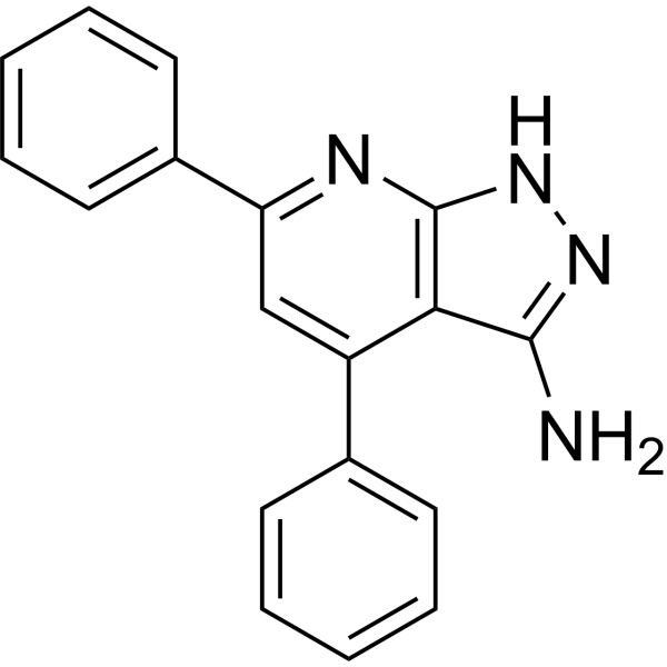
EB1 is the inhibitor of kinases MNK with IC50s of 0.69 μM (MNK1) and 9.4 μM (MNK2). EB1 selectively inhibits the growth of cancer cells, but not normal cells. EB1 also increases cell apoptosis and suppresses eIF4E phosphorylation.
商品は「研究用試薬」です。人や動物の医療用・臨床診断用・食品用の製品ではありません。
研究用途以外に使用した場合、当社は一切の責任を負いかねます。
- 純度 : 99.91%
- CAS 番号: 42951-68-8
- 分子式: C18H14N4
- 分子量:286.33
-
保管条件:Powder -20°C, 3 years , 4°C, 2 years ; In solvent -80°C, 6 months , -20°C, 1 month

To place orders, for customer services and technical support, please contact: MedChemExpress USA
Tel: 609-228-6898 E-mail: [email protected] [email protected]


-
 生物活性
生物活性
-
化学情報
-
溶剤 & 溶解度
- プロトコル
- 純度とドキュメンテーション
- 参考文献
-
 Help & FAQs


-
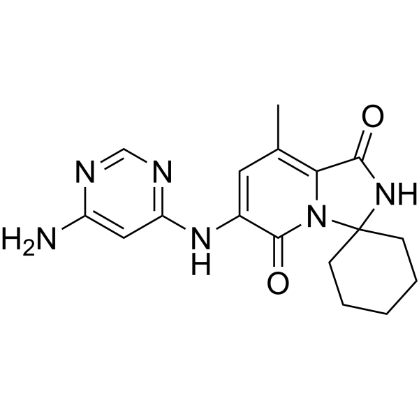
HY-100022
TomivosertibMNK1/2 Inhibitor -
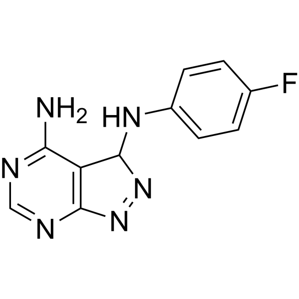
HY-10520
CGP 57380Mnk1 Inhibitor -
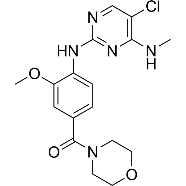
HY-13488
HG-10-102-01LRRK2 Inhibitor -
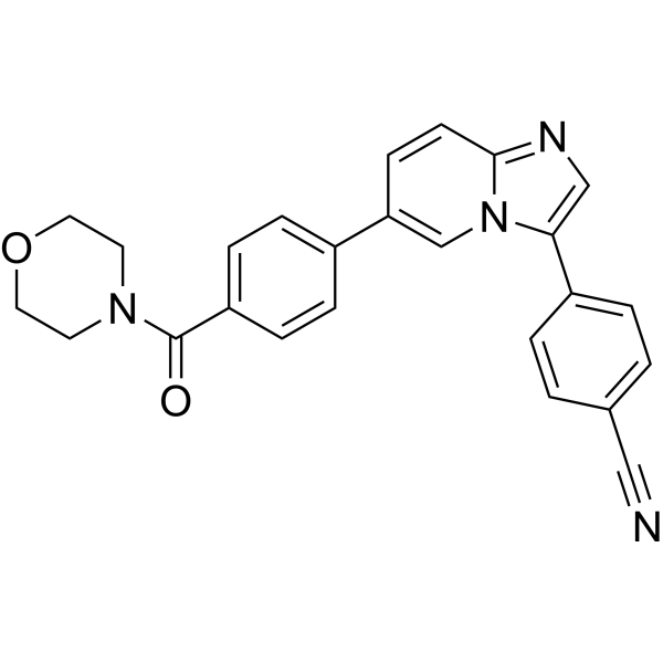
HY-112424
TinodasertibMNK1/2 Inhibitor -
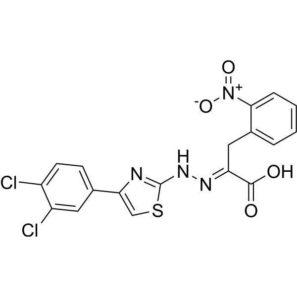
HY-19831
4EGI-1eIF4E-eIF4G Inhibitor -
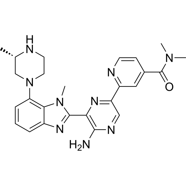
HY-124638
MNK inhibitor 9MNK1/2 Inhibitor -
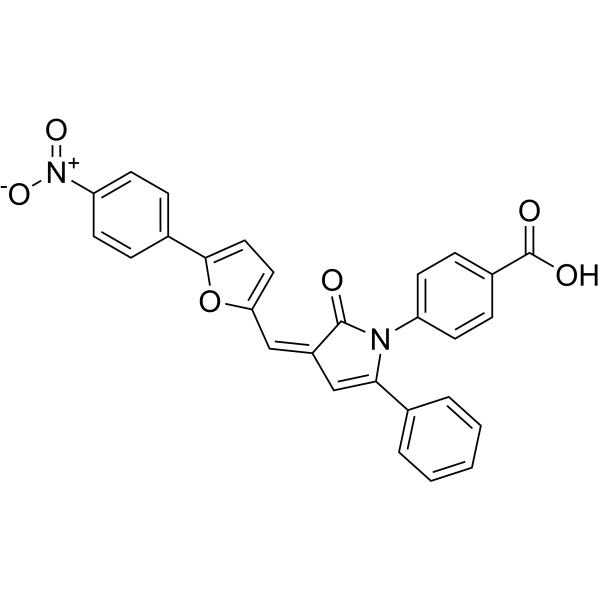
HY-14427
4E1RCateIF Inhibitor -
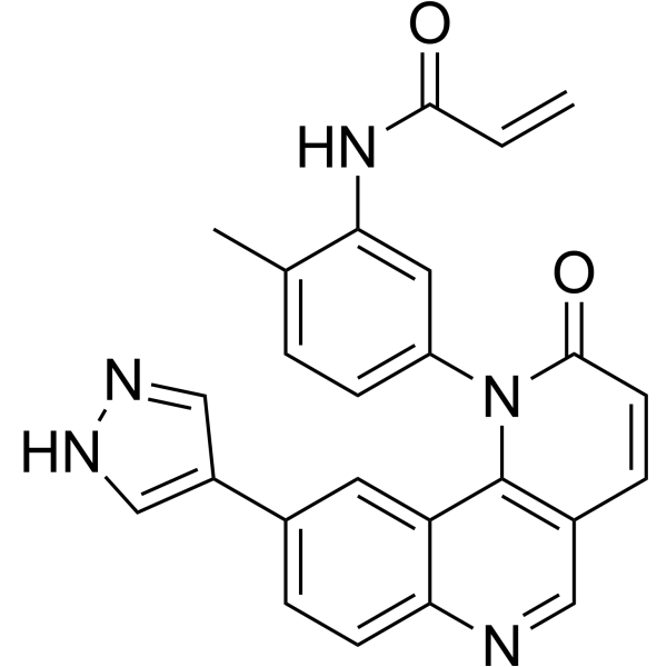
HY-124645
QL-X-138BTK/MNK Dual Inhibitor


生物活性
製品説明
IC50 & Target
[1]|
MNK1 0.69 μM (IC50) |
MNK2 9.4 μM (IC50) |
eIF4E |
Cellular Effect
|
Cell Line

|
Type | Value | Description | References |
|---|---|---|---|---|
| IMR-90 | GI50 |
>40 μM
Compound: 6b; EB1
|
Cytotoxicity against human IMR-90 cells assessed as reduction in cell viability measured after 72 hrs by crystal violet staining based assay
Cytotoxicity against human IMR-90 cells assessed as reduction in cell viability measured after 72 hrs by crystal violet staining based assay
|
[PMID: 35417652] |
| MCF7 | GI50 |
8.46 μM
Compound: 6b; EB1
|
Growth inhibition of human MCF7 cells measured after 72 hrs by crystal violet staining based assay
Growth inhibition of human MCF7 cells measured after 72 hrs by crystal violet staining based assay
|
[PMID: 35417652] |
| MDA-MB-231 | GI50 |
15.08 μM
Compound: 6b; EB1
|
Growth inhibition of human MDA-MB-231 cells measured after 72 hrs by crystal violet staining based assay
Growth inhibition of human MDA-MB-231 cells measured after 72 hrs by crystal violet staining based assay
|
[PMID: 35417652] |
| MDA-MB-468 | GI50 |
14.46 μM
Compound: 6b; EB1
|
Growth inhibition of human MDA-MB-468 cells measured after 72 hrs by crystal violet staining based assay
Growth inhibition of human MDA-MB-468 cells measured after 72 hrs by crystal violet staining based assay
|
[PMID: 35417652] |
体外実験
EB1 (1.3-40 μM; 24 hr) inhibits the phosphorylation of eIF4E dose-dependently[1].
EB1 (2.5-40 μM; 72 hr) shows dose-dependent cytotoxicity on tumor cells, and induces apoptosis[1].
EB1 (5 μM, 10 μM, and 20 μM; 24 hr) acts directly on MNK kinases without perturbing activating upstream signaling, such as activation of p38 (p-p38) and phosphorylation of its downstream effector HSP27[1].
EB1 (compound 14) inhibits the growth of HepG2 and MCF-7 cancer cells with IC50s of 0.74 μM and 5.18 μM, respectively[2].
MedChemExpress (MCE) has not independently confirmed the accuracy of these methods. They are for reference only.
-
Cell Line:MDA-MB-231, MDA-MB-468, MCF7, and IMR90 cells
-
Concentration:1.3 μM, 2.5 μM, 5 μM, 10 μM, 20 μM, 40 μM
-
Incubation Time:24 hours
-
Result:Blocked the phosphorylation of eIF4E in a dose-dependent manner.
-
Cell Line:MDA-MB-231, MDA-MB-468, MCF7, and IMR90 cells
-
Concentration:2.5 μM, 5 μM, 10 μM, 20 μM, 40 μM
-
Incubation Time:72 hours
-
Result:Increased the percentage of apoptosis cells among tumor cells.


化学情報
-
CAS 番号 42951-68-8
-
性状 Solid
-
分子量 286.33
-
分子式 C18H14N4
-
Color Light yellow to yellow
-
SMILES
NC1=NNC2=C1C(C3=CC=CC=C3)=CC(C4=CC=CC=C4)=N2
-
輸送条件
Room temperature in continental US; may vary elsewhere.
-
保管条件
Powder -20°C 3 years 4°C 2 years In solvent -80°C 6 months -20°C 1 month


溶剤 & 溶解度
体外:
DMSO : 8.33 mg/mL (29.09 mM; ultrasonic and warming and heat to 60°C; Hygroscopic DMSO has a significant impact on the solubility of product, please use newly opened DMSO)
Please refer to the solubility information to select the appropriate solvent. Once prepared, please aliquot and store the solution to prevent product inactivation from repeated freeze-thaw cycles.
Storage method and period of stock solution: -80°C, 6 months; -20°C, 1 month. When stored at -80°C, please use it within 6 months. When stored at -20°C, please use it within 1 month.
Please refer to the solubility information to select the appropriate solvent. Once prepared, please aliquot and store the solution to prevent product inactivation from repeated freeze-thaw cycles.
Storage method and period of stock solution: -80°C, 6 months; -20°C, 1 month. When stored at -80°C, please use it within 6 months. When stored at -20°C, please use it within 1 month.
濃度 (開始) × 体積 (開始) = 濃度 (終了) × 体積 (終了)


純度とドキュメンテーション
-
データシート (278 KB)
-
SDS (251 KB)
- English - EN (251 KB)
- Français - FR (251 KB)
- Deutsch - DE (251 KB)
- Norwegian - NO (251 KB)
- Español - ES (251 KB)
- Swedish - SV (251 KB)
- Italian - IT (251 KB)
- Korean - KR (251 KB)
- Portuguese - PT (251 KB)
-
取扱説明書 (2659 KB)
参考文献
[1]. Bou-Petit E, et al. Overcoming Paradoxical Kinase Priming by a Novel MNK1 Inhibitor. J Med Chem. 2022 Apr 28;65(8):6070-6087. [Content Brief]
[2]. Aboukhatwa SM, et al. Nicotinonitrile-derived apoptotic inducers: Design, synthesis, X-ray crystal structure and Pim kinase inhibition. Bioorg Chem. 2022 Dec;129:106126. [Content Brief]


Complete Stock Solution Preparation Table
Please refer to the solubility information to select the appropriate solvent. Once prepared, please aliquot and store the solution to prevent product inactivation from repeated freeze-thaw cycles.
Storage method and period of stock solution: -80°C, 6 months; -20°C, 1 month. When stored at -80°C, please use it within 6 months. When stored at -20°C, please use it within 1 month.
| Optional Solvent | Concentration Solvent Mass | 1 mg | 5 mg | 10 mg | 25 mg |
|---|---|---|---|---|---|
| DMSO | 1 mM | 3.4925 mL | 17.4624 mL | 34.9247 mL | 87.3118 mL |
| 5 mM | 0.6985 mL | 3.4925 mL | 6.9849 mL | 17.4624 mL | |
| 10 mM | 0.3492 mL | 1.7462 mL | 3.4925 mL | 8.7312 mL | |
| 15 mM | 0.2328 mL | 1.1642 mL | 2.3283 mL | 5.8208 mL | |
| 20 mM | 0.1746 mL | 0.8731 mL | 1.7462 mL | 4.3656 mL | |
| 25 mM | 0.1397 mL | 0.6985 mL | 1.3970 mL | 3.4925 mL |


EB1 関連分類
HY-146805 関連分類
 Powered by Bioz