



Propidium Iodide (solution)

Based on 1 Customer Validation
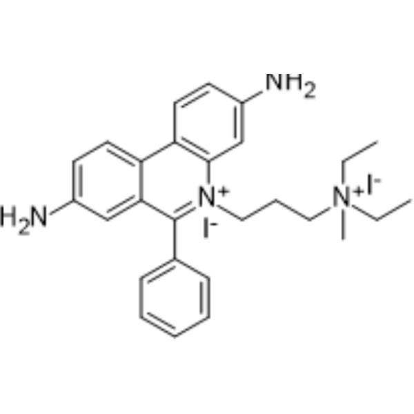
Propidium Iodide (PI) (solution) is a nuclear staining agent that stains DNA. Propidium Iodide is an analogue of ethidine bromide that emits red fluorescence upon embedding in double-stranded DNA. Propidium Iodide cannot pass through living cell membranes, but it can pass through damaged cell membranes to stain the nucleus. Propidium Iodide has a fluorescence wavelength of 493/617 nm and a wavelength of 536/635 nm after Mosaic with DNA. Propidium Iodide is commonly used in the detection of apoptosis (apoptosis) or necrosis (necrosis) , and is often used in flow cytometry analysis.
Solvent and Concentration: Sterile water: 1 mg/mL
The 1 mL volume is defined as the base specification. All larger sizes correspond to incremental volumes of this base.
For research use only. We do not sell to patients.
- CAS No.: 25535-16-4
- Formula: C27H34I2N4
- Molecular Weight:668.41
-
Storage:
Please store the product under the recommended conditions in the Certificate of Analysis.


To place orders, for customer services and technical support, please contact: MedChemExpress USA
Tel: 609-228-6898 E-mail: [email protected] [email protected]


-
 Biological Activity
-
Chemical Information
- Protocols
- Purity & Documentation
- References
-
 Help & FAQs


-

HY-DY1005
Hoechst 33342 trihydrochloride (solution)DNA Fluorescent Stain -
HY-D0815
Propidium IodideFluorochrome -
HY-D0814
DAPI dihydrochlorideFluorescent DNA Stain -
HY-101879
Acridine Orange hydrochlorideNucleic Acid dye -
HY-15559
Hoechst 33342DNA Fluorescent Stain -
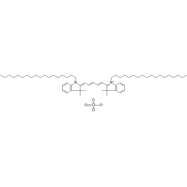
HY-D1028
DiD perchlorateDye -
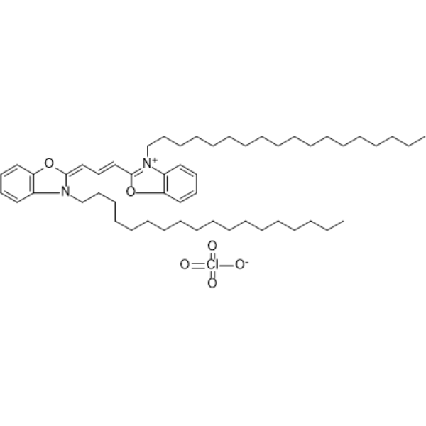
HY-D0969
DiOFluorescent Probe -

HY-DY1081
DAPI dihydrochloride (solution)Fluorescent DNA Stain -
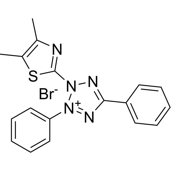
HY-15924
Thiazolyl BlueCytotoxicity Detection Reagent -
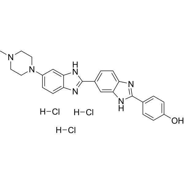
HY-15558A
Hoechst 33258 trihydrochlorideDNA Staining -
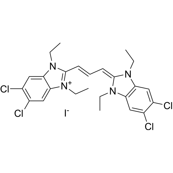
HY-15534
JC-1Mitochondrial Membrane Potential Probe -
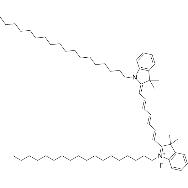
HY-D1048
DiRMembrane Dye
All DNA/RNA Synthesis Isoforms
More

Biological Activity
Description
Solvent and Concentration: Sterile water: 1 mg/mL
The 1 mL volume is defined as the base specification. All larger sizes correspond to incremental volumes of this base.
Cellular Effect
|
Cell Line

|
Type | Value | Description | References |
|---|---|---|---|---|
| HEK293 | IC50 |
1.1 μM
Compound: propidium iodide
|
Inhibition of wild type mouse AChE expressed in HEK293 cells using ATCh as substrate
Inhibition of wild type mouse AChE expressed in HEK293 cells using ATCh as substrate
|
[PMID: 23984975] |
In Vitro
Guidelines (Following is our recommended protocol. This protocol only provides a guideline, and should be modified according to your specific needs).
1. Preparation of Propidium Iodide working solution
The corresponding stock solution can be diluted according to the actual situation. Note that if the solvent is DMSO, the cytotoxicity of DMSO must be considered, and a solvent control should be prepared; if the solvent is pure water, the working solution needs to be filtered and sterilized before adding cells.
Note: Please adjust the concentration of Propidium Iodide working solution according to the actual situation.
2. Cell staining (Suspension cells)
2.1 Centrifuge at 1000 g at 4°C for 3-5 minutes and then discard the supernatant. Wash twice with PBS, 5 minutes each time. The cell density is 1×10⁶/mL.
2.2 Add 1 mL of working solution, and then incubate at room temperature for 5-10 minutes.
2.3 Centrifuge at 400 g at 4°C for 3-4 minutes and then discard the supernatant.
2.4 Wash twice with PBS, 5 minutes each time.
2.5 Resuspend cells with serum-free cell culture medium or PBS. Observation by fluorescence microscopy or flow cytometry.
3. Cell staining (Adherent cells)
3.1 Culture adherent cells on sterile coverslips.
3.2 Remove the coverslip from the medium and aspirate excess medium.
3.3 Add 100 μL of working solution, gently shake it to completely cover the cells, and then incubate at room temperature for 5-10 minutes.
3.4 Wash twice with medium, 5 minutes each time. Observation by fluorescence microscopy or flow cytometry.
Precautions
1. Please adjust the concentration of Propidium Iodide working solution according to the actual situation.
2. This product is for R&D use only, not for drug, household, or other uses.
3. Propidium Iodide is carcinogenic. For your safety and health, please wear a lab coat and disposable gloves to operate.
MedChemExpress (MCE) has not independently confirmed the accuracy of these methods. They are for reference only.
- Detection of Apoptosis
- TUNEL staining for apoptotic DNA fragmentation
- Flow Cytometry
- Flow cytometric DNA-content cell-cycle staining
- Annexin V plus membrane-impermeant dye apoptosis staining
- Nuclear DNA counterstaining and nuclear morphology staining
- Protocol for Phospho-flow cytometry
- Apoptosis Solutions


Chemical Information
-
CAS No. 25535-16-4
-
Appearance Liquid (Density: 1.5072 g/cm3)
-
Molecular Weight 668.41
-
Formula C27H34I2N4
-
Color Brown to red
-
SMILES
C[N+](CCC[N+]1=C(C2=CC=CC=C2)C3=CC(N)=CC=C3C4=C1C=C(N)C=C4)(CC)CC.[I-].[I-]
-
Shipping
Room temperature in continental US; may vary elsewhere.
-
Storage
Please store the product under the recommended conditions in the Certificate of Analysis.


Purity & Documentation
-
Data Sheet (272 KB)
-
SDS (252 KB)
- English - EN (252 KB)
- Français - FR (252 KB)
- Deutsch - DE (252 KB)
- Norwegian - NO (252 KB)
- Español - ES (252 KB)
- Swedish - SV (252 KB)
- Italian - IT (252 KB)
- Korean - KR (252 KB)
- Portuguese - PT (252 KB)
-
Handling Instructions (2659 KB)
References
[1]. Hezel M, et al. Propidium iodide staining: a new application in fluorescence microscopy for analysis of cytoarchitecture in adult and developing rodent brain. Micron. 2012 Oct;43(10):1031-8. [Content Brief]
[2]. A rapid and simple method for measuring thymocyte apoptosis by propidium iodidestaining and flow cytometry. J Immunol Methods. 1991 Jun 3;139(2):271-9. [Content Brief]
[3]. Nicoletti I, et al. A rapid and simple method for measuring thymocyte apoptosis by propidium iodide staining and flow cytometry. J Immunol Methods. 1991 Jun 3;139(2):271-9. [Content Brief]

Calculators
Concentration (start) × Volume (start) = Concentration (final) × Volume (final)


Propidium Iodide (solution) Related Classifications
HY-DY1006 Related Classifications
 Powered by Bioz