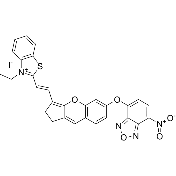
HS-CyBz is a Fluorescent probe for H₂S detection, enabling ratiometric optical/photoacoustic dual-modality in/ex vivo imaging. Its detection mechanism relies on nucleophilic substitution of its benzoic ester group by HS⁻, which releases an enolic meso-hydroxyltricarboheptamethine cyanine that then undergoes keto-enol tautomerization to form Cy-ketone; this tautomerization causes distinct shifts in absorption and emission spectra, producing a ratiometric response that reduces interferences from tissue scattering, autofluorescence, and probe concentration. In its initial state, HS-CyBz has an excitation wavelength of 595 nm, with emission bands centered at 805 nm (main) and 630 nm (minor); upon reaction with H₂S, the 805 nm emission band decreases while the 630 nm band drastically increases, and its absorption spectrum shows a sharp band at 775 nm and a shoulder band at 708 nm, which decrease upon H₂S reaction with a minor increase at 850 nm and an isosbestic point at 825 nm. For in vivo optical imaging, excitation at 560 nm is used with emission channels at 620 nm and 790 nm, while in vivo photoacoustic imaging uses excitation at 775 nm and 825 nm. The detection limit of HS-CyBz for H₂S is 0.5 μM, and it shows high selectivity, with only H₂S inducing a distinct enhancement of the emission ratio F₆₃₀/F₈₀₅ and PA ratio PA₈₂₅/PA₇₇₅, while other biochemical species including cations, anions, reactive oxygen species, biothiols, and carboxylesterase trigger only minor changes and do not interfere with H₂S sensing. Tail intravenous injection of HS-CyBz leads to accumulation in the liver of mice, and it can be used to verify endogenous H₂S upregulation triggered by S-adenosyl-L-methionine via ratiometric optical/photoacoustic imaging .

