



GPR43 Antibody
(Synonyms: GPCR GPR43; FFA2R; Ffar2; FFAR2_HUMAN; free fatty acid activated receptor 2; Free fatty acid receptor 2; G protein coupled receptor 43; G protein-coupled receptor 43; G-protein coupled receptor 43; GPR 43; GPCR 43.)
Based on 1 Customer Validation
GPR43 Antibody is a Rabbit-derived and non-conjugated IgG polyclonal antibody, targeting to GPR43.
-
Host:
Rabbit
-
Isotype:
IgG
-
Application:
IHC-P, IHC-F, ICC/IF, ELISA, IF-Tissue
-
Reactivity :
Human, Mouse
-
Formulation:
1.Supplied in 0.01M TBS(pH7.4) with 1% BSA, 0.03% Proclin300 and 50% Glycerol.
2.Supplied in 0.01M TBS(pH7.4) with 1% BSA, 0.02% Proclin300 and 50% Glycerol.
Please refer to the lot-specific COA for specific buffer information.
-
Conjugation:
Non-conjugated


To place orders, for customer services and technical support, please contact: MedChemExpress USA
Tel: 609-228-6898 E-mail: [email protected] [email protected]





Applications
| Application |
ELISA

ELISA: Enzyme Linked Immunosorbent Assay
|
IHC-P
IHC-P: Immunohistochemistry-Paraffin
|
IHC-F
IHC-F: Immunohistochemistry-Frozen
|
ICC/IF
ICC/IF: Immunocytochemistry/
Immunofluorescence |
IF-Tissue
IF-Tissue: Immunofluorescence-Tissue
|
|---|---|---|---|---|---|
| Dilution Ratio | 1:5000-10000 | 1:100-500 | 1:100-500 | 1:100-500 | 1:100-500 |


Product Details
GPR43 Antibody is a Rabbit-derived and non-conjugated IgG polyclonal antibody, targeting to GPR43.
-
Host Rabbit

-
Clonality Polyclonal

-
Species ReactivityHuman, Mouse Predicted Reactivity: Rat
Note: The predicted reactivity is for reference only and should not be considered a guarantee of product performance.
-
Observed Molecular WeightObserved band size: 37 kDa
Note: Due to possible protein modifications or aggregation, the molecular weight should be confirmed by actual measurement, and the predicted value is for reference only.

-
Calculated Molecular Weight Predicted band size: 37 kDa

Entrez Gene: 2867 Human ; 233079 Mouse ;
SwissProt: O15552 Human ; Q8VCK6 Mouse ;
OMIM: 603823 Human
KLH conjugated synthetic peptide derived from human GPR43: 41-140/330
Endogenous
affinity purified
Non-conjugated
Unmodified
IgG


Product Properties
-
Appearance
Liquid
-
Formulation
1.Supplied in 0.01M TBS(pH7.4) with 1% BSA, 0.03% Proclin300 and 50% Glycerol.
2.Supplied in 0.01M TBS(pH7.4) with 1% BSA, 0.02% Proclin300 and 50% Glycerol.
Please refer to the lot-specific COA for specific buffer information. -
Concentration
Batch-dependent, Please check the COA for the concentration of each lot. Check Lot Concentration
-
Storage & Stability
Stored at -20°C for 1 year. Avoid repeated freeze / thaw cycles.
-
Shipping
Shipping with blue ice.


Verification Images
-
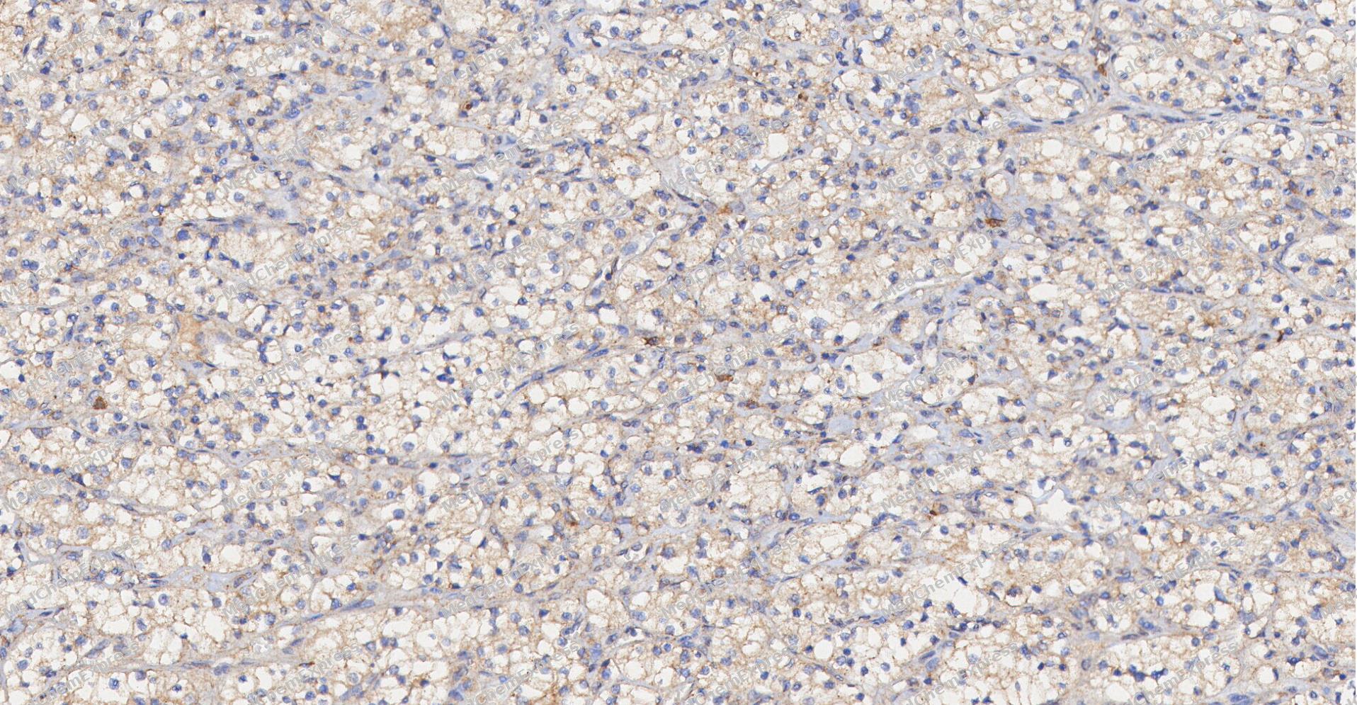 Immunohistochemical analysis of paraffin-embedded human Kidney cancer tissue using GPR43 antibody was performed. The section was pretreated using high-temperature and high-pressure mediated EDTA antigen retrieval buffer (pH 9.0), for 5 minutes. The tissues were incubated with primary antibody (HY-P81225, 1:200 dilution) at room temperature for 60 minutes. Detection was performed using an HRP conjugated compact polymer system. DAB was used as the chromogen. The tissues were counterstained with hematoxylin and mounted with neutral balsam mounting medium.
 -
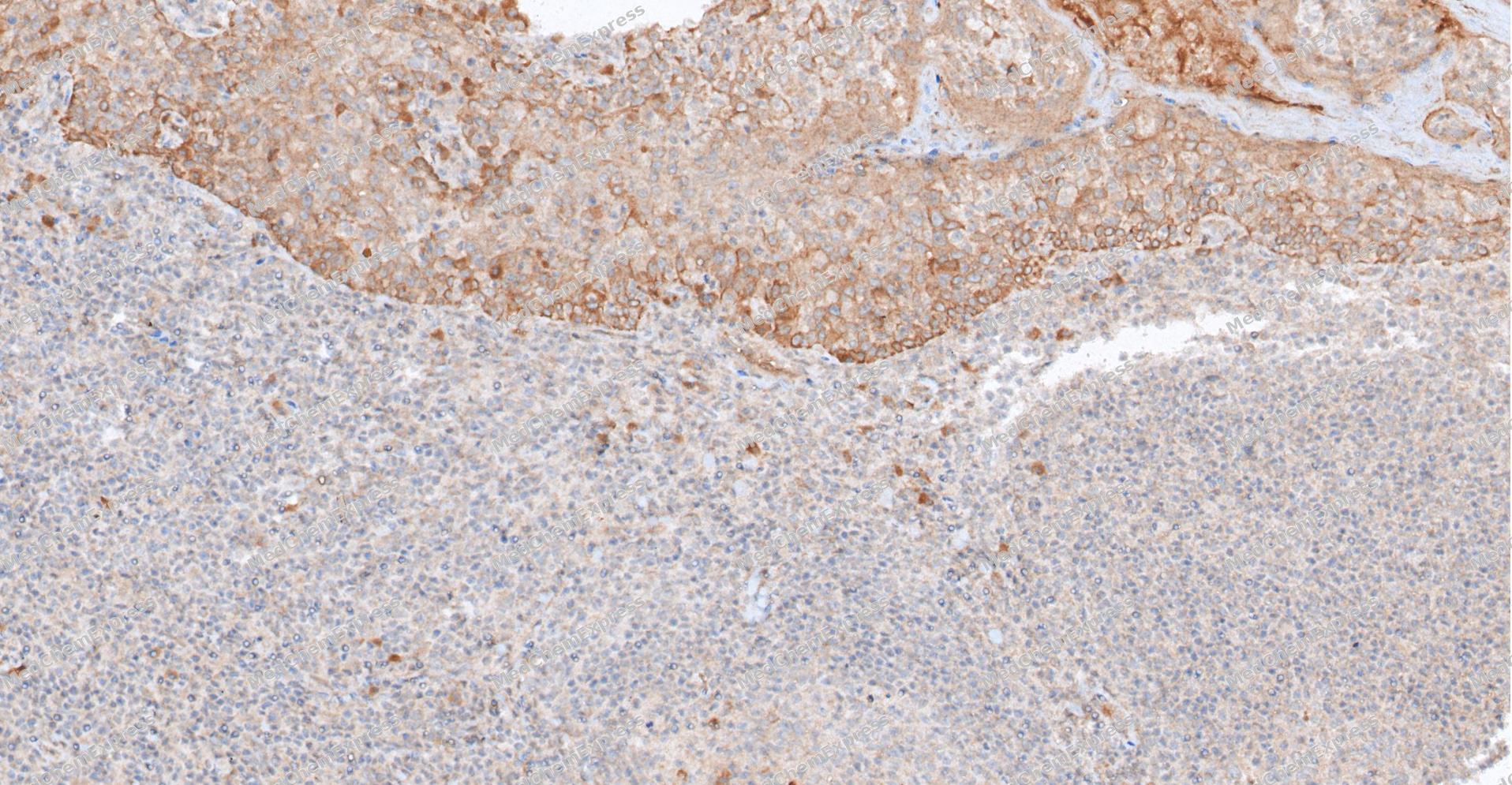 Immunohistochemical analysis of paraffin-embedded human Tonsil tissue using GPR43 antibody was performed. The section was pretreated using high-temperature and high-pressure mediated EDTA antigen retrieval buffer (pH 9.0), for 5 minutes. The tissues were incubated with primary antibody (HY-P81225, 1:200 dilution) at room temperature for 60 minutes. Detection was performed using an HRP conjugated compact polymer system. DAB was used as the chromogen. The tissues were counterstained with hematoxylin and mounted with neutral balsam mounting medium.
 -
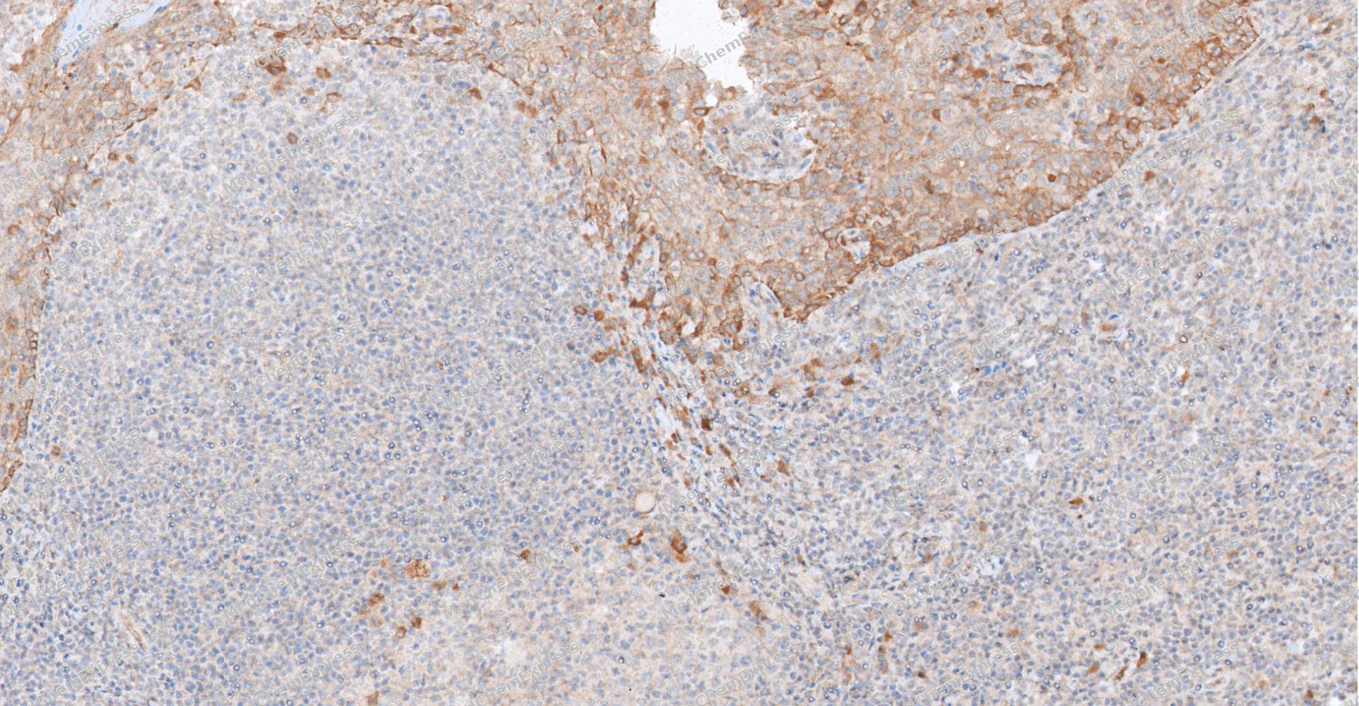 Immunohistochemical analysis of paraffin-embedded human Tonsil tissue using GPR43 antibody was performed. The section was pretreated using high-temperature and high-pressure mediated EDTA antigen retrieval buffer (pH 9.0), for 5 minutes. The tissues were incubated with primary antibody (HY-P81225, 1:200 dilution) at room temperature for 60 minutes. Detection was performed using an HRP conjugated compact polymer system. DAB was used as the chromogen. The tissues were counterstained with hematoxylin and mounted with neutral balsam mounting medium.
 -
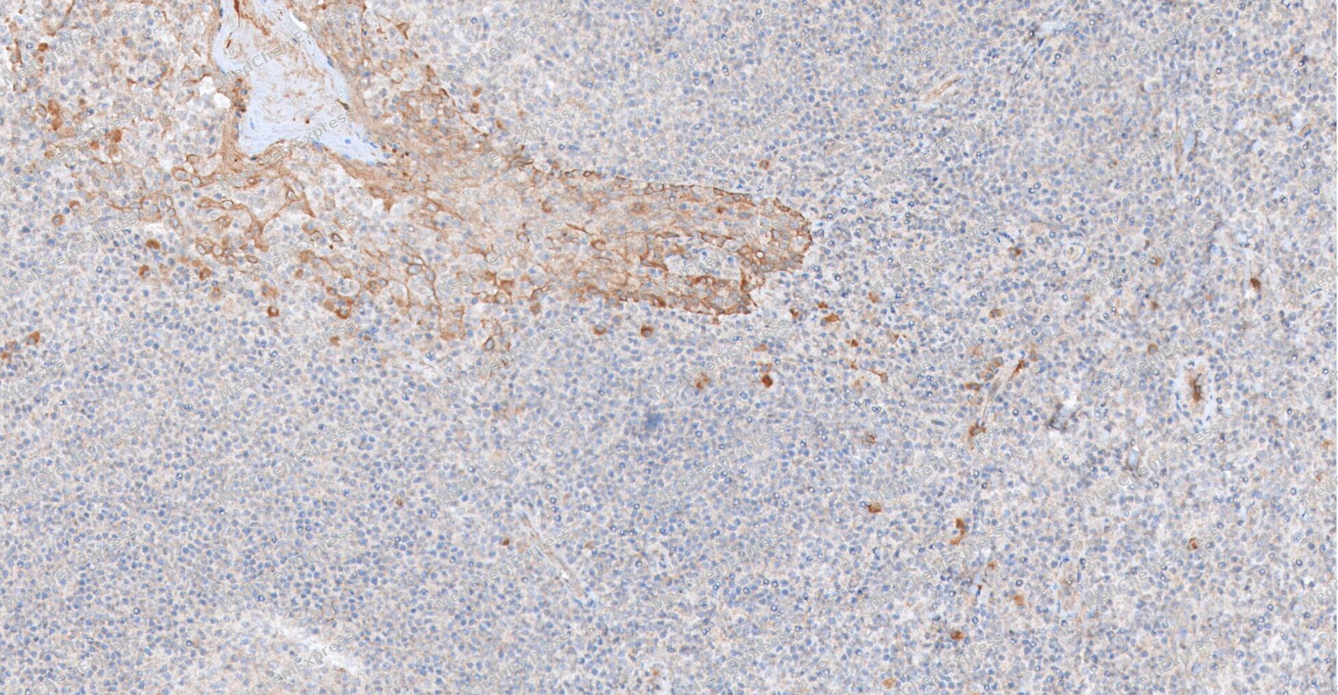 Immunohistochemical analysis of paraffin-embedded human Tonsil tissue using GPR43 antibody was performed. The section was pretreated using high-temperature and high-pressure mediated EDTA antigen retrieval buffer (pH 9.0), for 5 minutes. The tissues were incubated with primary antibody (HY-P81225, 1:200 dilution) at room temperature for 60 minutes. Detection was performed using an HRP conjugated compact polymer system. DAB was used as the chromogen. The tissues were counterstained with hematoxylin and mounted with neutral balsam mounting medium.
 -
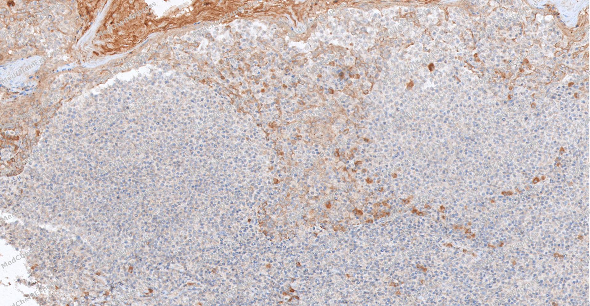 Immunohistochemical analysis of paraffin-embedded human Tonsil tissue using GPR43 antibody was performed. The section was pretreated using high-temperature and high-pressure mediated EDTA antigen retrieval buffer (pH 9.0), for 5 minutes. The tissues were incubated with primary antibody (HY-P81225, 1:200 dilution) at room temperature for 60 minutes. Detection was performed using an HRP conjugated compact polymer system. DAB was used as the chromogen. The tissues were counterstained with hematoxylin and mounted with neutral balsam mounting medium.
 -
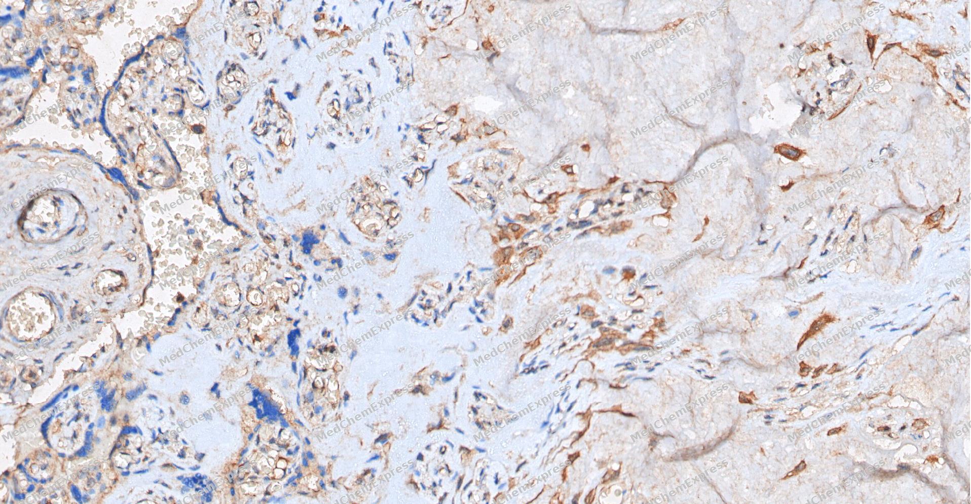 Immunohistochemical analysis of paraffin-embedded human placenta tissue using GPR43 antibody was performed. The section was pretreated using high-temperature and high-pressure mediated EDTA antigen retrieval buffer (pH 9.0), for 5 minutes. The tissues were incubated with primary antibody (HY-P81225, 1:200 dilution) at room temperature for 60 minutes. Detection was performed using an HRP conjugated compact polymer system. DAB was used as the chromogen. The tissues were counterstained with hematoxylin and mounted with neutral balsam mounting medium.
 -
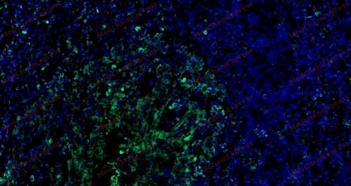 Tyramide signaling amplification based immunofluorescence was performed on paraffin-embedded human Tonsil tissue using GPR43 antibody. Antigen retrieval was performed in EDTA buffer pH 9.0 (95 °C, 20 min) followed by cooling to RT. Then incubated with primary antibody (HY-P81225, 1:200 dilution) at room temperature for 60 minutes and HRP conjugated secondary antibody for 10 minutes. Fluorescence was then developed with Vari Fluor 532 TSA (200×)(HY-D1832). The tissues were counterstained with DAPI and mounted with Anti-fade mounting medium.
 -
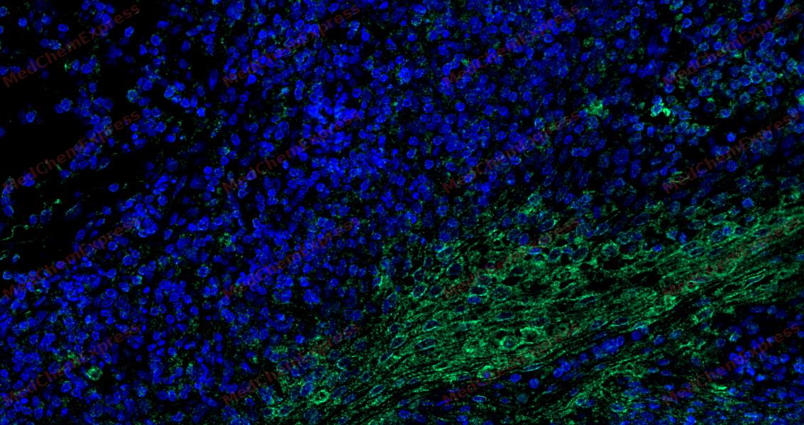 Tyramide signaling amplification based immunofluorescence was performed on paraffin-embedded human Tonsil tissue using GPR43 antibody. Antigen retrieval was performed in EDTA buffer pH 9.0 (95 °C, 20 min) followed by cooling to RT. Then incubated with primary antibody (HY-P81225, 1:200 dilution) at room temperature for 60 minutes and HRP conjugated secondary antibody for 10 minutes. Fluorescence was then developed with Vari Fluor 532 TSA (200×)(HY-D1832). The tissues were counterstained with DAPI and mounted with Anti-fade mounting medium.
 -
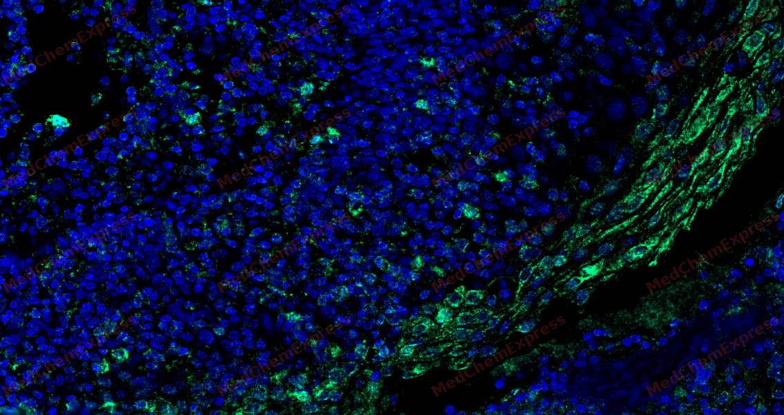 Tyramide signaling amplification based immunofluorescence was performed on paraffin-embedded human Tonsil tissue using GPR43 antibody. Antigen retrieval was performed in EDTA buffer pH 9.0 (95 °C, 20 min) followed by cooling to RT. Then incubated with primary antibody (HY-P81225, 1:200 dilution) at room temperature for 60 minutes and HRP conjugated secondary antibody for 10 minutes. Fluorescence was then developed with Vari Fluor 532 TSA (200×)(HY-D1832). The tissues were counterstained with DAPI and mounted with Anti-fade mounting medium.
 -
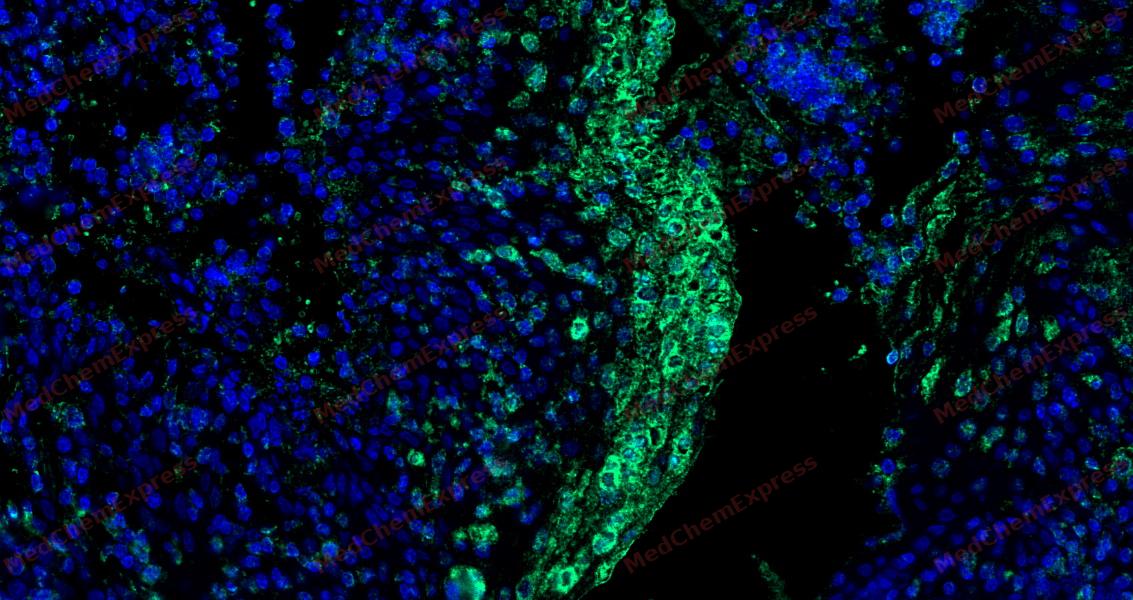 Tyramide signaling amplification based immunofluorescence was performed on paraffin-embedded human Tonsil tissue using GPR43 antibody. Antigen retrieval was performed in EDTA buffer pH 9.0 (95 °C, 20 min) followed by cooling to RT. Then incubated with primary antibody (HY-P81225, 1:200 dilution) at room temperature for 60 minutes and HRP conjugated secondary antibody for 10 minutes. Fluorescence was then developed with Vari Fluor 532 TSA (200×)(HY-D1832). The tissues were counterstained with DAPI and mounted with Anti-fade mounting medium.
 -
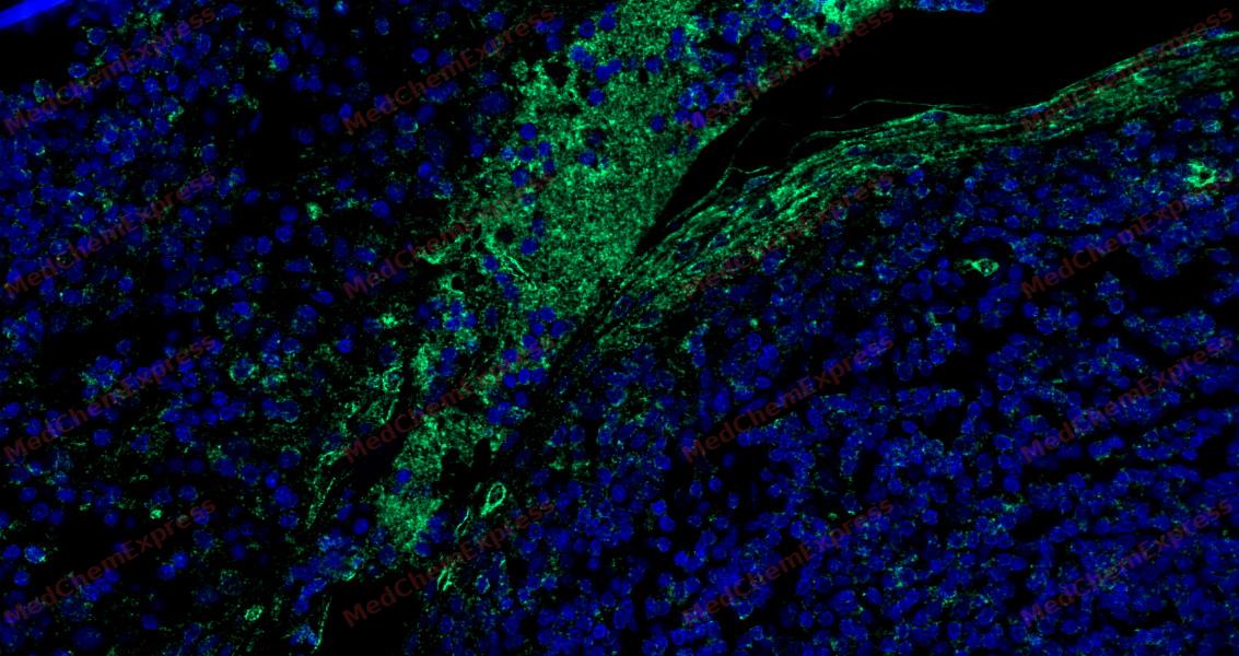 Tyramide signaling amplification based immunofluorescence was performed on paraffin-embedded human Tonsil tissue using GPR43 antibody. Antigen retrieval was performed in EDTA buffer pH 9.0 (95 °C, 20 min) followed by cooling to RT. Then incubated with primary antibody (HY-P81225, 1:200 dilution) at room temperature for 60 minutes and HRP conjugated secondary antibody for 10 minutes. Fluorescence was then developed with Vari Fluor 532 TSA (200×)(HY-D1832). The tissues were counterstained with DAPI and mounted with Anti-fade mounting medium.
 -
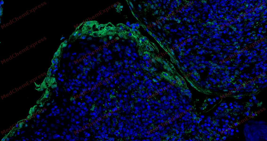 Tyramide signaling amplification based immunofluorescence was performed on paraffin-embedded human Tonsil tissue using GPR43 antibody. Antigen retrieval was performed in EDTA buffer pH 9.0 (95 °C, 20 min) followed by cooling to RT. Then incubated with primary antibody (HY-P81225, 1:200 dilution) at room temperature for 60 minutes and HRP conjugated secondary antibody for 10 minutes. Fluorescence was then developed with Vari Fluor 532 TSA (200×)(HY-D1832). The tissues were counterstained with DAPI and mounted with Anti-fade mounting medium.


Background
-
Function
GPR43 is a G protein-coupled receptor that is activated by a major product of dietary fiber digestion, the short chain fatty acids (SCFAs), and that plays a role in the regulation of whole-body energy homeostasis and in intestinal immunity. In omnivorous mammals, the short chain fatty acids acetate, propionate and butyrate are produced primarily by the gut microbiome that metabolizes dietary fibers. SCFAs serve as a source of energy but also act as signaling molecules. That G protein-coupled receptor is probably coupled to the pertussis toxin-sensitive, G(i/o)-alpha family of G proteins but also to the Gq family. Its activation results in the formation of inositol 1,4,5-trisphosphate, the mobilization of intracellular calcium, the phosphorylation of the MAPK3/ERK1 and MAPK1/ERK2 kinases and the inhibition of intracellular cAMP accumulation. May play a role in glucose homeostasis by regulating the secretion of GLP-1, in response to short-chain fatty acids accumulating in the intestine. May also regulate the production of LEP/Leptin, a hormone acting on the central nervous system to inhibit food intake. Finally, may also regulate whole-body energy homeostasis through adipogenesis regulating both differentiation and lipid storage of adipocytes. In parallel to its role in energy homeostasis, may also mediate the activation of the inflammatory and immune responses by SCFA in the intestine, regulating the rapid production of chemokines and cytokines. May also play a role in the resolution of the inflammatory response and control chemotaxis in neutrophils. In addition to SCFAs, may also be activated by the extracellular lectin FCN1 in a process leading to activation of monocytes and inducing the secretion of interleukin-8/IL-8 in response to the presence of microbes. Among SCFAs, the fatty acids containing less than 6 carbons, the most potent activators are probably acetate, propionate and butyrate. Exhibits a SCFA-independent constitutive G protein-coupled receptor activity[1][2][3][4][5].
-
Subcellular Localization
Cell membrane; Multi-pass membrane protein
-
Expression
Tissue_specificity:Expressed at relatively high levels in peripheral blood leukocytes and, to lesser extent, in spleen -
Subunit
Interacts with FCN1 (via Fibrinogen C-terminal domain)
-
SwissProt ID
-
Synonyms
GPCR GPR43; FFA2R; Ffar2; FFAR2_HUMAN; free fatty acid activated receptor 2; Free fatty acid receptor 2; G protein coupled receptor 43; G protein-coupled receptor 43; G-protein coupled receptor 43; GPR 43; GPCR 43.


Documentation
-
Data Sheet (234 KB)
-
SDS (252 KB)
- English - EN (252 KB)
- Français - FR (252 KB)
- Deutsch - DE (252 KB)
- Norwegian - NO (252 KB)
- Español - ES (252 KB)
- Swedish - SV (252 KB)
- Italian - IT (252 KB)
- Korean - KR (252 KB)
- Portuguese - PT (252 KB)
-
User Guide for Antibodies (1077 KB)

 Powered by Bioz
