



Ficerafusp alfa

Based on 1 Customer Validation
Ficerafusp alfa (BCA-101) is a bispecific antibody targeting EGFR and TGFβ, with a Kd of 2.58 nM against EGFR and a Kd of 61.3 nM against TGFβ1. Ficerafusp alfa binds to EGFR, inhibits EGFR phosphorylation, blocks EGF-dependent cell proliferation, and mediates antibody-dependent cellular cytotoxicity against EGFR-positive tumor cells. Ficerafusp alfa sequesters TGFβ via its TGFβRII ECD domain, neutralizes the activity of TGFβ and TGFβ1, and blocks TGFβ-dependent processes, including epithelial-mesenchymal transition, cell invasion, and differentiation of inducible regulatory T cells. Ficerafusp alfa is applicable to research related to head and neck squamous cell carcinoma, advanced solid tumors, squamous non-small cell lung cancer, anal squamous cell carcinoma, colorectal cancer, and pancreatic cancer.
Nur für Forschungszwecke. Wir verkaufen nicht an Patienten.
- Reinheit : 99.71%
- CAS. Nr.: 2764727-44-6
- Molecular Weight:178.105 kDa
-
Speicherung:
Please store the product under the recommended conditions in the Certificate of Analysis.


To place orders, for customer services and technical support, please contact: MedChemExpress USA
Tel: 609-228-6898 E-mail: [email protected] [email protected]


-
 Biologische Aktivität
-
Chemical Information
- Protokoll
- Reinheit & Dokumentation
- Verweise
-
 Help & FAQs



Alle EGFR Isoform-spezifische Produkte anzeigen
More

Biologische Aktivität
Beschreibung
Isotype
IgG1-kappa-[PROTEIN]2-in-VK
Recommend Isotype Controls
Species Reactivity
Human
IC50 & Target
ERBB1/EGFR/HER1
In Vitro
Ficerafusp alfa (BCA-101) (56 nM; 120 hours) inhibits and reverts TGFβ-induced epithelial-to-mesenchymal transition in A549 cells, as measured by reduced IL11 secretion[1].
In ELISA, Ficerafusp alfa is captured by plate-bound EGFR and the ability of Ficerafusp alfa to bind TGFβ as measured by spiking TGFβ followed by detection by a biotinylated anti-TGFβ antibody. Ficerafusp alfa shows a dose-dependent binding with an EC50 of 0.09 nM, indicating both the anti-EGFR and TGFβ trap arms are simultaneously functional[1].
Ficerafusp alfa could neutralize SMAD-mediated luciferase expression induced by TGFβ1 (EC50 of 1.49 nM) and TGFβ3 (EC50 of 3.51 nM), and not neutralize TGFβ2 in HEK-SMAD reporter assay[1].
MedChemExpress (MCE) has not independently confirmed the accuracy of these methods. They are for reference only.
-
Cell Line:A549 cells
-
Concentration:56 nM
-
Incubation Time:120 hours
-
Result:Significantly reduced TGFβ-induced IL11 secretion, with IL11 concentrations lower than those in cetuximab and isotype control groups.\nSignificantly reduced IL11 secretion in A549 cells with established TGFβ-induced EMT, with IL11 concentrations lower than those in cetuximab and isotype control groups.
In Vivo
Ficerafusp alfa (10 mg/kg; i.p.; twice a week) induces durable tumor regression and delayed relapse in head and neck squamous cell carcinoma patient-derived xenograft models[1].
Ficerafusp alfa (50 mg/kg; i.p.; twice a week) enhances the antitumor efficacy of anti-PD1 immunotherapy in a syngeneic human EGFR-expressing melanoma model, producing superior event-free survival compared with monotherapies[1].
Ficerafusp alfa (10 mg/kg; i.p.; twice a week) synergizes with Pembrolizumab (HY-P9902) to produce enhanced antitumor efficacy in a humanized prostate cancer xenograft model, with a TGI of 78% on day 18[1].
Ficerafusp alfa (5 mg/kg; i.v.; single dose) systemically neutralizes TGFβ1 in BALB/c mice, suppressing levels below detection for 4 days[1].
Ficerafusp alfa (10 mg/kg; i.p.; thrice weekly; 27 days) delivers sustained tumor growth delay in head and neck squamous cell carcinoma PDX models in female NOG mice and neutralizes 90% of tumor TGFβ[3].
Ficerafusp alfa (i.p.; six total doses) enhances the antitumor efficacy of PD-1 blockade (Pembrolizumab dosed at 10 mg/kg Q5Dx5) in PC-3 xenografts in humanized Hu-NOG-EXL mice[3].
MedChemExpress (MCE) has not independently confirmed the accuracy of these methods. They are for reference only.
-
Animal Model:Athymic nude mice; BALB/c mice (female)[1]
-
Dosage:1-50 mg/kg (FaDu xenografts; A431 xenografts)
-
Administration:i.p.; twice a week; seven doses (for 1, 5, 10 mg/kg)
-
Result:Exhibited superior tumor growth inhibition compared with cetuximab, TGFβRII-Fc, and cetuximab plus TGFβRII-Fc in FaDu xenografts, with efficacy evident as early as day 4 and sustained throughout the experiment.\nProduced a significantly higher apoptotic-to-mitotic index ratio in FaDu xenograft tumor tissues compared with vehicle, cetuximab, and TGFβRII-Fc.\nSuppressed tumor TGFβ1 levels by an average of 90% (10 mg/kg), 83% (5 mg/kg), and 56% (1 mg/kg) in A431 xenografts, superior to the 54% suppression achieved with equimolar TGFβRII-Fc (5 mg/kg).
-
Animal Model:NOG mice[1]
-
Dosage:10 mg/kg
-
Administration:i.p.; twice a week; 27 days (PDX-1); 34 days (PDX-2); 32 days (PDX-3)
-
Result:In PDX-1, after 27 days of treatment and 52 days of follow-up, only 1 of 9 mice had tumor regrowth beyond initial 130 mm3 volume, compared with 6 of 10 mice in cetuximab group; mean tumor volume on day 79 was 67 mm3 vs. 146 mm3 for cetuximab.\nIn PDX-2, produced statistically significant lower mean tumor volume at treatment cessation (134 mm3 vs. 210 mm3 for cetuximab) and longer tumor doubling time (68 days vs. 47 days for cetuximab) after 34 days of treatment and 74 days of follow-up.\nIn PDX-3 (recurrent metastatic model), no tumors relapsed, compared with 2 of 10 mice in cetuximab group.
-
Animal Model:C57BL/6 mice[1]
-
Dosage:50 mg/kg (monotherapy; in combination with anti-PD1 mAb 10 mg/kg)
-
Administration:i.p.; twice a week
-
Result:As monotherapy, reduced the number of mice reaching a tumor volume ≥300 mm3 to 4 of 9, compared with 8 of 9 in the vehicle group.\nIn combination with anti-PD1 mAb, only 1 of 9 mice reached a tumor volume ≥300 mm3, superior to anti-PD1 monotherapy (3 of 9 mice) and cetuximab plus anti-PD1 combination (3 of 9 mice).\nThe combination produced a significant improvement in event-free survival (P=0.0034 vs. vehicle).
-
Animal Model:HuNOG-EXL humanized mice[1]
-
Dosage:10 mg/kg (monotherapy; in combination with pembrolizumab 10 mg/kg)
-
Administration:i.p.; twice a week
-
Result:As monotherapy, produced a 40% tumor growth inhibition (TGI) on day 18.\nIn combination with pembrolizumab, TGI increased to 78% on day 18, superior to the 52% TGI achieved with pembrolizumab monotherapy.\nAll treatments were associated with acceptable weight loss (≤15%).
- Somatic Cell Culture
- Cell Cytotoxicity Assay
- Organotypic 3D Invasion Assay
- Cell invasion
- Matrigel/ECM Transwell Invasion Assay
- MTT Cell Proliferation Assay
- 3D Collagen/Hydrogel Matrix Invasion Assay
- Spheroid/Tumor Organoid Invasion Assay
- Western Blot
- Ex Vivo Tissue Slice/Explant Invasion Assay
- Patient-Derived Organoid Invasion Assay
- Mammalian live/dead viability and cytotoxicity staining
- BrdU Incorporation Assay
- CFSE Dye Dilution Proliferation Assay
- Dye-dilution cell tracking and proliferation staining
- CCK-8/WST-8 Cell Proliferation Assay
- Protocol for Cell Cycle
- Cell Counting-Based Growth Curve Assay
- Colony Formation (Clonogenic) Assay
- 3D Tumor Spheroid Invasion Assay
- Cell Viability Determination by MTT Colorimetric Assay
- Matrigel Transwell/Boyden Chamber Invasion Assay
- Kinase activity and phosphorylation assays
- Invadopodia/Fluorescent Gelatin Degradation Assay
- EdU Incorporation Assay (Click Chemistry-Based DNA Synthesis Measurement)
- Ki-67 Immunostaining Proliferation Assay
- PCNA Immunodetection Proliferation Assay
- Protocol for Cell Counting and Cell Density Analysis
- Protocol for Kinase activity and phosphorylation assays
Gene ID
Accession
Conjugated
Unconjugated
Reconsititution
The product can be reconstituted/diluted with sterile PBS or saline.
Format
-
IgG1-kappa-[PROTEIN]2-in-VK

Anwendung
ELISA, FACS, Functional assay
Verified Bioactivity
-
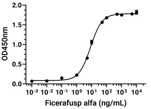 Immobilized EGFR Protein, Human (621a.a, HEK293, His, HY-P70613) can bind Ficerafusp alfa. The EC50 for this effect is 8.521 ng/mL.


Chemical Information
-
CAS. Nr. 2764727-44-6
-
Appearance Liquid
-
Molecular Weight 178.105 kDa
-
Color Colorless to light yellow
-
SMILES
[Ficerafusp alfa]
-
Synonyms
BCA-101; FMAB2
-
Versand
Shipping with dry ice.
-
Formulation
Please refer to the lot-specific COA for specific buffer information.
-
Speicherung
Please store the product under the recommended conditions in the Certificate of Analysis.


Reinheit & Dokumentation
-
Data Sheet (268 KB)
-
SDS (251 KB)
- English - EN (251 KB)
- Français - FR (251 KB)
- Deutsch - DE (251 KB)
- Norwegian - NO (251 KB)
- Español - ES (251 KB)
- Swedish - SV (251 KB)
- Italian - IT (251 KB)
- Korean - KR (251 KB)
- Portuguese - PT (251 KB)
-
Inhibitory Antibodies User Guide (603 KB)
Verweise
[1]. Boreddy SR, et al. BCA101 Is a Tumor-Targeted Bifunctional Fusion Antibody That Simultaneously Inhibits EGFR and TGFβ Signaling to Durably Suppress Tumor Growth. Cancer Res. 2023;83(11):1883-1904. [Content Brief]
[2]. Hernando-Calvo A, et al. Phase I Clinical Trial of the Bifunctional EGFR/TGF-β Fusion Protein Ficerafusp Alfa (BCA101) Alone and in Combination with Pembrolizumab for Advanced Solid Tumors. Clin Cancer Res. 2025;31(22):4623-4632. [Content Brief]

Calculators
Konzentration (Stammlösung) × Volumen (Stammlösung) = Konzentration (Ziellösung) × Volumen (Ziellösung)


Ficerafusp alfa Related Classifications
HY-P990957 Related Classifications
 Powered by Bioz
Keywords
- Ficerafusp alfa
- 2764727-44-6
- BCA-101
- FMAB2
- BCA101
- BCA 101
- FMAB2
- FMAB 2
- FMAB-2
- EGFR
- TGF-beta/Smad
- epithelial to mesenchymal transition
- TGFβ1
- TGFβ
- head and neck squamous cell carcinoma
- colorectal cancer
- A549 cells
- regulatory T cell
- squamous non-small cell lung cancer
- peripheral blood mononuclear cells
- Inhibitor
- inhibitor
- inhibit