



PAD4-IN-2
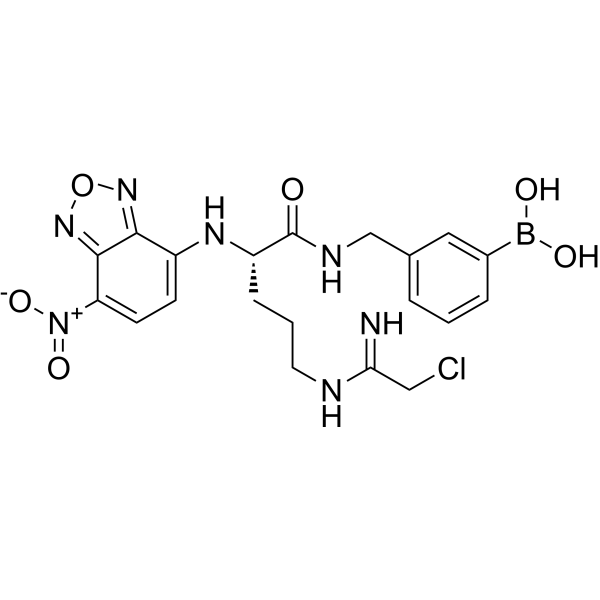
PAD4-IN-2 is a highly tumor-targeted and irreversible PAD4 inhibitor with an IC50 of 1.94 μM. It selectively recognizes sialic acid on tumor surfaces and accumulates in tumor tissues, with distribution in the cytoplasm of tumor cells and nuclei of neutrophils. PAD4-IN-2 inhibits the PAD4-H3cit-NETs (Neutrophil Extracellular Traps) pathway, reduces senescent tumor-associated neutrophils, and promotes M1 macrophage polarization. The compound exhibits potent anti-tumor and anti-metastatic activities and is suitable for breast cancer research.
For research use only. We do not sell to patients.
- CAS No.: 2642327-52-2
- Formula: C20H23BClN7O6
- Molecular Weight:503.70
-
Storage:
Please store the product under the recommended conditions in the Certificate of Analysis.

To place orders, for customer services and technical support, please contact: MedChemExpress USA
Tel: 609-228-6898 E-mail: [email protected] [email protected]


-
 Biological Activity
-
Chemical Information
- Protocols
- Purity & Documentation
- References
-
 Help & FAQs




Biological Activity
Description
IC50 & Target
IC50: 1.94 μM (PAD4)[1].
In Vitro
PAD4-IN-2 (compound 5i) inhibits PAD4 enzyme activity in a colorimetric assay (IC50 = 1.94 μM) and exhibits negligible cytotoxicity in LLC, 4T1, and normal HL7702 cells after 48 h treatment (IC50 > 100 μM)[1].
PAD4-IN-2 (25-100 μM; 48 h) effectively inhibits the monoclonal proliferation and dose-dependently suppresses the migration of 4T1 cells[1].
PAD4-IN-2 (50–100 μM; 1–48 h) is selectively taken up by 4T1 cells in a time-dependent manner but not by normal HL7702 cells, induces mitochondrial shrinkage and pseudopodia reduction, localizes to neutrophil nuclei to decrease histone H3 citrullination (H3cit) levels, and inhibits neutrophil extracellular trap (NET) formation[1].
MedChemExpress (MCE) has not independently confirmed the accuracy of these methods. They are for reference only.
-
Cell Line:LLC, 4T1, and HL7702 cells
-
Concentration:3.125, 6.25, 12.5, 25, 50, and 100 μM
-
Incubation Time:48 h
-
Result:Showed no significant lethal effect on either tumor or normal cells, with an IC50 value greater than 100 μM.
-
Cell Line:4T1 cells
-
Concentration:25, 50, and 100 μM
-
Incubation Time:48 h
-
Result:Reduced the number of single cell proliferation significantly and effectively inhibited the monoclonal proliferation of tumor cells.
-
Cell Line:4T1 cells
-
Concentration:25, 50, and 100 μM
-
Incubation Time:48 h
-
Result:Inhibited tumor cell migration in a dose-dependent manner, and significantly decreased both the number of migrated cells and the migration distance.
-
Cell Line:4T1 cells and bone marrow neutrophils
-
Concentration:50 μM and 100 μM
-
Incubation Time:48 h
-
Result:Localized to the cytoplasm of tumor cells and the nucleus of neutrophils, decreased intracellular histone 3 citrullination (H3cit) levels, and significantly inhibited the formation of neutrophil extracellular traps (NETs).
In Vivo
PAD4-IN-2 (5.04 mg/kg; i.p. ; once daily for 7 days) shows initial antitumor efficacy in an S180 sarcoma mouse model[1].
PAD4-IN-2 (1.01-5.04 mg/kg; i.v. ; every day for 21 days) decreases NETs formation, alters immune cells infiltration in the tumor microenvironment, and exhibits no significant hepatotoxicity or nephrotoxicity in vivo[1].
MedChemExpress (MCE) has not independently confirmed the accuracy of these methods. They are for reference only.
-
Animal Model:S180 sarcoma mouse model (Kunming mice)[1]
-
Dosage:10 μmol/kg
-
Administration:Intraperitoneal injection (i.p.), once daily for 7 days
-
Result:Inhibited tumor growth with an in vivo tumor inhibition rate (TIR) of 49.2%.
-
Animal Model:Orthotopic 4T1 breast cancer mouse model (BALB/c female mice)[1]
-
Dosage:2, 5, and 10 μmol/kg
-
Administration:Tail vein intravenous injection (i.v.), once daily for 21 days
-
Result:Reduced primary tumor volume and tumor weight substantially at the dose of 10 μmol/kg.
Suppressed the number of metastatic tumor nodules in the lungs markedly in a dose-dependent manner.
Maintained stable body weight in treated mice throughout the administration period.
-
Animal Model:Orthotopic 4T1 breast cancer mouse model (BALB/c female mice)[1]
-
Dosage:10 μmol/kg
-
Administration:Tail vein intravenous injection (i.v.), once daily for 21 days
-
Result:Showed intact alveolar structures and absence of tumor cell lesions in H&E-stained lung sections.
Decreased the number of Ly6G-positive neutrophils and H3cit-positive cells in immunofluorescence-stained tumor sections.
Caused no significant variations in serum levels of Cr, BUN, AST, and ALT compared with the normal saline control group.
Exhibited no apparent histopathological damage or organic lesions in H&E-stained sections of the heart, liver, spleen, and kidney tissues.
Reduced the proportion of aged neutrophils (Naged CD194hi CD62Llo) in tumor tissues.Increased the ratio of M1-type macrophages in tumor tissues.


Chemical Information
-
CAS No. 2642327-52-2
-
Molecular Weight 503.70
-
Formula C20H23BClN7O6
-
SMILES
O=C(NCC1=CC=CC(B(O)O)=C1)[C@H](CCCNC(CCl)=N)NC2=CC=C([N+]([O-])=O)C3=NON=C32
-
Shipping
Room temperature in continental US; may vary elsewhere.
-
Storage
Please store the product under the recommended conditions in the Certificate of Analysis.


Purity & Documentation
References

Calculators
Concentration (start) × Volume (start) = Concentration (final) × Volume (final)


PAD4-IN-2 Related Classifications
HY-155052 Related Classifications
 Powered by Bioz