



RM-581
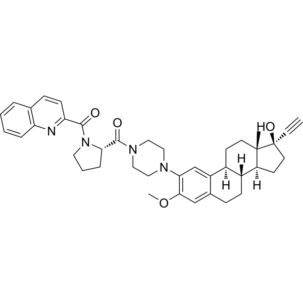
RM-581, aminosteroid, is an orally active anticancer agent. RM-581 shows anticancer potency against pancreatic, breast, prostate, and ovarian cancers. RM-581 induces endoplasmic reticulum stress and apoptosis via disturbance of cancer cell cholesterol homeostasis. RM-581 can be used for the research of cancer, such as prostate cancer and breast cancer.
For research use only. We do not sell to patients.
- CAS No.: 2086809-59-6
- Formula: C40H46N4O4
- Molecular Weight:646.82
-
Storage:
Please store the product under the recommended conditions in the Certificate of Analysis.

To place orders, for customer services and technical support, please contact: MedChemExpress USA
Tel: 609-228-6898 E-mail: [email protected] [email protected]


-
 Biological Activity
-
Chemical Information
- Protocols
- Purity & Documentation
- References
-
 Help & FAQs




Biological Activity
Description
Cellular Effect
|
Cell Line

|
Type | Value | Description | References |
|---|---|---|---|---|
| LAPC4 | IC50 |
0.6 μM
Compound: RM-581
|
Antiproliferative activity against human LAPC4 cells assessed as reduction in cell viability after 72 hrs by MTS assay
Antiproliferative activity against human LAPC4 cells assessed as reduction in cell viability after 72 hrs by MTS assay
|
[PMID: 31893547] |
| LNCaP | IC50 |
1.2 μM
Compound: RM-581
|
Antiproliferative activity against human LNCaP cells assessed as reduction in cell viability after 72 hrs by MTS assay
Antiproliferative activity against human LNCaP cells assessed as reduction in cell viability after 72 hrs by MTS assay
|
[PMID: 31893547] |
| MCF7 | IC50 |
2.6 μM
Compound: RM-581
|
Antiproliferative activity against human MCF7 cells assessed as reduction in cell viability after 72 hrs by MTS assay
Antiproliferative activity against human MCF7 cells assessed as reduction in cell viability after 72 hrs by MTS assay
|
[PMID: 31893547] |
| OVCAR-3 | IC50 |
5 μM
Compound: RM-581
|
Antiproliferative activity against human OVCAR-3 cells assessed as reduction in cell viability after 72 hrs by MTS assay
Antiproliferative activity against human OVCAR-3 cells assessed as reduction in cell viability after 72 hrs by MTS assay
|
[PMID: 31893547] |
| PANC-1 | IC50 |
3.9 μM
Compound: RM-581
|
Antiproliferative activity against human PANC-1 cells assessed as reduction in cell viability after 72 hrs by MTS assay
Antiproliferative activity against human PANC-1 cells assessed as reduction in cell viability after 72 hrs by MTS assay
|
[PMID: 31893547] |
| PC-3 | IC50 |
1.6 μM
Compound: RM-581
|
Antiproliferative activity against human PC-3 cells assessed as reduction in cell viability after 72 hrs by MTS assay
Antiproliferative activity against human PC-3 cells assessed as reduction in cell viability after 72 hrs by MTS assay
|
[PMID: 31893547] |
| T47D | IC50 |
0.5 μM
Compound: RM-581
|
Antiproliferative activity against human T47D cells assessed as reduction in cell viability after 72 hrs by MTS assay
Antiproliferative activity against human T47D cells assessed as reduction in cell viability after 72 hrs by MTS assay
|
[PMID: 31893547] |
In Vitro
RM-581 (1-5 μM; 3 days) inhibits proliferation of prostate (PC-3, LAPC-4, LNCaP), breast (MCF-7, T-47D), pancreatic (PANC-1), and ovarian (OVCAR-3) cancer cell lines with mean IC50 values ranging from 1.1 μM to 5.0 μM[1].
RM-581 (10 μM; 1 h) shows moderate metabolic stability in human liver S9 fraction[1].
MedChemExpress (MCE) has not independently confirmed the accuracy of these methods. They are for reference only.
-
Cell Line:LAPC-4, PC-3, LNCaP, MCF-7, T-47D, PANC-1, OVCAR-3
-
Concentration:1 μM, 5 μM
-
Incubation Time:3 days
-
Result:Inhibited cell proliferation with IC50 values: 1.6 μM (PC-3), 0.6 μM (LAPC-4), 1.2 μM (LNCaP), 2.6 μM (MCF-7), 0.5 μM (T-47D), 3.9 μM (PANC-1), 5.0 μM (OVCAR-3).
Exhibited mean IC50 values of 1.1 μM (prostate cancer cells), 1.5 μM (breast cancer cells), 3.9 μM (pancreatic cancer cells), and 5.0 μM (ovarian cancer cells).
In Vivo
RM-581 (10-80 mg/kg, i.g., daily for 5 weeks) inhibits tumor growth in a PANC-1-Luc cell-derived orthotopic xenograft model in nu/nu mice[3].
MedChemExpress (MCE) has not independently confirmed the accuracy of these methods. They are for reference only.
-
Animal Model:17β-estradiol (E2)-induced growth of human MCF-7 breast
tumors in ovariectomized (OVX) nude mice[2] -
Dosage:60 mg/kg
-
Administration:s.c., 6 days/week for 28 days
-
Result:Reduced tumor size and weight.
-
Animal Model:PANC-1-Luc cell-derived orthotopic xenograft model in nu/nu mice[3]
-
Dosage:10, 40, 80 mg/kg
-
Administration:i.g., daily for 5 weeks
-
Result:Showed inhibited tumor growth of 11, 54 and 76% at 10, 40 and 80 mg/kg, respectively.


Chemical Information
-
CAS No. 2086809-59-6
-
Molecular Weight 646.82
-
Formula C40H46N4O4
-
SMILES
C[C@@]12[C@](CC[C@@]2(O)C#C)([H])[C@@]3([H])[C@@](CC1)([H])C4=CC(N5CCN(CC5)C([C@H]6N(CCC6)C(C7=NC8=C(C=CC=C8)C=C7)=O)=O)=C(OC)C=C4CC3
-
Shipping
Room temperature in continental US; may vary elsewhere.
-
Storage
Please store the product under the recommended conditions in the Certificate of Analysis.


Purity & Documentation
References
[1]. Maltais R, et al. Minor chemical modifications of the aminosteroid derivative RM-581 lead to major impact on its anticancer activity, metabolic stability and aqueous solubility. Eur J Med Chem. 2020;188:111990. [Content Brief]
[2]. Perreault M, et al. Design of a Mestranol 2-N-Piperazino-Substituted Derivative Showing Potent and Selective in vitro and in vivo Activities in MCF-7 Breast Cancer Models. ChemMedChem. 2017 Jan 20;12(2):177-182. [Content Brief]

Calculators
Concentration (start) × Volume (start) = Concentration (final) × Volume (final)


RM-581 Related Classifications
HY-147526 Related Classifications
 Powered by Bioz