8 Results for "

yellow light

" in MedChemExpress (MCE) Product Catalog:
Products (8)

8 Results for "yellow light" in MCE Product Catalog:

2
2 Cited Publications
Cat. No.: HY-W034953
CAS No.: 52698-84-7
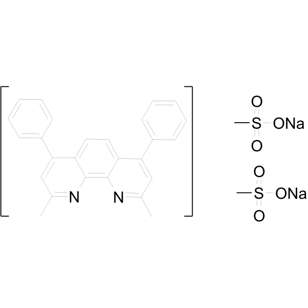
Bathocuproine disulfonate disodium (BCS) is an organic compound used as a highly sensitive colorimetric reagent for copper ions in biochemical and analytical applications. It has a bright yellow color and absorbs light at specific wavelengths, so it can be used to detect and quantify trace copper. In biochemical applications, BCS is commonly used to study the role of copper ions in various biological processes. Copper is an essential nutrient for many organisms, but it can also be toxic in high concentrations, so accurate measurement of copper levels is important to understand its impact on living systems. In terms of analysis, BCS is often used in environmental monitoring and water quality testing to detect copper pollution. It can detect copper even at very low concentrations, making it an invaluable tool for identifying potential sources of pollution and assessing the impact of industrial activities on aquatic ecosystems.
loading...
    loading...
1
1 Cited Publications
Cat. No.: HY-D1416
CAS No.: 1287651-36-8
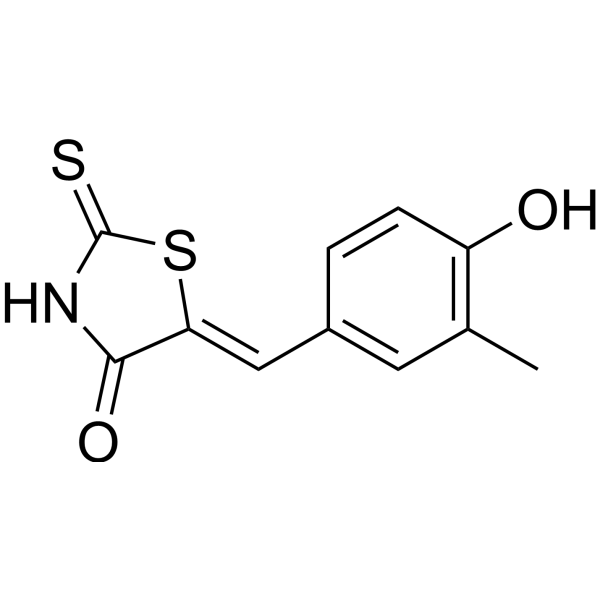
HMBR is an analogue with an additional methyl group on the aromatic ring and is non-fluorescent. HMBR conjugated with Y-FAST emits yellow fluorescence under blue light excitation (Ex= 419 nm; Em= 525–539 nm). HMBR is non-toxic to zebrafish embryos. HMBR has high cell permeability .
loading...
    loading...
Cat. No.: HY-121694
CAS No.: 80072-86-2
CENTA is a colorimetric cephalosporin substrate for β-lactamases. Upon hydrolysis by β-lacatamases, CENTA turns from light yellow to chrome yellow, which can be quantified by colorimetric detection at 405 nm as a measure of β-lactamase activity.
loading...
    loading...
Cat. No.: HY-D1343
CAS No.: 223920-67-0
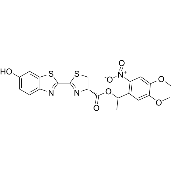
DMNPE-caged D-luciferin is a heterocyclic luminescent compound that is a natural ligand for luciferase, an enzyme used to detect cell activity. Its reaction requires ATP and emits yellow-green light with a peak wavelength of about 530 nm. The luciferin in the DMNPE cage easily crosses the cell membrane.
loading...
    loading...
Cat. No.: HY-D1189
CAS No.: 1471982-33-8
Synonyms: Lucifer yellow 3-amino-D-alanine
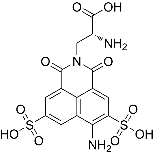
YADA (Lucifer Yellow 3-amino-D-alanine) is a conjugate of the fluorescent dyes Lucifer yellow and D-alanine, which is a green-yellow fluorescent dye. YADA is suitable for labeling peptidoglycans in living bacteria that can be incorporated into the cell wall where they are being synthesized. YADA has a large Stokes shift and a wide emission spectrum, allowing excitation through a purple light source and detection using a green filter. YADA showed good water solubility, light stability and thermal stability.
loading...
    loading...
Cat. No.: HY-W087913
CAS No.: 596-01-0
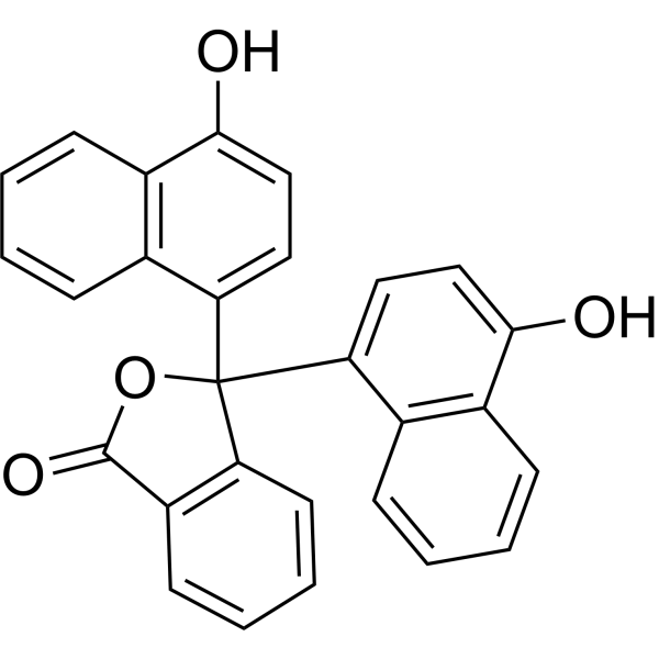
alpha-Naphtholphthalein is an indicator that is light yellow at pH 6 and gradually shows a clear color change with increasing pH to light green-green-blue. alpha-Naphtholphthalein can be impregnated into cotton-blend fabrics and used to develop medical supplies for wound pH monitoring, such as medical gauze, hospital gowns and compression bandages .
loading...
    loading...
Cat. No.: HY-D3356
CAS No.: 327174-91-4
Target:  

Fluorescent Dye

Research Areas:  

Others

VIC is a yellow-green fluorescent dye belonging to the xanthracene class of fluorescent dyes. It is mainly used in research on fluorescently labeled oligonucleotides (such as 5' end labeling), TaqMan qPCR probes, and nucleic acid detection. VIC dye can directly generate a stable fluorescent signal upon light excitation, without relying on enzyme catalysis, pH, or membrane potential activation mechanisms.
loading...
    loading...
Cat. No.: HY-K0612

MCE Weigert Elastin Staining Kit combines Weigert Resorcinol Fuchsin Staining Solution with Van Gieson (VG) Staining Solution. Weigert Resorcinol Fuchsin Solution is mainly used for staining elastic fibers, Van Gieson (VG) Solution is used for collagen fiber staining. The principle of VG staining is based on differences in the size of anionic dye molecules and the permeability of tissues. Picric acid (PA) has the smallest molecular weight and preferentially enters dense structures. Ponceau Red or acid fuchsin has a relatively larger molecular weight and primarily binds to collagen fibers, while light green has the largest molecular weight and stains other tissue components. After VG staining, collagen fibers appear red, while muscle fibers and cytoplasm appear yellow, enabling a clear distinction between tissue components.

loading...
    loading...

