
Unoprostone
Based on 1 Customer Validation
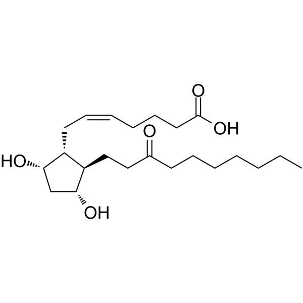
Unoprostone, a prostaglandin F2α analog, is a large conductance Ca2+-activated K+ (BK) channels and ClC-2 type chloride channels activator. Unoprostone reduces oxidative stress- and light-induced retinal cell death, and phagocytotic dysfunction. Unoprostone reduces intraocular pressure and can be used for the study of glaucoma, ocular hypertension and retinitis pigmentosa.
For research use only. We do not sell to patients.
- Purity : 99.0%
- CAS No.: 120373-36-6
- Formula: C22H38O5
- Molecular Weight:382.53
-
Storage:
Solution, -20°C, 2 years
Biological Activity
Description
In Vitro
Pretreatment with Unoprostone (0.01, 0.1, 1 μM; 1 h before H2O2 treatment) protects against H2O2-induced cell death in a concentration-dependent manner and the effect is significant at 0.1 μM and 1 μM concentrations[1].
Pretreatment with Unoprostone at concentrations of 0.1 to 3 μM protects against light-induced cell death in a concentration-dependent manner; the effect is significant at the 1 and 3 μM concentrations. Unoprostone reduces the morphological change, and it significantly inhibits the low mitochondrial membrane potential and cell death induced by light irradiation[1].
Unoprostone has prostaglandin F2α receptors (FP) binding affinity with a Ki of 3.86 μM[2].
MedChemExpress (MCE) has not independently confirmed the accuracy of these methods. They are for reference only. Further protocols information, click here.
-
Cell Line:H2O2-induced photoreceptor cell
-
Concentration:0.01, 0.1, 1 μM
-
Incubation Time:1 hour before H2O2 treatment
-
Result:Protected against H2O2-induced cell death in a concentration-dependent manner.
In Vivo
MedChemExpress (MCE) has not independently confirmed the accuracy of these methods. They are for reference only.
-
Animal Model:Male mice (6 weeks old)[3]
-
Dosage:0.06%, 0.12%
-
Administration:Topical instillation; 3 µL
-
Result:Reduced mouse IOP in a dose-dependent manner.
Clinical Trial
| NCT Number | Sponsor | Condition | Start Date |
Phase
|
|---|---|---|---|---|
| NCT01329991 | Plexxikon| | 2011-05 | PHASE1 |
Chemical Information
-
CAS No. 120373-36-6
-
Appearance Liquid
-
Molecular Weight 382.53
-
Formula C22H38O5
-
Color Colorless to light yellow
-
SMILES
OC(CCC/C=C\C[C@@H]1[C@H]([C@H](O)C[C@@H]1O)CCC(CCCCCCC)=O)=O
-
Shipping
Room temperature in continental US; may vary elsewhere.
-
Storage
Solution, -20°C, 2 years
Protocols
-
Cell Cytotoxicity Assay
Cytotoxicity assays are usually based on the assessment of cell membrane damage, which can also be indirectly detected by measuring cell viability. Detection methods include MTT assay, CKK-8 assay, LDH assay and ATP assay, etc.
-
Mammalian live/dead viability and cytotoxicity staining
Live/dead viability and cytotoxicity staining assays are based on the simultaneous detection of intracellular esterase activity in metabolically active (viable) cells and membrane integrity loss in non-viable cells. In commonly used dual-staining approaches, membrane-permeant fluorogenic substrates are converted by intracellular esterases into fluorescent products in live cells, while impermeant DNA-binding dyes selectively enter cells with compromised plasma membranes and label nucleic acids in dead or dying cells, enabling discrimination between viable and non-viable populations by fluorescence microscopy or flow cytometry.
-
ROS/oxidative-stress fluorescent staining
ROS/oxidative-stress fluorescent staining uses cell-permeant fluorogenic probes that become fluorescent after oxidation inside cells or tissues; commonly used examples include DCFH-DA/DCFDA for broad cellular oxidant detection, DHE for superoxide-related signal detection, MitoSOX for mitochondrial superoxide-related signal detection, and CellROX probes for oxidative-stress-associated fluorescence readouts. The assay detects probe oxidation rather than a single ROS species unless the probe and analysis method have been chemically validated for that species. DCFH-DA enters cells, is deacetylated by intracellular esterases to DCFH, and produces fluorescent DCF after oxidation, so the readout is used as an operational measure of total cellular oxidative stress rather than a species-specific ROS measurement. DHE and MitoSOX can report superoxide-related oxidation, but red fluorescence alone can include non-specific ethidium-like oxidation products; HPLC or optimized spectral approaches are
-
Apoptosis Solutions
Apoptosis is a regulated, generally non-lytic cell-death pathway that removes unwanted, damaged, infected, or abnormal cells through coordinated morphological changes, caspase activation, DNA fragmentation, and membrane remodeling. The intrinsic apoptosis pathway is controlled mainly by mitochondrial outer membrane permeabilization, BCL-2 family proteins, cytochrome c release, apoptosome formation, caspase-9 activation, and downstream executioner caspase-3/7 activation. The extrinsic apoptosis pathway is initiated by death receptors such as Fas, TNFR, and TRAIL receptors, which recruit adaptor proteins and activate caspase-8 before engaging executioner caspases or mitochondrial amplification through BID cleavage. Apoptosis is linked to many phenotypes, including cancer cell killing, tissue homeostasis, immune regulation, neurodegeneration, infection response, and treatment-induced cytotoxicity; unresolved questions include how apoptosis interacts with necroptosis, pyroptosis, ferroptos
-
Research Protocol for Cardiovascular Diseases
Cardiovascular disease can be modeled as maladaptive cardiac remodeling, where ischemic injury or pressure overload activates inflammatory signaling, fibroblast activation, extracellular-matrix deposition, cardiomyocyte hypertrophy, vascular remodeling, and progressive ventricular dysfunction. The TGF-β/SMAD axis is a central profibrotic pathway after myocardial injury and pressure overload, while innate immune and cytokine pathways regulate leukocyte recruitment, scar formation, and adverse remodeling. Key unresolved questions include which inflammatory signals are reparative versus harmful, when fibrosis is protective versus maladaptive, and whether pathway inhibition improves function without weakening necessary infarct healing or compensatory remodeling.
-
Ca2+ Staining Technique
Ca2+ staining is an experimental technique that utilizes specific fluorescent probes (such as Fluo-4 AM, Fura-2, etc.) to qualitatively or quantitatively detect dynamic changes in intracellular Ca2+ concentrations; this is achieved by monitoring the changes in fluorescent signals generated when these probes bind to free intracellular calcium ions. The underlying principle relies primarily on the presence of chelating groups within the probe's molecular structure that possess high affinity for calcium ions.
Purity & Documentation
-
Data Sheet (270 KB)
-
SDS (393 KB)
- English - EN (393 KB)
- Français - FR (393 KB)
- Deutsch - DE (393 KB)
- Norwegian - NO (393 KB)
- Español - ES (393 KB)
- Swedish - SV (393 KB)
- Italian - IT (393 KB)
- Korean - KR (393 KB)
- Portuguese - PT (393 KB)
-
Handling Instructions (2659 KB)
References
[1]. Tsuruma K, et al. Unoprostone reduces oxidative stress- and light-induced retinal cell death, and phagocytoticdysfunction, by activating BK channels. Mol Vis. 2011;17:3556-65. Epub 2011 Dec 30. [Content Brief]
[2]. Kelly CR, et al. Real-time intracellular Ca2+ mobilization by travoprost acid, bimatoprost, unoprostone, and otheranalogs via endogenous mouse, rat, and cloned human FP prostaglandin receptors. J Pharmacol Exp Ther. 2003 Jan;304(1):238-45. [Content Brief]
[3]. Ota T, et al. The effects of prostaglandin analogues on IOP in prostanoid FP-receptor-deficient mice. Invest Ophthalmol Vis Sci. 2005 Nov;46(11):4159-63. [Content Brief]
Calculators
Concentration (start) × Volume (start) = Concentration (final) × Volume (final)

