8 Results for "

real-time visualization

" in MedChemExpress (MCE) Product Catalog:
Products (8)

8 Results for "real-time visualization" in MCE Product Catalog:

1
1 Cited Publications
Cat. No.: HY-157765
CAS No.: 3065420-66-5
Purity:  97.55%
Research Areas:  

Cancer

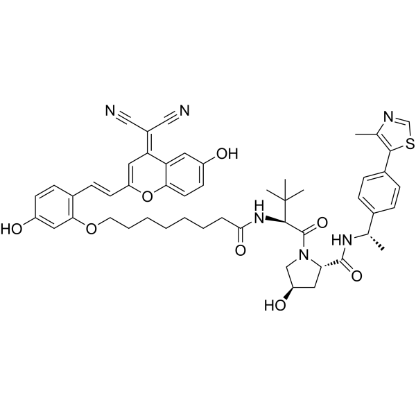
PROTAC ERα Degrader-6 (compound A2) is a potent PROTAC degrader of ERα, with IC50 of 0.11 μM in MCE-7 cells. PROTAC ERα Degrader-6 has anti-tumor effect. PROTAC ERα Degrader-6 is a fluorescent probes with Em of 582 nm that enable real-time visualization of ERα protein degradation .
loading...
    loading...
Cat. No.: HY-D2381
AF 488 maleimide is a thiol-reactive dye used to label SH groups of proteins, which can attach the AF 488 fluorophore to cysteine residue-containing proteins and peptides as well as other thiolated molecules. AF 488 maleimide enables real-time visualization of dynamic pilus extension and retraction in live bacterial cells via epifluorescence microscopy (Ex/Em = 470/520 nm) .
loading...
    loading...
Cat. No.: HY-W115721
CAS No.: 523-21-7
Synonyms: Sodium rhodizonate dibasic
Rhodizonic acid disodium (Sodium rhodizonate dibasic) is a transition metal-dependent pro-oxidant and lead detection agent that induces reactive oxygen species generation, DNA damage, and inhibits Aconitase activity. Rhodizonic acid disodium generates superoxide anion radicals in an iron (II)-dependent manner, leading to aconitase inactivation. Rhodizonic acid disodium also triggers hydroxyl radical-mediated DNA strand breaks and 8-OHdG formation via copper ion reduction. Rhodizonic acid disodium reacts with lead to form a scarlet precipitate, with the color intensity proportional to lead content, enabling qualitative or quantitative analysis of lead. Rhodizonic acid disodium can also be used for real-time visualization of the dynamic process of lead sequestration in the plant rhizosphere and evaluation of the effects of environmental factors such as soil type on the stability of lead-sequestering structures .
loading...
    loading...
Cat. No.: HY-D1556
CAS No.: 384832-91-1
Synonyms: 1,2-dioleoyl-sn-glycero-3-phosphoethanolamine-N-(carboxyfluorescein) ammonium
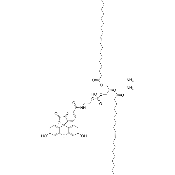
18:1 PE CF (1,2-dioleoyl-sn-glycero-3-phosphoethanolamine-N-(carboxyfluorescein) (ammonium)) is a pH-sensitive fluorescent indicator. 18:1 PE CF exhibits changes in fluorescence intensity in response to pH variations and reflects the electrostatic states of cationic liposomes and lipoplexes. 18:1 PE CF supports real-time visualization of pH distribution and membrane deformation in giant unilamellar vesicles and is suitable for steady-state fluorescence testing in large unilamellar vesicle membranes. 18:1 PE CF is capable of monitoring the surface electrical potential of cationic liposomes and lipoplexes and can effectively label lipid bilayers and nanomicelles. 18:1 PE CF can be used for the research of the investigation of lung cancer (Ex = 495 nm; Em = 520 nm) .
loading...
    loading...
Cat. No.: HY-D3198
CAS No.: 2351937-34-1
NIR-fluorescent ceramide is a neutral near-infrared (NIF) fluorescent probe (λem=650 nm) based on the C-bridged nitrobenzoxadiazole SCOTfluor core, which enables visualization of lipid metabolic processes such as sphingolipid trafficking. The emission signal of NIR-fluorescent ceramide increases significantly in liposomal environments. NIR-fluorescent ceramide acts as an intracellular trafficking tracer, localizing to regions surrounding the endoplasmic reticulum and Golgi apparatus within a short period, and then translocating to recycling lysosomes over a longer period, thus enabling real-time visualization of sphingolipid trafficking and biological lipid functions in living cells. NIR-fluorescent ceramide also generates metabolic uptake profiles and provides multiple metabolic readouts in human cancer cell lines .
loading...
    loading...
Cat. No.: HY-D3195
CAS No.: 2028316-19-8
CDr15 is a deep-red fluorescent probe (Ex=733 nm) that can selectively intercalate into and label bacterial extracellular DNA (eDNA). CDr15 exhibits extremely high specificity for biofilm imaging and cannot effectively bind to mammalian nuclear DNA. CDr15 enables real-time visualization of the microcolony structure and developmental process of three-dimensional *Pseudomonas aeruginosa* biofilms, and accurately localizes biofilm-forming regions of microorganisms in a mouse corneal infection model. With low background interference signals, CDr15 serves as an ideal diagnostic tool for research fields including bacterial biofilms and corneal infections .
loading...
    loading...
Cat. No.: HY-D3311
M1219 is a GSH/ATP dual near-infrared activated fluorescent probe that enables independent real-time monitoring of dynamic changes in intracellular GSH and ATP without spectral crosstalk (GSH: Ex=640 nm, Em=740~800 nm; ATP: Ex=594 nm/610 nm, Em=650~700 nm). M1219 not only visualizes the metabolic regulatory mechanism of TNBC under single/dual-target inhibition of SLC7A11/GLUT1 and accurately evaluates its in vivo efficacy, but also achieves precise localization of the TNBC tumor invasion boundary. M1219 can be used for the research of triple-negative breast cancer .
loading...
    loading...
Cat. No.: HY-D3186
CAS No.: 2163840-29-5
HMRef-βGal is a fluorescent probe and a substrate responsive to β-galactosidase (β-galactosidase) (Ex/Em=498 nm/505-600 nm). After being cleaved by β-galactosidase, HMRef-βGal triggers significant fluorescence enhancement via intramolecular spirocyclic function regulation. HMRef-βGal generates bright fluorescence in cancer cells with elevated β-galactosidase activity, enabling visualization of tiny peritoneal metastases in mouse models. HMRef-βGal exhibits low in vitro cytotoxicity and low acute in vivo toxicity in mice. HMRef-βGal can be used for preclinical fluorescence-guided diagnosis and cytoreductive surgery of peritoneal metastases, including compatibility with real-time naked-eye detection and endoscopic imaging, as well as for studies related to peritoneal metastases of ovarian cancer .
loading...
    loading...

