



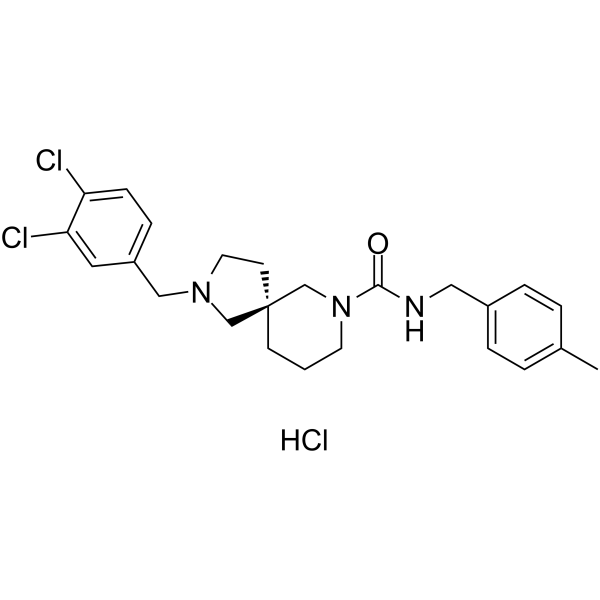
GSK2850163 hydrochloride
Based on 9 publication(s) in Google Scholar
GSK2850163 hydrochloride is a novel inhibitor of inositol-requiring enzyme-1 alpha (IRE1α) which can inhibit IRE1α kinase activity and RNase activity with IC50s of 20 and 200 nM, respectively.
For research use only. We do not sell to patients.
- CAS No.: 2319838-09-8
- Formula: C24H30Cl3N3O
- Molecular Weight:482.87
-
Storage:
Please store the product under the recommended conditions in the Certificate of Analysis.

To place orders, for customer services and technical support, please contact: MedChemExpress USA
Tel: 609-228-6898 E-mail: [email protected] [email protected]


-
 Biological Activity
-
Chemical Information
- Protocols
- Purity & Documentation
- References
-
 Help & FAQs


-

HY-19707
4μ8CIRE1 Inhibitor -
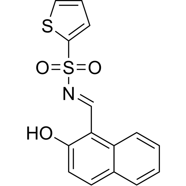
HY-15845
STF-083010IRE1 Inhibitor -
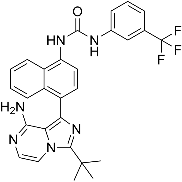
HY-19708
KIRA6IRE1α RNase Inhibitor -
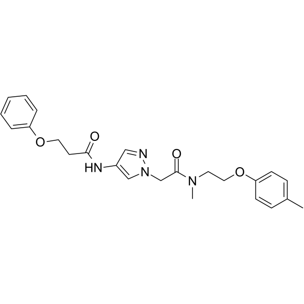
HY-139214
IXA4IRE1/XBP1s Activator -
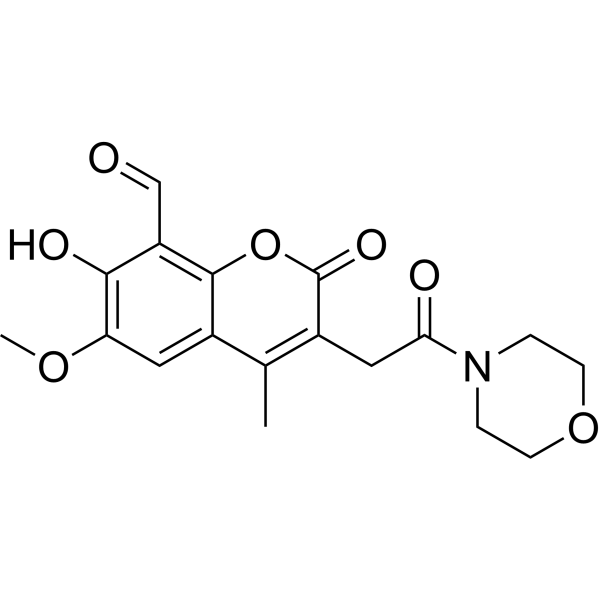
HY-104040
MKC8866IRE1α RNase Inhibitor -
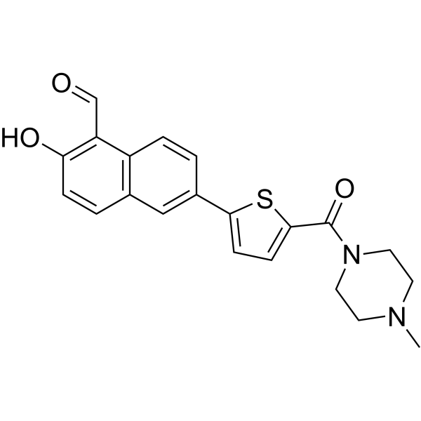
HY-19710
MKC3946IRE1 Inhibitor -
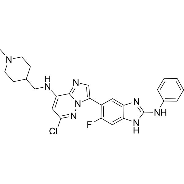
HY-136735
IRE1α kinase-IN-1IRE1α Inhibitor -
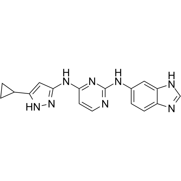
HY-17537
APY29IRE1 Inhibitor -
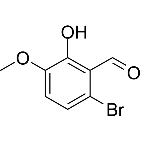
HY-107371
6-Bromo-2-hydroxy-3-methoxybenzaldehydeIRE1α Inhibitor -
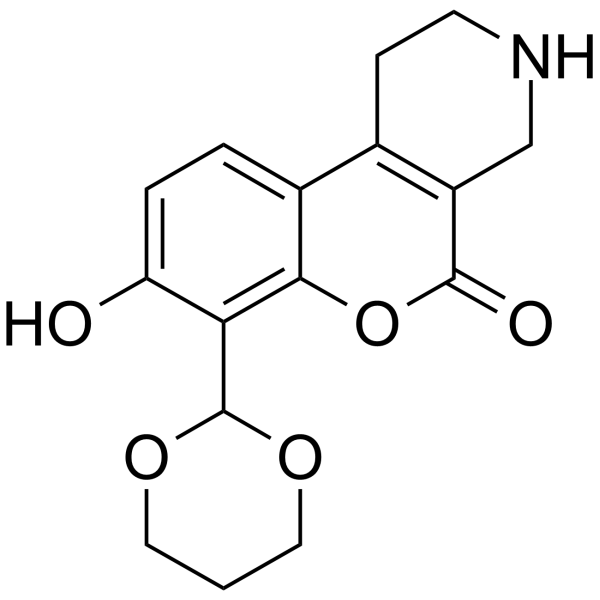
HY-107400
B I09IRE-1 Inhibitor -
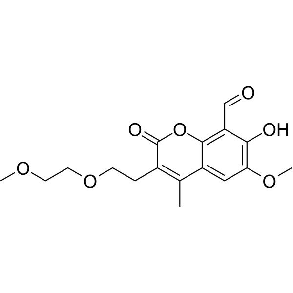
HY-12399
MKC9989HAA Inhibitor -
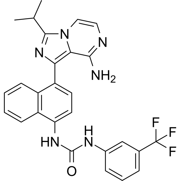
HY-18509
IRE1α kinase-IN-2IRE1α Knase Inhibitor
Publications Citing Use of MedChemExpress (MCE) GSK2850163 hydrochloride
More- Cell. 2026 Sep 14:S0092-8674(26)01003-2.
- Sci Signal. 2021 Jan 26;14(667):eabb3616. [Abstract]
- Mucosal Immunol. 2024 Apr;17(2):288-302. [Abstract]
- Microb Pathog. 2026 Jan 21:108313. [Abstract]
- Microbiologyopen. 2026 Feb;15(1):e70219. [Abstract]
- Cell Biochem Biophys. 2026 Sep 18.
- Vet Microbiol. 2026 May:316:110968. [Abstract]
- Vet Microbiol. 2020 May:244:108648. [Abstract]
- Res Sq. 2025 May 22.
 Customer Validation & Images

-
WB
-
ELISA
-
RT-PCR


Biological Activity
Description
IC50 & Target
IC50: 20 nM (IRE1α kinase activity), 200 nM (IRE1α RNase activity)[1]
In Vitro
GSK2850163 hydrochloride is a novel inhibitor of inositol-requiring enzyme-1 alpha (IRE1α) which can inhibit IRE1α kinase activity and RNase activity with IC50s of 20 and 200 nM, respectively. The increased autophosphorylation of IRE1α can be reduced in a dose-dependent manner by GSK2850163 hydrochloride. Increasing concentrations of GSK2850163 hydrochloride are capable of reducing the increased XBP 1 transcriptional activity. Two additional kinases are weakly inhibited by GSK2850163 hydrochloride: Ron (IC50=4.4 μM) and FGFR1 V561M (IC50=17 μM)[1].
MedChemExpress (MCE) has not independently confirmed the accuracy of these methods. They are for reference only. Further protocols information, click here.


Chemical Information
-
CAS No. 2319838-09-8
-
Molecular Weight 482.87
-
Formula C24H30Cl3N3O
-
SMILES
O=C(N(CCC1)C[C@]21CCN(CC3=CC=C(Cl)C(Cl)=C3)C2)NCC4=CC=C(C)C=C4.Cl
-
Synonyms
GSK163 hydrochloride
-
Shipping
Room temperature in continental US; may vary elsewhere.
-
Storage
Please store the product under the recommended conditions in the Certificate of Analysis.


Publications (9)
-
Journal Impact Factor
-
Most Recent
-
-
Sci Signal
PERK inhibition attenuates vascular remodeling in pulmonary arterial hypertension caused by BMPR2 mutation. [Abstract]2021 Jan 26;14(667):eabb3616. PMID: 33500333 -
Mucosal Immunol
Deficiency of immunoglobulin IgSF6 enhances antibacterial effects by promoting ER stress and the inflammatory response in intestinal macrophages. [Abstract]2024 Apr;17(2):288-302. PMID: 38387824 -
Microb Pathog
BvrR of Brucella initiates microglial inflammation through the activation of the IRE1 pathway. [Abstract]2026 Jan 21:108313. PMID: 41577152 -
Microbiologyopen
BvrR From Brucella abortus Induces Neuroinflammation Through IRE1-Mediated Activation of ATF2 and NF-κB. [Abstract]2026 Feb;15(1):e70219. PMID: 41566528
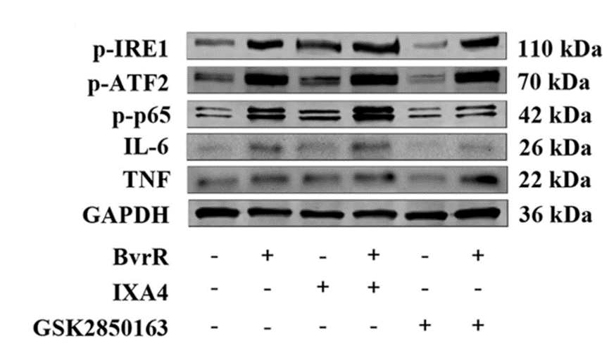
GSK2850163 hydrochloride purchased from MedChemExpress. Usage Cited in: Microbiologyopen. 2026 Feb;15(1):e70219. [Abstract]
Western blot analysis of p‐IRE1, p‐ATF2, p‐p65, IL‐6, and TNF‐α protein levels treated with GSK2850163 (8 μM).
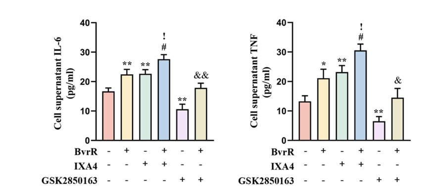
GSK2850163 hydrochloride purchased from MedChemExpress. Usage Cited in: Microbiologyopen. 2026 Feb;15(1):e70219. [Abstract]
ELISA measurement of IL‐6 and TNF‐α in supernatants treated with GSK2850163 (8 μM).
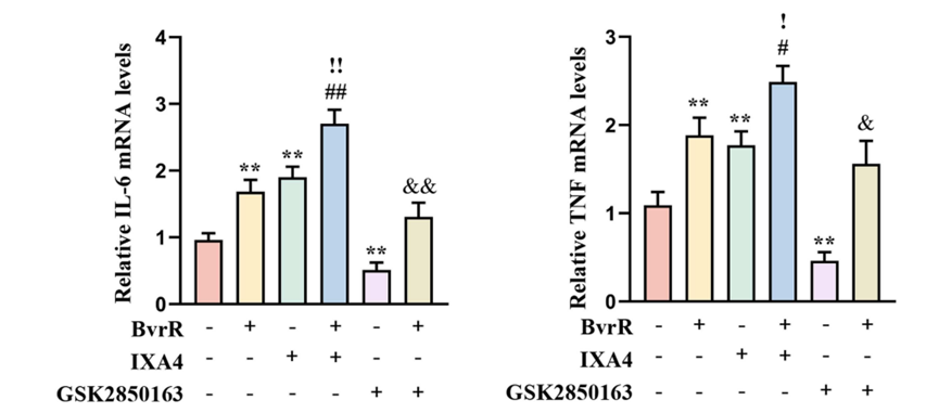
GSK2850163 hydrochloride purchased from MedChemExpress. Usage Cited in: Microbiologyopen. 2026 Feb;15(1):e70219. [Abstract]
RT‐qPCR analysis of IL6 and TNF‐α mRNA expression treated with GSK2850163 (8 μM).
-
-
Vet Microbiol
The PERK-eIF2α branch activates the NLRP3 inflammasome through the NF-κB signaling pathway to suppress NDV replication. [Abstract]2026 May:316:110968. PMID: 41793984 -
Vet Microbiol
A rough Brucella mutant induced macrophage death depends on secretion activity of T4SS, but not on cellular Txnip- and Caspase-2-mediated signaling pathway. [Abstract]2020 May:244:108648. PMID: 32402333 -

Protocols
-
Kinase activity and phosphorylation assays
Kinase activity assays measure the ability of kinases to transfer phosphate groups from ATP to specific substrates, while phosphorylation assays detect the presence and levels of phosphorylated proteins. Common methods include radiolabeled ATP incorporation (e. g. ,), ADP release detection via bioluminescence (e. g. ,[3]), enzyme-linked immunosorbent assays (ELISA) for phospho-specific epitopes (e. g. ,[6]), and microtiter-based formats for high-throughput screening (e. g. ,[8]). The ADP-Glo assay quantifies kinase activity by measuring ADP produced during phosphorylation using a luciferase-based system. Radiometric assays involve autoradiography or scintillation counting after incorporation of 32P-labeled ATP into substrate proteins. ELISA-based approaches rely on phospho-specific antibodies to detect activated kinases in cell lysates or purified samples.
-
Protocol for Kinase activity and phosphorylation assays
Kinase activity assays measure transfer of phosphate from ATP to a protein or peptide substrate, generating phosphorylated substrate, ADP, or incorporated radiolabeled phosphate as the readout; phosphorylation assays measure site-specific phosphorylation in cells or tissues as a proxy for kinase-pathway activation, inhibition, or substrate regulation. Phosphorylation can be detected by phospho-specific Western blot, immunoprecipitation kinase assay, phospho-immunofluorescence, phospho-flow cytometry, luminescent ADP detection, radiolabeled ATP incorporation, or reporter-based pathway assays, and these readouts can be applied to cancer cells, primary neurons, mouse tumors, organoids, inflammatory macrophages, ferroptosis studies, and mitophagy studies when the kinase target is biologically relevant.


Purity & Documentation
References

Calculators
Concentration (start) × Volume (start) = Concentration (final) × Volume (final)


GSK2850163 hydrochloride Related Classifications
HY-U00459B Related Classifications
 Powered by Bioz