



c-Met-IN-22
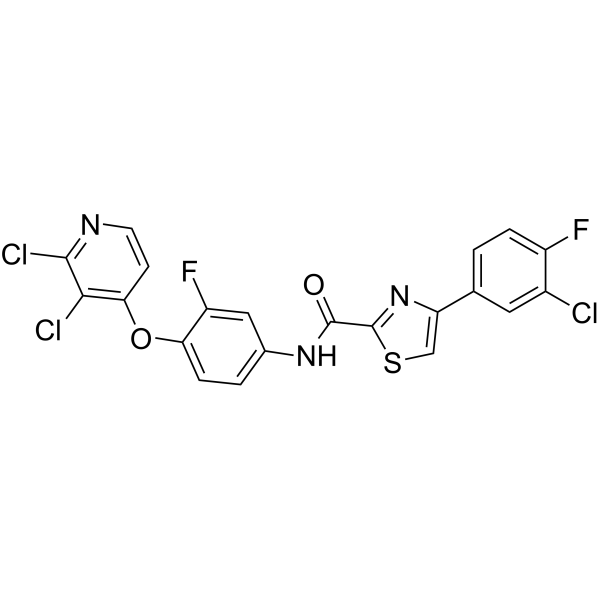
c-Met-IN-22 (compound 51am) is an orally active inhibitor against c-Met with an IC50 value of 2.54 nM. c-Met-IN-22 has antiproliferative and antitumor activities. c-Met-IN-22 induces cell apoptosis.
For research use only. We do not sell to patients.
- Formula: C21H10Cl3F2N3O2S
- Molecular Weight:512.74
-
Storage:
Please store the product under the recommended conditions in the Certificate of Analysis.

To place orders, for customer services and technical support, please contact: MedChemExpress USA
Tel: 609-228-6898 E-mail: [email protected] [email protected]


-
 Biological Activity
-
Chemical Information
- Protocols
- Purity & Documentation
- References
-
 Help & FAQs




Biological Activity
Description
IC50 & Target
[1]|
c-Metwild type 2.54 nM (IC50) |
c-MetH1094R 93.6 nM (IC50) |
c-MetD1228H 29.4 nM (IC50) |
c-MetY1230H 45.8 nM (IC50) |
c-MetY1235D 54.2 nM (IC50) |
c-MetM1250T 26.5 nM (IC50) |
c-Metkit 4.94 nM (IC50) |
c-MetRon 3.83 nM (IC50) |
c-MetPDGFRα 425 nM (IC50) |
c-MetPDGFRβ 513 nM (IC50) |
c-MetVEGFR-2 527 nM (IC50) |
c-MetFit-3 6.12 nM (IC50) |
c-MetFit-4 276 nM (IC50) |
In Vitro
c-Met-IN-22 shows antiproliferative activity against MKN-45, A-549, HT-29, MDA-MB-231, HUVEC and FHC with IC50s of 0.092, 0.83, 0.68, 3.94,2.54 and 8.63 μM, respectively[1].
c-Met-IN-22 shows anti-drug resistence against tH1094R, D1228H, Y1230H, Y1235D, and M1250T, with IC50 values of 93.6, 29.4, 45.8, 54.2 and 26.5 nM,respectively[1].
c-Met-IN-22 (0, 2.5, 5.0, and 10.0 μM, 24 h) inhibits c-Met phosphorylation in MKN-45 in a dose-dependent manner[1] .
c-Met-IN-22 (0.4, 0.8, and 1.2 μM, 24 h) induces cell cycle arrest at G2 phase and apotosis of MNK-45 in a dose-dependent manner[1].
MedChemExpress (MCE) has not independently confirmed the accuracy of these methods. They are for reference only.
-
Cell Line:MNK- 45 cells
-
Concentration:0.4, 0.8, 1.2 μM
-
Incubation Time:24 h
-
Result:Induced cell apoptosis in a dose-dependent manner.
-
Cell Line:MNK- 45 cells
-
Concentration:0.4, 0.8, 1.2 μM
-
Incubation Time:24 h
-
Result:Induced cell cycle arrest at G2 phase in a dose-dependent manner.
-
Cell Line:MNK- 45 cells
-
Concentration:0, 2.5, 5, 10 μM
-
Incubation Time:24 h
-
Result:Inhibited c-Met phosphorylation in a dose-dependent manner.
In Vivo
c-Met-IN-22 (1.5mg/kg;i.v.) exhibits an elimination half-time of 3.2h[1].
Pharmacokinetic Analysis of c-Met-IN-22 in BALB/c mice[1]
| Route | Dose (mg/kg) | AUC0→∞ (μg·h/mL) | T1/2 (h) | Tmax (h) | Cmax (ng/mL) | Cl (L/h·kg) | F (%) |
| i.v. | 1.5mg/kg | 2.5 | 3.2 | / | 552 | 0.6 | / |
| p.o. | 10mg/kg | 11.5 | 5.6 | 4.1 | 1756 | / | 69 |
MedChemExpress (MCE) has not independently confirmed the accuracy of these methods. They are for reference only.
-
Animal Model:Pharmacokinetic Analysis in BALB/c mice
-
Dosage:p.o.10 mg/kg; i.v. 1.5 mg/kg
-
Administration:Intravenous (i.v.) injection, Oral administration (p.o.)
-
Result:Characterised good maximum concentration, plasma exposure and elimination half-time in pharmacokinetics.


Chemical Information
-
Molecular Weight 512.74
-
Formula C21H10Cl3F2N3O2S
-
SMILES
O=C(C1=NC(C2=CC=C(F)C(Cl)=C2)=CS1)NC3=CC=C(OC4=C(Cl)C(Cl)=NC=C4)C(F)=C3
-
Shipping
Room temperature in continental US; may vary elsewhere.
-
Storage
Please store the product under the recommended conditions in the Certificate of Analysis.


Purity & Documentation
References

Calculators
Concentration (start) × Volume (start) = Concentration (final) × Volume (final)


c-Met-IN-22 Related Classifications
HY-157387 Related Classifications
 Powered by Bioz