



Camostat
Based on 49 publication(s) in Google Scholar
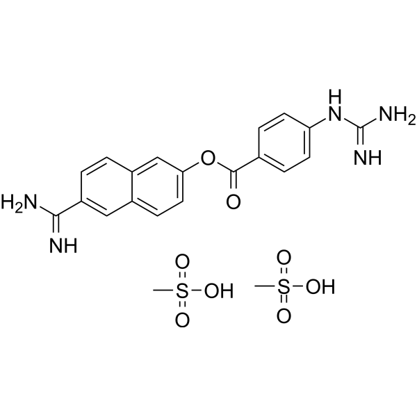
Camostat is an orally active trypsin inhibitor. Camostat can reduce pancreatic fibrosis induced by repeated administration of superoxide dismutase inhibitors in rats, and decrease the proliferation and activation of pancreatic stellate cells (PSCs).
Nos produits utilisent uniquement pour la recherche. Nous ne vendons pas aux patients.
- Pureté : 99.42%
- CAS No.: 59721-28-7
- Formule: C20H22N4O5
- Masse moléculaire:398.41
-
Stockage:Powder -20°C, 3 years ; In solvent -80°C, 6 months , -20°C, 1 month


To place orders, for customer services and technical support, please contact: MedChemExpress USA
Tel: 609-228-6898 E-mail: [email protected] [email protected]


-
 Activité biologique
-
Chemical Information
-
Solvant et solubilité
- Protocole
- Pureté et documentation
- Références
-
 Help & FAQs


Publications Citing Use of MedChemExpress (MCE) Camostat
More- Signal Transduct Target Ther. 2022 Mar 11;7(1):83. [Abstract]
- Nature. 2022 Mar;603(7902):693-699. [Abstract]
- Cell. 2023 Feb 16;186(4):850-863.e16. [Abstract]
- Nat Microbiol. 2025 Nov;10(11):2860-2874. [Abstract]
- Nat Microbiol. 2024 Sep;9(9):2383-2394. [Abstract]
- Nat Commun. 2025 Dec 11;16(1):11002. [Abstract]
- Nat Commun. 2025 Jul 7;16(1):6241. [Abstract]
- Nat Commun. 2024 Oct 9;15(1):8728. [Abstract]
- Nat Commun. 2024 Jul 3;15(1):5606. [Abstract]
- Nat Commun. 2022 Jul 26;13(1):4331. [Abstract]
- Nat Commun. 2022 Mar 17;13(1):1444. [Abstract]
- Cell Discov. 2021 Dec 14;7(1):119. [Abstract]
- Nat Chem Biol. 2022 Sep;18(9):963-971. [Abstract]
- Nucleic Acids Res. 2021 Jan 8;49(D1):D1113-D1121. [Abstract]
- Cell Rep Med. 2022 Sep 20;3(9):100743. [Abstract]
- Sci Adv. 2023 Jan 20;9(3):eadd3867. [Abstract]
- J Control Release. 2026 Jul 8:397:115167.
- EBioMedicine. 2023 Sep:95:104753. [Abstract]
- Sci China Life Sci. 2025 Oct 24. [Abstract]
- Proc Natl Acad Sci U S A. 2022 Jan 25;119(4):e2117576119. [Abstract]
- Cell Prolif. 2025 Dec;58(12):e70060. [Abstract]
- Sci Signal. 2024 Aug 20;17(850):eadn3785. [Abstract]
- Emerg Microbes Infect. 2022 Dec;11(1):2275-2287. [Abstract]
- Virulence. 2026 Dec;17(1):2691344. [Abstract]
- Eur J Med Chem. 2021 Nov 15:224:113683. [Abstract]
- Biochem Pharmacol. 2023 Jul:213:115617. [Abstract]
- Eur J Pharmacol. 2023 Jan 5:938:175394. [Abstract]
- Eur J Pharmacol. 2022 Mar 15:919:174795. [Abstract]
- mBio. 2022 Dec 20;13(6):e0256622. [Abstract]
- PLoS Pathog. 2025 Oct 14;21(10):e1013593. [Abstract]
- Antiviral Res. 2023 Jun:214:105606. [Abstract]
- J Virol. 2026 Aug 18;100(8):e0061426.
- J Virol. 2026 Apr 21;100(4):e0027026. [Abstract]
- J Virol. 2025 Nov 18:e0127425. [Abstract]
- J Virol. 2025 Aug 11:e0105525. [Abstract]
- J Virol. 2023 May 31;97(5):e0032423. [Abstract]
- Viruses. 2022 Feb 8;14(2):353. [Abstract]
- Am J Physiol Renal Physiol. 2024 Aug 1;327(2):F265-F276. [Abstract]
- Biosaf Health. 2022 Feb;4(1):38-44. [Abstract]
- bioRxiv. 2026 May 7.
- Vita. 2026 Apr 7.
- Res Sq. 2024 Nov 21:rs.3.rs-5454588. [Abstract]
- SSRN. 2024 Jul 11.
- SSRN. 2024 Jan 26.
- Res Sq. 2023 Sep 11:rs.3.rs-3220157. [Abstract]
- Utrecht University. 2022 Jul.
- Research Square Preprint. 2022 Jan.
- Universidade de São Paulo. 2020 Sep.
- SSRN. 2020 Jun.
 Customer Validation & Images

-
Bio/Physico-chemical Assay
-
Bio/Physico-chemical Assay
-
WB
-
Bio/Physico-chemical Assay
-
RT-PCR


Activité biologique
Description
Cellular Effect
|
Cell Line

|
Type | Value | Description | References |
|---|---|---|---|---|
| Calu-3 | CC50 |
>500 mM
Compound: Camostat
|
Cytotoxicity against human Calu-3 cells assessed as reduction in cell viability
Cytotoxicity against human Calu-3 cells assessed as reduction in cell viability
|
[PMID: 37284689] |
| Calu-3 | CC50 |
200 μM
Compound: Camostat
|
Cytotoxicity against human Calu-3 cells infected with SARS-COV-2 by celltiter-glo luminescent cell viability assay
Cytotoxicity against human Calu-3 cells infected with SARS-COV-2 by celltiter-glo luminescent cell viability assay
|
[PMID: 34273661] |
| Calu-3 | CC50 |
230 nM
Compound: Camostat
|
Cytotoxicity against human Calu-3 cells
Cytotoxicity against human Calu-3 cells
|
[PMID: 39004019] |
| HEK293 | IC50 |
12.7 μM
Compound: camostat
|
Inhibition of human MATE2K-mediated ASP+ uptake expressed in HEK293 cells after 1.5 mins by fluorescence assay
Inhibition of human MATE2K-mediated ASP+ uptake expressed in HEK293 cells after 1.5 mins by fluorescence assay
|
[PMID: 23241029] |
| HEK293 | IC50 |
16.5 μM
Compound: camostat
|
Inhibition of human OCT2-mediated ASP+ uptake expressed in HEK293 cells after 3 mins by fluorescence assay
Inhibition of human OCT2-mediated ASP+ uptake expressed in HEK293 cells after 3 mins by fluorescence assay
|
[PMID: 23241029] |
| HEK293 | IC50 |
2.9 μM
Compound: camostat
|
Inhibition of human MATE1-mediated ASP+ uptake expressed in HEK293 cells after 1.5 mins by fluorescence assay
Inhibition of human MATE1-mediated ASP+ uptake expressed in HEK293 cells after 1.5 mins by fluorescence assay
|
[PMID: 23241029] |
| Sf9 | IC50 |
32 μM
Compound: 7
|
Inhibition of recombinant N-terminal His-tagged HGFA (unknown origin) expressed in baculovirus-infected Sf9 cells incubated for 30 mins prior to cromogenic substrate addition by spectrophotometry
Inhibition of recombinant N-terminal His-tagged HGFA (unknown origin) expressed in baculovirus-infected Sf9 cells incubated for 30 mins prior to cromogenic substrate addition by spectrophotometry
|
[PMID: 25882520] |
| Vero | CC50 |
>50 μM
Compound: Camostat
|
Cell viability measured by CellTiter-Glo assay in Vero cells at MOI 0.05 after 72hr
Cell viability measured by CellTiter-Glo assay in Vero cells at MOI 0.05 after 72hr
|
10.1101/2020.03.20.999730 |
| Vero | IC50 |
>50 μM
Compound: Camostat
|
Antiviral activity against SARS-CoV-2 (viral titer) measured by plaque assay in Vero cells at MOI 0.0125 after 24 hr
Antiviral activity against SARS-CoV-2 (viral titer) measured by plaque assay in Vero cells at MOI 0.0125 after 24 hr
|
10.1101/2020.03.20.999730 |


Chemical Information
-
CAS No. 59721-28-7
-
Appearance Solid
-
Masse moléculaire 398.41
-
Formule C20H22N4O5
-
Color White to off-white
-
SMILES
O=C(C1=CC=C(NC(N)=N)C=C1)OC2=CC=C(CC(OCC(N(C)C)=O)=O)C=C2
-
Livraison
Room temperature in continental US; may vary elsewhere.
-
Stockage
Powder -20°C 3 years In solvent -80°C 6 months -20°C 1 month


Publications (49)
-
Journal Impact Factor
-
Most Recent
-
Signal Transduct Target Ther
2022 Mar 11;7(1):83. PMID: 35277473
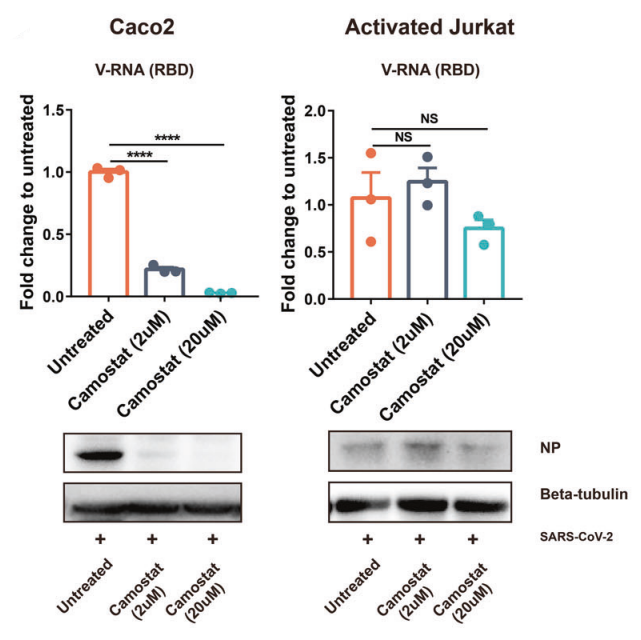
Camostat purchased from MedChemExpress. Usage Cited in: Signal Transduct Target Ther. 2022 Mar 11;7(1):83. [Abstract]
Camostat mesylate (2-20 μM; 1 h) inhibited SARS-CoV-2 infection of Caco2 cells in a dose-dependent manner.
-
Nature
2022 Mar;603(7902):693-699. PMID: 35062016
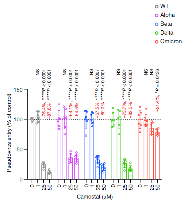
Camostat purchased from MedChemExpress. Usage Cited in: Nature. 2022 Mar;603(7902):693-699. [Abstract]
VeroE6-TMPRSS2 cells were pretreated with Camostat mesylate (1–50 μM; 2 h) followed by transduction with pseudoviruses carrying the spike protein of SARS-CoV-2 WT, Alpha, Beta, Delta, or Omicron. Pseudovirus entry was quantified by measuring the luciferase signal of the cell lysates at 24 h.p.i.
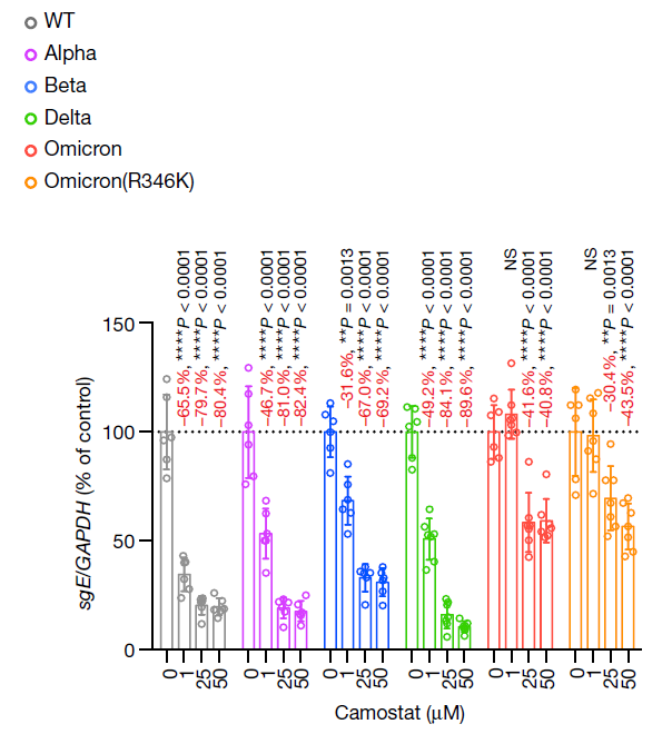
Camostat purchased from MedChemExpress. Usage Cited in: Nature. 2022 Mar;603(7902):693-699. [Abstract]
VeroE6-TMPRSS2 cells were pretreated with Camostat mesylate (1-50 μM; 2 h) and challenged with the indicated authentic SARS-CoV-2 variants at 0.1 m.o.i.. The amount of viral subgenomic envelope RNA in the collected cell lysates at 24 h.p.i. was determined using RT–qPCR.
-
Cell
A bat MERS-like coronavirus circulates in pangolins and utilizes human DPP4 and host proteases for cell entry. [Abstract]2023 Feb 16;186(4):850-863.e16. PMID: 36803605 -
Nat Microbiol
2025 Nov;10(11):2860-2874. PMID: 41168429
Camostat purchased from MedChemExpress. Usage Cited in: Nat Microbiol. 2025 Nov;10(11):2860-2874. [Abstract]
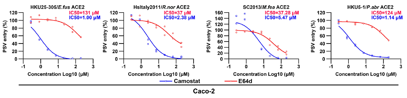
TMPRSS2 inhibitor Camostat mesylate (66.7 μM; 1 h; 37 ℃) dose-dependently inhibited pseudovirus entry of HKU25 clade or HKU5-1 in Caco-2 cells overexpressing the indicated ACE2s.
-
Nat Microbiol
Evolution of SARS-CoV-2 in the murine central nervous system drives viral diversification. [Abstract]2024 Sep;9(9):2383-2394. PMID: 39179693
Camostat purchased from MedChemExpress. Usage Cited in: Nat Microbiol. 2024 Sep;9(9):2383-2394. [Abstract]
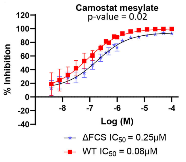
TMPRSS2 inhibitor Camostat mesylate (0.01-100 μM) inhibited the WT pseudovirus approximately 3-fold better than the ΔFCS pseudovirus in VeroE6 cells overexpressing hACE2 and TMPRSS2 (VAT cells).
-
Nat Commun
Pathogenicity, virological features, and immune evasion of SARS-CoV-2 JN.1-derived variants including JN.1.7, KP.2, KP.3, and KP.3.1.1. [Abstract]2025 Dec 11;16(1):11002. PMID: 41381428 -
Nat Commun
Vesicle-associated membrane protein 5 is an intrinsic defense factor for embryonic stem cells against coronaviruses. [Abstract]2025 Jul 7;16(1):6241. PMID: 40624080 -
Nat Commun
Lineage-specific pathogenicity, immune evasion, and virological features of SARS-CoV-2 BA.2.86/JN.1 and EG.5.1/HK.3. [Abstract]2024 Oct 9;15(1):8728. PMID: 39379369 -
Nat Commun
Recreating the biological steps of viral infection on a cell-free bioelectronic platform to profile viral variants of concern. [Abstract]2024 Jul 3;15(1):5606. PMID: 38961055 -
Nat Commun
Paeniclostridium sordellii hemorrhagic toxin targets TMPRSS2 to induce colonic epithelial lesions. [Abstract]2022 Jul 26;13(1):4331. PMID: 35882856 -
Nat Commun
2022 Mar 17;13(1):1444. PMID: 35301316 -
Cell Discov
SARS-CoV-2 uses metabotropic glutamate receptor subtype 2 as an internalization factor to infect cells. [Abstract]2021 Dec 14;7(1):119. PMID: 34903715 -
Nat Chem Biol
2022 Sep;18(9):963-971. PMID: 35676539 -
Nucleic Acids Res
COVID19 Drug Repository: text-mining the literature in search of putative COVID19 therapeutics. [Abstract]2021 Jan 8;49(D1):D1113-D1121. PMID: 33166390 -
Cell Rep Med
2022 Sep 20;3(9):100743. PMID: 36084644 -
Sci Adv
2023 Jan 20;9(3):eadd3867. PMID: 36662861 -
-
EBioMedicine
The viral fitness and intrinsic pathogenicity of dominant SARS-CoV-2 Omicron sublineages BA.1, BA.2, and BA.5. [Abstract]2023 Sep:95:104753. PMID: 37579626 -
Sci China Life Sci
Receptor affinity-selective differential dynamics of membrane fusion initiation govern deltacoronavirus cross-species transmission. [Abstract]2025 Oct 24. PMID: 41144156 -
Proc Natl Acad Sci U S A
2022 Jan 25;119(4):e2117576119. PMID: 35022217 -
Cell Prolif
LL-37 Inhibits TMPRSS2-Mediated S2' Site Cleavage and SARS-CoV-2 Infection but Not Omicron Variants. [Abstract]2025 Dec;58(12):e70060. PMID: 40375579 -
Sci Signal
The host protease KLK5 primes and activates spike proteins to promote human betacoronavirus replication and lung inflammation. [Abstract]2024 Aug 20;17(850):eadn3785. PMID: 39163389 -
Emerg Microbes Infect
Spike mutations contributing to the altered entry preference of SARS-CoV-2 omicron BA.1 and BA.2. [Abstract]2022 Dec;11(1):2275-2287. PMID: 36039901 -
Virulence
Bovine coronavirus enters PBIECs via membrane fusion and clathrin-, caveolin-mediated endocytosis, macropinocytosis. [Abstract]2026 Dec;17(1):2691344. PMID: 42334091 -
Eur J Med Chem
System-oriented optimization of multi-target 2,6-diaminopurine derivatives: Easily accessible broad-spectrum antivirals active against flaviviruses, influenza virus and SARS-CoV-2. [Abstract]2021 Nov 15:224:113683. PMID: 34273661 -
Biochem Pharmacol
2023 Jul:213:115617. PMID: 37211174 -
Eur J Pharmacol
Mechanisms of acid-sensing ion channels inhibition by nafamostat, sepimostat and diminazene. [Abstract]2023 Jan 5:938:175394. PMID: 36403685 -
Eur J Pharmacol
2022 Mar 15:919:174795. PMID: 35122868 -
mBio
2022 Dec 20;13(6):e0256622. PMID: 36409074 -
PLoS Pathog
UBXN7 facilitates SARS-CoV-2 replication via inhibiting the K48-linked ubiquitination of viral N protein. [Abstract]2025 Oct 14;21(10):e1013593. PMID: 41086194 -
Antiviral Res
Omicsynin B4 potently blocks coronavirus infection by inhibiting host proteases cathepsin L and TMPRSS2. [Abstract]2023 Jun:214:105606. PMID: 37076089 -
-
J Virol
Disruption of spike protein N-glycosylation induces its endoplasmic reticulum retention and attenuates SARS-CoV-2 infectivity. [Abstract]2026 Apr 21;100(4):e0027026. PMID: 41910405 -
J Virol
Bovine coronavirus enters HRT-18 cells via membrane fusion and clathrin-mediated endocytosis in a low pH-, dynamin-, cholesterol-, microtubule-, Rab7-, and Rab11-dependent manner. [Abstract]2025 Nov 18:e0127425. PMID: 41251346 -
J Virol
Cathepsin L and transmembrane serine protease 11E mediate trypsin-independent entry of porcine deltacoronavirus into Huh7 cells. [Abstract]2025 Aug 11:e0105525. PMID: 40787987 -
J Virol
Identification of Embryonic Chicken Proteases Activating Newcastle Disease Virus and Their Roles in the Pathogenicity of Virus Used as In Ovo Vaccine. [Abstract]2023 May 31;97(5):e0032423. PMID: 37042750 -
Viruses
Screening of Botanical Drugs against SARS-CoV-2 Entry Reveals Novel Therapeutic Agents to Treat COVID-19. [Abstract]2022 Feb 8;14(2):353. PMID: 35215943 -
Am J Physiol Renal Physiol
2024 Aug 1;327(2):F265-F276. PMID: 38867672 -
Biosaf Health
Establishment of a pseudovirus neutralization assay based on SARS-CoV-2 S protein incorporated into lentiviral particles. [Abstract]2022 Feb;4(1):38-44. PMID: 35005601 -
-
-
Res Sq
An orally available Mpro/TMPRSS2 bispecific inhibitor with potent anti-coronavirus efficacy in vivo. [Abstract]2024 Nov 21:rs.3.rs-5454588. PMID: 39606435 -
-
-
Res Sq
2023 Sep 11:rs.3.rs-3220157. PMID: 37790412 -
-
-
-


Solvant et solubilité
In Vitro:
DMSO : 175 mg/mL (439.25 mM; Need ultrasonic; Hygroscopic DMSO has a significant impact on the solubility of product, please use newly opened DMSO)
Please refer to the solubility information to select the appropriate solvent. Once prepared, please aliquot and store the solution to prevent product inactivation from repeated freeze-thaw cycles.
Storage method and period of stock solution: -80°C, 6 months; -20°C, 1 month. When stored at -80°C, please use it within 6 months. When stored at -20°C, please use it within 1 month.
Please refer to the solubility information to select the appropriate solvent. Once prepared, please aliquot and store the solution to prevent product inactivation from repeated freeze-thaw cycles.
Storage method and period of stock solution: -80°C, 6 months; -20°C, 1 month. When stored at -80°C, please use it within 6 months. When stored at -20°C, please use it within 1 month.
Concentration (start) × Volume (start) = Concentration (final) × Volume (final)


Pureté et documentation
-
Fiche technique (268 KB)
-
SDS (251 KB)
- English - EN (251 KB)
- Français - FR (251 KB)
- Deutsch - DE (251 KB)
- Norwegian - NO (251 KB)
- Español - ES (251 KB)
- Swedish - SV (251 KB)
- Italian - IT (251 KB)
- Korean - KR (251 KB)
- Portuguese - PT (251 KB)
-
Instruction de manipulation (2659 KB)
Références


Complete Stock Solution Preparation Table
Please refer to the solubility information to select the appropriate solvent. Once prepared, please aliquot and store the solution to prevent product inactivation from repeated freeze-thaw cycles.
Storage method and period of stock solution: -80°C, 6 months; -20°C, 1 month. When stored at -80°C, please use it within 6 months. When stored at -20°C, please use it within 1 month.
| Optional Solvent | Concentration Solvent Mass | 1 mg | 5 mg | 10 mg | 25 mg |
|---|---|---|---|---|---|
| DMSO | 1 mM | 2.5100 mL | 12.5499 mL | 25.0998 mL | 62.7494 mL |
| 5 mM | 0.5020 mL | 2.5100 mL | 5.0200 mL | 12.5499 mL | |
| 10 mM | 0.2510 mL | 1.2550 mL | 2.5100 mL | 6.2749 mL | |
| 15 mM | 0.1673 mL | 0.8367 mL | 1.6733 mL | 4.1833 mL | |
| 20 mM | 0.1255 mL | 0.6275 mL | 1.2550 mL | 3.1375 mL | |
| 25 mM | 0.1004 mL | 0.5020 mL | 1.0040 mL | 2.5100 mL | |
| 30 mM | 0.0837 mL | 0.4183 mL | 0.8367 mL | 2.0916 mL | |
| 40 mM | 0.0627 mL | 0.3137 mL | 0.6275 mL | 1.5687 mL | |
| 50 mM | 0.0502 mL | 0.2510 mL | 0.5020 mL | 1.2550 mL | |
| 60 mM | 0.0418 mL | 0.2092 mL | 0.4183 mL | 1.0458 mL | |
| 80 mM | 0.0314 mL | 0.1569 mL | 0.3137 mL | 0.7844 mL | |
| 100 mM | 0.0251 mL | 0.1255 mL | 0.2510 mL | 0.6275 mL |


Camostat Related Classifications
HY-165529 Related Classifications
 Powered by Bioz