



E7130
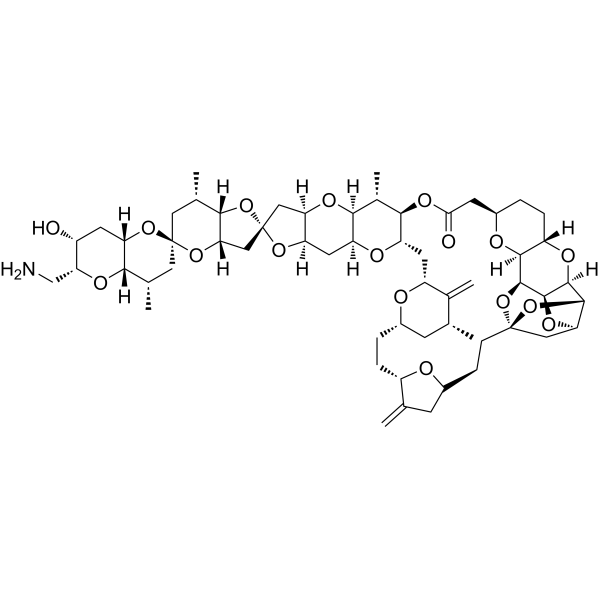
E7130 is a microtubule inhibitor, which ameliorates the tumor microenvironment through suppression of cancer-associated fibroblasts (CAF) and promotion of tumor vasculature remodeling.
For research use only. We do not sell to patients.
- Formula: C58H83NO17
- Molecular Weight:1066.28
-
Storage:
Please store the product under the recommended conditions in the Certificate of Analysis.

To place orders, for customer services and technical support, please contact: MedChemExpress USA
Tel: 609-228-6898 E-mail: [email protected] [email protected]


-
 Biological Activity
-
Chemical Information
- Protocols
- Purity & Documentation
- References
-
 Help & FAQs




Biological Activity
Description
In Vitro
E7130 inhibits microtubule dynamics and exhibits anti-proliferative efficacy in cancer cells KPL-4, OSC-19, FaDu and HSC-2, with IC50s of 0.01-0.1 nM[1].
E7130 (0.15 nM) inihibits TGF-β-induced myofibroblast transdifferentiation through disruption of microtubule network formation, and thereby deactivates the PI3K/AKT/mTOR pathway[1].
MedChemExpress (MCE) has not independently confirmed the accuracy of these methods. They are for reference only.
-
Cell Line:BJ cells
-
Concentration:0.15 nM
-
Incubation Time:48 h
-
Result:Inhibited TGF-β-induced α-SMA expression in BJ cells without growth inhibitory activity.
-
Cell Line:BJ cells
-
Concentration:0.15 nM
-
Incubation Time:48 h
-
Result:Decreased levels of pAKT and pS6.
In Vivo
E7130 (45-180 μg/kg, i.v.) reduces the α-SMA-positive CAFs, the E7130-CTX combination modulates the phenotypes of the fibroblasts in FaDu SCCHN xenograft BALB/c mice[1].
MedChemExpress (MCE) has not independently confirmed the accuracy of these methods. They are for reference only.
-
Animal Model:FaDu SCCHN xenograft BALB/c mice[1]
-
Dosage:45-180 μg/kg
-
Administration:i.v.
-
Result:Reduced the α-SMA-positive CAFs, modulated the phenotypes of the fibroblasts with combination of CTX.
-
Animal Model:HSC-2 SCCHN xenograft BALB/c mice[1]
-
Dosage:90 μg/kg
-
Administration:i.v.
-
Result:Increased MVD, inhibited tumor growth.
Increased survival rate with combination of CTX.
Clinical Trial
| NCT Number | Sponsor | Condition | Start Date |
Phase

|
|---|---|---|---|---|
| NCT01329991 | 2011-05 | PHASE1 |


Chemical Information
-
Molecular Weight 1066.28
-
Formula C58H83NO17
-
SMILES
O=C(C[C@@H]1O[C@@]([C@H](O[C@]2(CC3)O[C@H]4[C@H]5C2)[C@H](O5)[C@@]4([H])O6)([H])[C@]6([H])CC1)O[C@H]([C@H]7C)[C@H](C[C@H]8O[C@@H](CC[C@H]9C(C[C@@H]3O9)=C)C[C@@H](C)C8=C)O[C@@]([C@@]7([H])O%10)([H])C[C@@]([C@@]%10([H])C%11)([H])O[C@@]%11(O%12)C[C@]([C@]%12([H])[C@@H](C)C%13)([H])O[C@@]%14%13C[C@H](C)[C@]%15([H])O[C@H](CN)[C@H](O)C[C@]%15([H])O%14
-
Shipping
Room temperature in continental US; may vary elsewhere.
-
Storage
Please store the product under the recommended conditions in the Certificate of Analysis.


Purity & Documentation
References

Calculators
Concentration (start) × Volume (start) = Concentration (final) × Volume (final)


E7130 Related Classifications
HY-161248 Related Classifications
 Powered by Bioz