



Fulvic Acid
Based on 1 publication(s) in Google Scholar
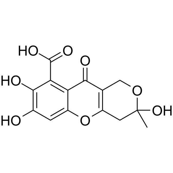
Fulvic Acid is a natural product, which comes from humic substances produced by microorganisms in soil. Fulvic Acid can modulate the immune system, influence the oxidative state of cells, and improve gastrointestinal function. Fulvic Acid has the potential for researching chronic inflammatory diseases, including diabetes.
연구목적의 판매만을 진행합니다. 환자를 대상으로 한 판매는 하지 않습니다.
- Purity : 98.18%
- CAS No.: 479-66-3
- 화학식: C14H12O8
- 분자량:308.24
-
보관:Powder -20°C, 3 years ; In solvent -80°C, 6 months , -20°C, 1 month

To place orders, for customer services and technical support, please contact: MedChemExpress USA
Tel: 609-228-6898 E-mail: [email protected] [email protected]


-
 Biological Activity
-
Chemical Information
- Protocol
- 순도&문서
- References
-
 Help & FAQs




Publications Citing Use of MedChemExpress (MCE) Fulvic Acid
More

Biological Activity
제품 설명
In Vitro
Fulvic acid (200 μg/mL) reduces TNF-α expression in LPS-exposed differentiated human U937 monocytes[1].
Fulvic acid reduces proinflammatory mediator release (B-hexosaminidase, histamine, TNF−α, IL-4, IL-13) from immunoglobulin-E-sensitized mast cells and basophil cells[1].
Fulvic acid increases oxidative stress in isolated cartilage cells from 12-day-old embryonic chicks[1].
Fulvic acid increases oxidative markers and induces apoptosis in hepatic cancer cell lines[1].
Fulvic acid (100 μg/mL) increases NO and ROS production and reduces cell viability in isolated murine peritoneal macrophages[1].
Fulvic acid increases copper absorption and reduces copper toxicity in porcine oviductal epithelial cells[1].
MedChemExpress (MCE) has not independently confirmed the accuracy of these methods. They are for reference only. Further protocols information, click here.
In Vivo
Fulvic acid (100 mg/kg; oral) reduces carrageenan-induced paw edema in rats to levels similar to nonsteroidal anti-inflammatory drugs[1].
Fulvic acid (300 mg/kg/day; daily; 4 weeks) reduces isoproterenol-induced myocardial oxidative damage in rats by decreasing lipid peroxidation and increasing antioxidant enzyme levels[1].
Fulvic acid (1.5% w/w in feed; daily; 60 days) modulates intestinal microbiota and increases digestive enzyme activity in juvenile loach[1].
Fulvic acid (211 p.p.m.; oral; continuously from parental generation through second generation) supplementation induces lysine overhydroxylation in bone and cartilage collagens, reduces collagen thermal stability, and decreases bone mechanical strength in Mus musculus[2].
MedChemExpress (MCE) has not independently confirmed the accuracy of these methods. They are for reference only.
-
Animal Model:unspecified[1]
-
Dosage:100 mg/kg
-
Administration:oral
-
Result:Reduced paw edema to levels similar to nonsteroidal anti-inflammatory drugs.
-
Animal Model:unspecified[1]
-
Dosage:300 mg/kg/day
-
Administration:daily; 4 weeks
-
Result:Decreased lipid peroxidation and myocardial damage markers; significantly increased glutathione, superoxide dismutase, and catalase levels.
-
Animal Model:Paramisgurnus dabryanus (juvenile)[1]
-
Dosage:1.5% (w/w in feed)
-
Administration:daily; 60 days
-
Result:Decreased abundance of Proteobacteria phyla and increased Firmicute levels in the intestine; increased abundance of Variovorax, Lactococcus, and Lactobacillus genera and decreased abundance of Serratia and Acinetobacter genera; increased activity of digestive enzymes (lysozyme, proteases, acid/alkaline phosphatases).
-
Animal Model:NMRI (50 days old at study start; second-generation studied at 49 days old)[2]
-
Dosage:211 p.p.m.
-
Administration:oral (in drinking water; continuously from parental generation through second generation)
-
Result:Detected overhydroxylation of lysine residues in bone collagen I (Hyl/(Hyl + Lys) value of 0.434 ± 0.005 vs. 0.277 ± 0.005 in controls) and cartilage collagen II (Hyl/(Hyl + Lys) value of 0.552 vs. 0.482 in controls); Reduced melting temperature of bone collagen I by 1°C compared to controls; Lowered preload force at tibial breakage point (treated groups including FA had breakage forces of 2.75-5 N vs. 8.5-11.5 N in controls); Markedly affected reproduction, with no significant influence on growth.


Chemical Information
-
CAS No. 479-66-3
-
Appearance Solid
-
분자량 308.24
-
화학식 C14H12O8
-
Color White to off-white
-
SMILES
O=C(C1=C2C(C(COC(C)(O)C3)=C3OC2=CC(O)=C1O)=O)O
-
Structure Classification
-
Initial Source
microorganisms in soil
-
선적
Room temperature in continental US; may vary elsewhere.
-
보관
Powder -20°C 3 years In solvent -80°C 6 months -20°C 1 month


Publications (1)
-
Journal Impact Factor
-
Most Recent

Protocol
-
Research Protocol for Metabolic Diseases
AMP-activated protein kinase, AMPK, is a conserved cellular energy sensor that responds to reduced cellular energy status and coordinates metabolism by increasing ATP-generating catabolic pathways while suppressing ATP-consuming anabolic processes. In metabolic disease research, the AMPK pathway is experimentally relevant because it regulates hepatic lipid synthesis, fatty acid oxidation, glucose production, skeletal-muscle glucose disposal, mTORC1-linked biosynthesis, autophagy, mitochondrial homeostasis, and whole-body energy balance. The central pathway logic is that energy stress, metformin, exercise-like stimulation, or direct AMPK activators increase AMPKα Thr172 phosphorylation and downstream substrate phosphorylation, including ACC and RAPTOR. Phosphorylation of ACC suppresses lipogenesis and supports fatty acid oxidation, whereas phosphorylation of RAPTOR suppresses mTORC1 signaling and links cellular energy status to growth and protein synthesis control. The pathway is linked


순도&문서
-
Data Sheet (278 KB)
-
SDS (393 KB)
- English - EN (393 KB)
- Français - FR (393 KB)
- Deutsch - DE (393 KB)
- Norwegian - NO (393 KB)
- Español - ES (393 KB)
- Swedish - SV (393 KB)
- Italian - IT (393 KB)
- Korean - KR (393 KB)
- Portuguese - PT (393 KB)
-
Handling Instructions (2659 KB)
References

Calculators
Concentration (start) × Volume (start) = Concentration (final) × Volume (final)


Fulvic Acid Related Classifications
HY-122515 Related Classifications
 Powered by Bioz
Keywords