



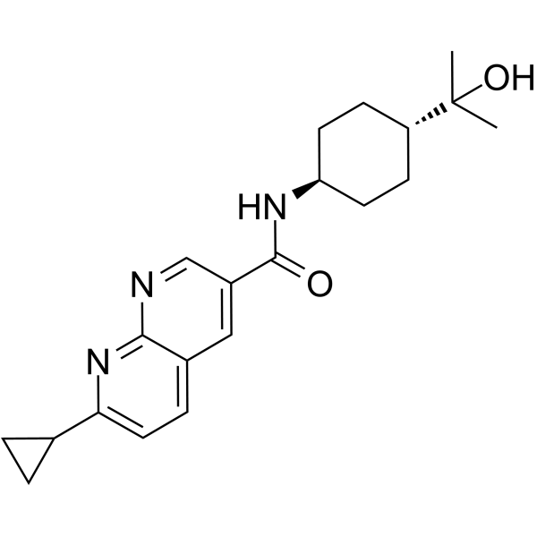
HPGDS inhibitor 3
HPGDS inhibitor 3 is an orally active and highly potent peripherally restricted hematopoietic prostaglandin D synthase (H-PGDS) inhibitor with IC50 value of 9.4 nM and EC50 of 42 nM, respectively. HPGDS inhibitor 3 exhibits good selectivity, good pharmacokinetic parameters in mouse, rat, and dog, and no CNS toxicity. HPGDS inhibitor 3 has anti-inflammatory activity.
For research use only. We do not sell to patients.
- CAS No.: 2255311-93-2
- Formula: C21H27N3O2
- Molecular Weight:353.46
-
Storage:
Please store the product under the recommended conditions in the Certificate of Analysis.

To place orders, for customer services and technical support, please contact: MedChemExpress USA
Tel: 609-228-6898 E-mail: [email protected] [email protected]


-
 Biological Activity
-
Chemical Information
- Protocols
- Purity & Documentation
- References
-
 Help & FAQs




Biological Activity
Description
IC50 & Target
Cellular Effect
|
Cell Line

|
Type | Value | Description | References |
|---|---|---|---|---|
| Mast cell | EC50 |
3.4 nM
Compound: 1y
|
Anti-inflammatory activity against mast cell degranulation inflammatory C57BL/6J mouse model assessed as blood EC50 required for reduction in compound 48/80-induced PGD2 release into peritoneal lavage fluid administered orally with compound and 1 hr later
Anti-inflammatory activity against mast cell degranulation inflammatory C57BL/6J mouse model assessed as blood EC50 required for reduction in compound 48/80-induced PGD2 release into peritoneal lavage fluid administered orally with compound and 1 hr later
|
[PMID: 33059303] |
| Mast cell | ED50 |
0.009 mg/kg
Compound: 1y
|
Anti-inflammatory activity against mast cell degranulation inflammatory C57BL/6J mouse model assessed as reduction in compound 48/80-induced PGD2 release into peritoneal lavage fluid administered orally with compound and 1 hr later treated with compound 4
Anti-inflammatory activity against mast cell degranulation inflammatory C57BL/6J mouse model assessed as reduction in compound 48/80-induced PGD2 release into peritoneal lavage fluid administered orally with compound and 1 hr later treated with compound 4
|
[PMID: 33059303] |
In Vivo
HPGDS inhibitor 3 (0.003-1 mg/kg; PO; single) attenuates PGD2 release to baseline levels in a dose-dependent manner; also inhibits LPS-induced PGD2 increase in plasma and skeletal muscle in a dose-dependent manner[1].
HPGDS inhibitor 3 (0.003-1 mg/kg; PO; single) [1].
HPGDS inhibitor 3 (1, 3, and 10 mg/kg; PO; q.d., for 16 days) significantly enhances functional recovery of injured limbs, and hastens the time to full functional recovery of injured limb muscles[1].
HPGDS inhibitor 3 (10, 30 and 100 mg/kg; PO; once daily, for 7 days or 4 days) exhibits well tolerated at 30 mg/kg/day in rat but not tolerated at 100 mg/kg/day; shows well tolerated at 30 mg/kg/day in dogs but not tolerated at 75 mg/kg/day[1].
Pharmacokinetic Parameters of HPGDS inhibitor 3 in mice, rats and dogs[1].
| Mouse IV, 1 mg/kg PO, 3 mg/kg |
Rat IV, 0.4 mg/kg PO, 2.4 mg/kg |
Dog IV, 0.5 mg/kg PO, 1 mg/kg |
|
| T1/2 (h) | 2.9 | 5.1 | 6.2 |
| CL (mL/min/kg) | 9.0 | 4.5 | 1.9 |
| Vss (L/kg) | 1.6 | 1.6 | 1.0 |
| F (%) | 71 | 100 | 92 |
| Brain:blood ratio | 0.06 |
MedChemExpress (MCE) has not independently confirmed the accuracy of these methods. They are for reference only.
-
Animal Model:Male C57BL/6J mice (murine mast cell degranulation model of inflammation)[1]
-
Dosage:0.003, 0.01, 0.03, 0.1, 0.3 and 1.0 mg/kg
-
Administration:PO; single (anesthetized 1 hour later, intraperitoneally injected with 0.2 mL PBS or 48/80 (0.75 mg/mL))
-
Result:Attenuated PGD2 release to baseline levels in a dose-dependent manner with an ED50 of 0.009 mg/kg (blood EC50 = 3.4 nM) in this acute inflammation model.
-
Animal Model:Male C57BL6/N mice (12 weeks, n=6)[1]
-
Dosage:0.003, 0.01, 0.03, 0.1, 0.3 and 1.0 mg/kg
-
Administration:PO; single (intraperitoneally injection of PBS or 20 ng/kg LPS 1 hour later)
-
Result:Inhibited LPS-induced PGD2 increase in plasma and skeletal muscle in a dose-dependent manner.
-
Animal Model:Male C57Bl/6 mice (10-12 weeks, n=7-8; chronic eccentric contraction-induced muscle injury models)[1]
-
Dosage:1, 3, and 10 mg/kg
-
Administration:PO; q.d., for 16 days
-
Result:Significantly enhanced functional recovery of injured limbs, and significantly hastened the time to full functional recovery of injured limb muscles, with maximal efficacy observed at ≥ 10 mg/kg q.d..
-
Animal Model:Mdx mouse (6-8 mouths, duchenne muscular dystrophy model)[1]
-
Dosage:0.1, 0.3, 1, 3, and 10 mg/kg
-
Administration:PO; q.d., for 43 days
-
Result:Significantly improved functional recovery (~90% to 100% restoration), following eccentric contraction-induced muscle injury in mdx mice.
-
Animal Model:Male Wistar Han rat and dog[1]
-
Dosage:10, 30 and 100 mg/kg for rat; 10, 30, and 75 mg/kg for dog
-
Administration:PO; once daily; for 7 days (rat) or for 4 days (dog)
-
Result:In rat, the AUC values at 10, 30, and 100 mg/kg/day were 120, 410, and 820 µg•hr/mL, respectively; respective Cmax values were 8.7, 24, and 57 μg/mL. In dog, it showed well tolerated at dose levels up to 30 mg/kg/day with no abnormal microscopic findings; but exhibited discoloration in the small intestine and esophagus (female) at 75 mg/kg/day.
-
Animal Model:Mice, rats, dongs[1]
-
Dosage:1 mg/kg IV and 3 mg/kg p.o in mice, 0.4 mg/kg IV and 2.4 mg/kg PO in rat, 0.5 mg/kg IV and 1 mg/kg PO in dog
-
Administration:IV and PO; single (Pharmacokinetics Analysis)
-
Result:Had a lower IV clearance, similar steady state volume of distribution, longer terminal half-life, and high oral bioavailability, as well as very low brain exposures in mouse, rat and dog.


Chemical Information
-
CAS No. 2255311-93-2
-
Molecular Weight 353.46
-
Formula C21H27N3O2
-
SMILES
CC(O)([C@H]1CC[C@@H](CC1)NC(C2=CC3=CC=C(N=C3N=C2)C4CC4)=O)C
-
Shipping
Room temperature in continental US; may vary elsewhere.
-
Storage
Please store the product under the recommended conditions in the Certificate of Analysis.

Protocols
-
Neurotoxicity Study
This protocol assesses in vitro neurotoxicity by combining neuronal viability, mitochondrial/metabolic activity, neurite outgrowth, and optional neuronal network function readouts. Calcein-AM or resazurin/PrestoBlue readouts estimate viable or metabolically active cells; βIII-tubulin immunofluorescence detects neuronal morphology and neurite networks; TMRE detects mitochondrial membrane potential; and MEA recordings detect functional changes in neuronal network activity.
-
Cell Cytotoxicity Assay
Cytotoxicity assays are usually based on the assessment of cell membrane damage, which can also be indirectly detected by measuring cell viability. Detection methods include MTT assay, CKK-8 assay, LDH assay and ATP assay, etc.
-
Cotton Pellet Granuloma
Cotton pellet granuloma is a classical in vivo chronic inflammation model used to evaluate the anti-inflammatory potential of test substances by measuring their ability to inhibit granuloma tissue formation around an implanted foreign body (cotton pellet) in rodents. The method is based on the biological response to a sterile implanted material, which induces proliferative phase inflammation characterized by fibroblast proliferation and collagen-rich granuloma formation, and the final readout reflects the extent of chronic inflammatory tissue growth surrounding the pellet. In multiple preclinical pharmacological evaluations, inhibition of cotton pellet-induced granuloma formation has been used as an indicator of anti-inflammatory activity in both synthetic and natural product screening contexts.
-
Carrageenan-Induced Paw Edema
Carrageenan-induced paw edema is an acute inflammation model in which intraplantar injection of carrageenan induces localized inflammatory swelling characterized by vascular permeability, leukocyte infiltration, and production of inflammatory mediators such as prostaglandins and cytokines, making it widely used to evaluate anti-inflammatory agents in vivo. The resulting paw volume or thickness increase is quantified over time as a direct readout of inflammatory intensity and drug efficacy, typically reflecting cyclooxygenase-mediated prostaglandin-driven edema formation and immune cell recruitment in peripheral tissue[20].
-
Research Protocol for Inflammation-related Diseases
The NLRP3 inflammasome is a cytosolic innate immune signaling platform that integrates priming signals and danger-signal activation to promote caspase-1 activation, maturation of IL-1β and IL-18, and gasdermin D-mediated pyroptotic cell death. The core experimental logic is to determine whether inflammatory disease phenotypes are driven by increased NLRP3 expression, ASC-containing inflammasome assembly, caspase-1 cleavage, GSDMD cleavage, and extracellular release of IL-1β/IL-18 rather than by nonspecific cell injury alone. The pathway is strongly linked to inflammation-related disease phenotypes because monosodium urate crystals activate NALP3/NLRP3 inflammasome signaling in gout-like crystal inflammation, cholesterol crystals activate NLRP3 inflammasomes in atherogenesis models, and DSS-induced intestinal inflammation has been reported to involve NLRP3 inflammasome activity. However, experimental colitis studies also show context-dependent protective effects of NLRP3 inflammasome co


Purity & Documentation
References

Calculators
Concentration (start) × Volume (start) = Concentration (final) × Volume (final)


HPGDS inhibitor 3 Related Classifications
HY-146662 Related Classifications
 Powered by Bioz