



MAO-B-IN-20
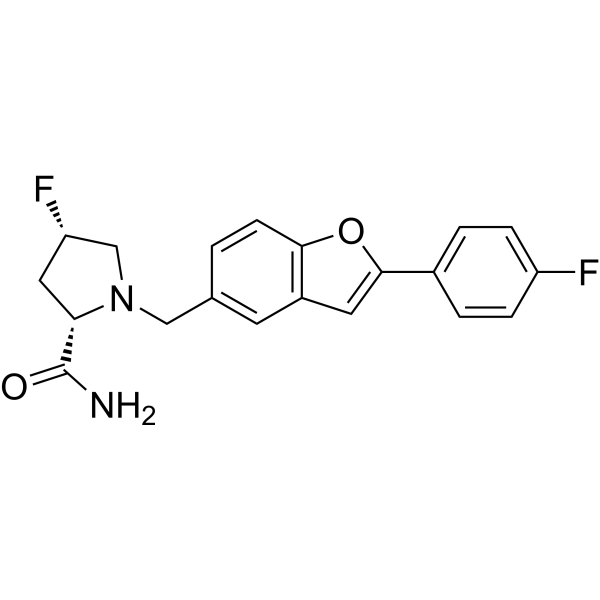
MAO-B-IN-20 (Compound C14) is a potent MAO-B inhibitor with an IC50 of 0.037 μM. MAO-B-IN-20 displays good metabolic stability and brain-blood barrier permeability. MAO-B-IN-20 can be used for the research of Parkinson's disease.
For research use only. We do not sell to patients.
- Formula: C20H18F2N2O2
- Molecular Weight:356.37
-
Storage:
Please store the product under the recommended conditions in the Certificate of Analysis.

To place orders, for customer services and technical support, please contact: MedChemExpress USA
Tel: 609-228-6898 E-mail: [email protected] [email protected]


-
 Biological Activity
-
Chemical Information
- Protocols
- Purity & Documentation
- References
-
 Help & FAQs




Biological Activity
Description
IC50 & Target
[1]|
MAO-B 0.037 μM (IC50) |
MAO-A >10 μM (IC50) |
In Vitro
MAO-B-IN-20 (Compound C14) can well bind into the active site of MAO-B and shares a similar binding mode with Safinamide (HY-70057)[1].
MedChemExpress (MCE) has not independently confirmed the accuracy of these methods. They are for reference only. Further protocols information, click here.
In Vivo
MAO-B-IN-20 (0.08-1.28 mg/kg; i.p.; once) prominently inhibits the MAO-B activity in a dose-dependent manner in the mouse brain[1].
MAO-B-IN-20 (0.3-3 mg/kg; i.p.; once) exhibits a potential efficacy for dopamine deficits in the MPTP (HY-15608)-induced mouse model and significantly increased dopamine concentration in the striatum[1].
Pharmacokinetic profile of MAO-B-IN-20 (Compound C14) in SD ratsa[1]
| Compound | Route | Dose (mg/kg) | Cmax (ng/mL) | AUCt (ng•h/mL) | T1/2 (h) | Vss (L/kg) | CL (mL/min/kg) | F (%) |
| MAO-B-IN-20 (Compound C14) | iv | 1 | 273 | 305 | 0.74 | 3.33 | 54.2 | / |
| ig | 5 | 436 | 2280 | 4.22 | / | / | 149.5 |
aFasted male SD rats. Dosing volumes: 5 mL/kg for ig and 1 mL/kg for iv. Cmax: Maximum Concentration, AUCt: Area under the plasma concentration-time curve from time 0 to last time of quantifiable concentration; T1/2: Elimination half time, Vss: Steady-state distribution volume, CL: plasma clearance, F: bioavailability.
Pharmacokinetic profile of MAO-B-IN-20 (Compound C14) in ICR micea[1]
| Compound | Route | Dose (mg/kg) | Cmax (ng/mL) | AUCt (ng•h/mL) | T1/2 (h) | Vss (L/kg) | CL (mL/min/kg) | F (%) |
| MAO-B-IN-20 (Compound C14) | iv | 2 | 900 | 2680 | 3.37 | 2.72 | 11 | / |
| ig | 5 | 913 | 7950 | 3.80 | / | / | 104.8 |
aFasted male ICR mice. Dosing volumes: 5 mL/kg for ig and 2 mL/kg for iv. Cmax: Maximum Concentration, AUCt: Area under the plasma concentration-time curve from time 0 to last time of quantifiable concentration; T1/2: Elimination half time, Vss: Steady-state distribution volume, CL: plasma clearance, F: bioavailability.
MedChemExpress (MCE) has not independently confirmed the accuracy of these methods. They are for reference only.
-
Animal Model:MPTP-induced acute mouse model of PD[1]
-
Dosage:0.3, 1.0, 3.0 mg/kg
-
Administration:Intraperitoneal injection, 30 min before MPTP (20 mg/kg, ip) injection
-
Result:Dopamine concentration in the striatum of mice significantly increased compared with the MPTP-alone-injected group.
-
Animal Model:SD rats and ICR mice[1]
-
Dosage:1, 2 and 5 mg/kg
-
Administration:IV and IG (Pharmacokinetic Analysis)
-
Result:Showed good pharmacokinetic profiles.


Chemical Information
-
Molecular Weight 356.37
-
Formula C20H18F2N2O2
-
SMILES
NC([C@@H]1C[C@@H](CN1CC2=CC3=C(OC(C4=CC=C(C=C4)F)=C3)C=C2)F)=O
-
Shipping
Room temperature in continental US; may vary elsewhere.
-
Storage
Please store the product under the recommended conditions in the Certificate of Analysis.

Protocols
-
Cell Cytotoxicity Assay
Cytotoxicity assays are usually based on the assessment of cell membrane damage, which can also be indirectly detected by measuring cell viability. Detection methods include MTT assay, CKK-8 assay, LDH assay and ATP assay, etc.
-
Human pluripotent stem cell midbrain dopaminergic neuron differentiation
Human pluripotent stem cells are directed toward midbrain dopaminergic neurons by first inducing a neural floor-plate-like progenitor state, then patterning cells with ventralizing SHH signaling and midbrain/WNT-FGF cues, and finally maturing progenitors into neurons expressing dopaminergic markers such as TH, NURR1/NR4A2, PITX3, DAT/SLC6A3, VMAT2/SLC18A2, GIRK2/KCNJ6, FOXA2, LMX1A, and EN1. The main readouts are loss of pluripotency, acquisition of FOXA2+/LMX1A+ midbrain floor-plate progenitors, emergence of βIII-tubulin+/MAP2+ neurons, and production of TH+ dopaminergic neurons with molecular, dopamine-release, and electrophysiological features of midbrain dopaminergic identity.
-
Transepithelial/transendothelial electrical resistance assay
TEER measures electrical resistance across epithelial or endothelial monolayers cultured on permeable supports, and the readout reflects ionic conductance through the cell barrier, especially the paracellular pathway regulated by junctional integrity. TEER can be measured without destroying the monolayer and is commonly used before or during transport, permeability, barrier-disruption, and barrier-maturation experiments. TEER values are influenced by biological maturation and technical conditions; reported factors include temperature, medium formulation, passage number, electrode geometry, membrane properties, and junctional length during early monolayer maturation. Therefore, TEER should be interpreted with blank-insert subtraction, area normalization, repeated readings, and, when possible, orthogonal barrier readouts such as FITC-dextran flux or tight-junction staining.


Purity & Documentation
References

Calculators
Concentration (start) × Volume (start) = Concentration (final) × Volume (final)


MAO-B-IN-20 Related Classifications
HY-149242 Related Classifications
 Powered by Bioz