



PCTR1
PCTR1 is a potent monocyte/macrophage agonist, regulating key anti-inflammatory and pro-resolving processes during bacterial infection. PCTR1 is a member of the protectin family of specialized pro-resolving mediators (SPMs).
Nur für Forschungszwecke. Wir verkaufen nicht an Patienten.
- CAS. Nr.: 1810710-59-8
- Formel: C32H47N3O9S
- Molecular Weight:649.80
-
Speicherung:
Please store the product under the recommended conditions in the Certificate of Analysis.

To place orders, for customer services and technical support, please contact: MedChemExpress USA
Tel: 609-228-6898 E-mail: [email protected] [email protected]


-
 Biologische Aktivität
-
Chemical Information
- Protokoll
- Reinheit & Dokumentation
- Verweise
-
 Help & FAQs


-
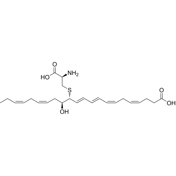
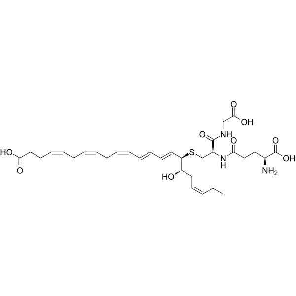
HY-125516
MCTR3Pro-resolving Mediator -
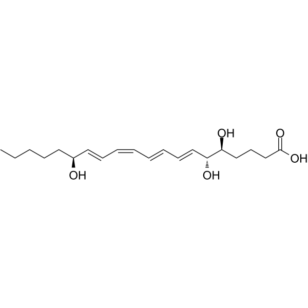
HY-113509
Lipoxin A4Anti-inflammatory Agent -
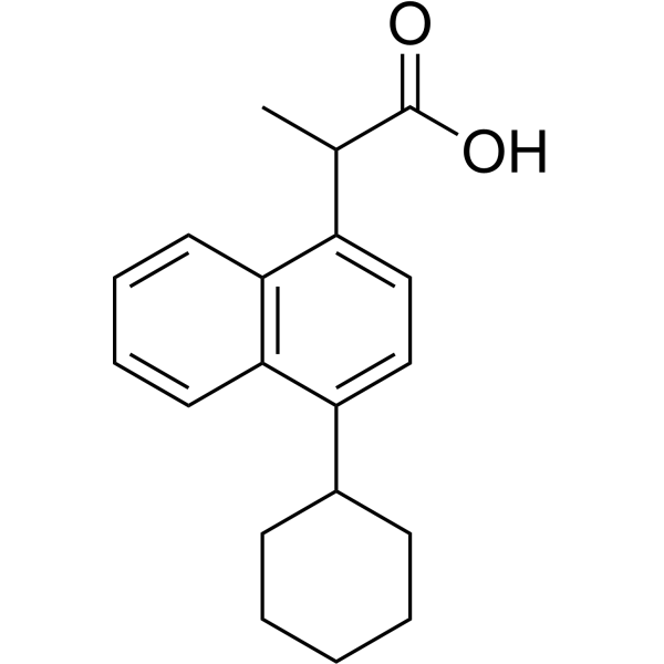
HY-118827
VedaprofenAnti-inflammatory Agent -
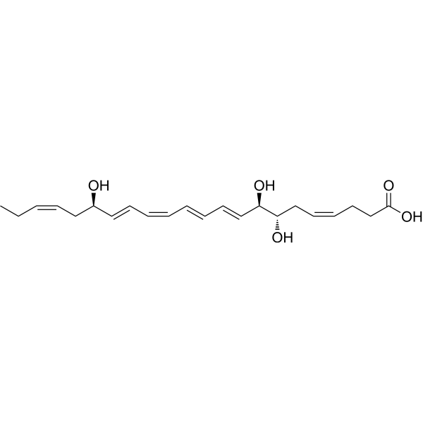
HY-125527A
17(R)-Resolvin D1TRPV3 Inhibitor -

HY-155286
(7S,8R,17S)-RCTR1Resolvin-Sulfido Conjugate -
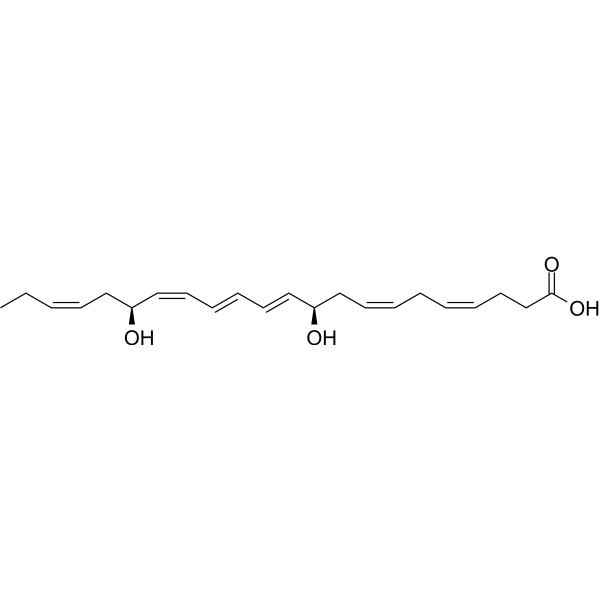
HY-130413
Protectin D1Neuroprotectin D1 -
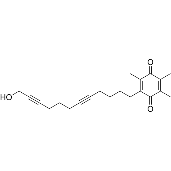
HY-12886
DocebenoneLipoxygenase Inhibitor -
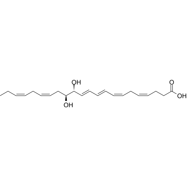
HY-119684
Maresin 2Anti-inflammatory And Pro-resolving Mediator -
HY-128171
DiflapolinFLAP/sEH Inhibitor -
HY-116124
17(S)-HpDHAHydrolyzate -

HY-118827R
Vedaprofen (Standard)Anti-inflammatory Agent -

HY-W708469
Resolvin D2 methyl ester


Biologische Aktivität
Beschreibung
In Vitro
PCTR1 is a peptide-containing lipid mediator. PCTR1 is temporally regulated during self-limited inflammation and promotes the resolution of bacterial infection. PCTR1 significantly decreases prostaglandin (PG)E2 (48%), PGD2 (64%), PGF2α (38%), and thromboxane B2 (40%). PCTR1 increases exudate macrophage levels, accelerates clearance of E. coli infection, decreases local inflammatory mediator production, and promotes resolution[1].
PCTR1 is an agonist of human keratinocyte migration and has unique structural features that impart this agonist activity. PCTR1 enhances human keratinocyte migration in a cAMP/PKA-dependent manner. Stimulation of keratinocytes with PCTR1 (10 nM; 15 minutes) significantly elevates levels of cAMP[2].
MedChemExpress (MCE) has not independently confirmed the accuracy of these methods. They are for reference only. Further protocols information, click here.
-
Cell Line:Primary human epidermal keratinocytes (HPEKp)
-
Concentration:0.1, 1, and 10 nM
-
Incubation Time:24 hours
-
Result:In comparison with untreated cells in which approximately 25% of the initially wounded area was covered by cells 24 hours after wounding, 10 nM significantly enhanced migration such that approximately 46% of the wound area was covered by cells at 24 hours after wounding.
In Vivo
PCTR1 (50?ng/mice) improves the survival rate in an LPS-induced acute inflammatory mouse model[3].
MedChemExpress (MCE) has not independently confirmed the accuracy of these methods. They are for reference only.
-
Animal Model:C57BL/6J wild-type male mice (8-10 weeks old, 22-25 g)[3]
-
Dosage:50 ng/mice
-
Administration:Administered via the tail vein 1 h after LPS (15 mg/kg) intraperitoneal injection. The survival rate was recorded every day for 7 days
-
Result:Compared with the LPS group, the survival rate was significantly higher in the LPS + PCTR1 group, indicating that PCTR1 markedly increases the survival rate.


Chemical Information
-
CAS. Nr. 1810710-59-8
-
Molecular Weight 649.80
-
Formel C32H47N3O9S
-
SMILES
CC/C=C\C[C@@H]([C@@H](/C=C/C=C/C=C\C/C=C\C/C=C\CCC(O)=O)SC[C@H](NC(CC[C@H](N)C(O)=O)=O)C(NCC(O)=O)=O)O
-
Versand
Room temperature in continental US; may vary elsewhere.
-
Speicherung
Please store the product under the recommended conditions in the Certificate of Analysis.

Protokoll
-
Phagocytosis Functional Assay
A phagocytosis functional assay measures the ability of phagocytic cells, such as neutrophils, macrophages, monocytes, or microglia/macrophages, to bind and internalize particulate targets including bacteria, yeast particles, beads, or myelin particles. Fluorescent flow-cytometry assays detect target uptake as fluorescence associated with gated phagocytes, while pH-sensitive dyes such as pHrodo increase signal in acidic phagosomal compartments and therefore preferentially report internalized particles rather than particles remaining outside the cell. Microscopy or high-content imaging can be used to confirm intracellular localization and, in some protocols, to follow uptake kinetics.
-
Research Protocol for Infectious Diseases
Infectious-disease experiments test how pathogens interact with host barriers, innate immune receptors, inflammatory signaling, pathogen replication, and tissue injury; pattern-recognition receptors such as TLRs, RIG-I-like receptors, NOD-like receptors, and inflammasomes detect microbial molecules and activate NF-κB, interferon, and cytokine responses. The central hypothesis is that infection severity reflects the balance between pathogen burden and host response: protective inflammation restricts pathogen growth, whereas excessive or mislocalized inflammation contributes to tissue damage and disease phenotype. Unresolved questions include which host pathways are protective versus pathogenic, why some infection models fail to translate to human disease, and which combined readouts best predict clinically relevant infection outcomes.
-
Research Protocol for Inflammation-related Diseases
The NLRP3 inflammasome is a cytosolic innate immune signaling platform that integrates priming signals and danger-signal activation to promote caspase-1 activation, maturation of IL-1β and IL-18, and gasdermin D-mediated pyroptotic cell death. The core experimental logic is to determine whether inflammatory disease phenotypes are driven by increased NLRP3 expression, ASC-containing inflammasome assembly, caspase-1 cleavage, GSDMD cleavage, and extracellular release of IL-1β/IL-18 rather than by nonspecific cell injury alone. The pathway is strongly linked to inflammation-related disease phenotypes because monosodium urate crystals activate NALP3/NLRP3 inflammasome signaling in gout-like crystal inflammation, cholesterol crystals activate NLRP3 inflammasomes in atherogenesis models, and DSS-induced intestinal inflammation has been reported to involve NLRP3 inflammasome activity. However, experimental colitis studies also show context-dependent protective effects of NLRP3 inflammasome co


Reinheit & Dokumentation
Verweise
[1]. Sesquile Ramon, et al. The Protectin PCTR1 Is Produced by Human M2 Macrophages and Enhances Resolution of Infectious Inflammation. Am J Pathol. 2016 Apr;186(4):962-73. [Content Brief]
[2]. Brian E Sansbury, et al. PCTR1 Enhances Repair and Bacterial Clearance in Skin Wounds. Am J Pathol. 2021 Jun;191(6):1049-1063. [Content Brief]
[3]. Yong-Jian Liu, et al. PCTR1 ameliorates lipopolysaccharide-induced acute inflammation and multiple organ damage via regulation of linoleic acid metabolism by promoting FADS1/FASDS2/ELOV2 expression and reducing PLA2 expression. Lab Invest. 2020 Jul;100(7):904-915. [Content Brief]

Calculators
Konzentration (Stammlösung) × Volumen (Stammlösung) = Konzentration (Ziellösung) × Volumen (Ziellösung)


PCTR1 Related Classifications
HY-125445 Related Classifications
 Powered by Bioz