



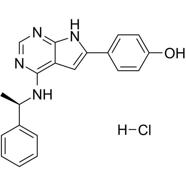
PKI-166 hydrochloride
Based on 1 publication(s) in Google Scholar
PKI-166 hydrochloride is a potent, selective and orally active EGFR tyrosine kinase inhibitor, with an IC50 of 0.7 nM.
For research use only. We do not sell to patients.
- CAS No.: 2230253-82-2
- Formula: C20H19ClN4O
- Molecular Weight:366.84
-
Storage:
Please store the product under the recommended conditions in the Certificate of Analysis.

To place orders, for customer services and technical support, please contact: MedChemExpress USA
Tel: 609-228-6898 E-mail: [email protected] [email protected]


-
 Biological Activity
-
Chemical Information
- Protocols
- Purity & Documentation
- References
-
 Help & FAQs


Publications Citing Use of MedChemExpress (MCE) PKI-166 hydrochloride
MoreAll EGFR Isoforms
More

Biological Activity
Description
IC50 & Target
IC50: 0.7 nM (EGFR tyrosine kinase)[1]
In Vitro
PKI-166 hydrochloride (0-0.5 μM; 1 hour; pretreatment) inhibits EGFR autophosphorylation in human pancreatic cancer cells[1].
PKI-166 hydrochloride (0.03 μM; 6 days) enhances the cytotoxicity mediated by gemcitabine[1].
MedChemExpress (MCE) has not independently confirmed the accuracy of these methods. They are for reference only. Further protocols information, click here.
-
Cell Line:L3.6pl cells
-
Concentration:0.01 μM, 0.05 μM, 0.5 μM
-
Incubation Time:1 hour
-
Result:Inhibited EGFR autophosphorylation in a dose-dependent manner.
-
Cell Line:L3.6pl cells
-
Concentration:0.03 μM
-
Incubation Time:6 days
-
Result:Enhanced the cytotoxicity mediated by gemcitabine.
In Vivo
MedChemExpress (MCE) has not independently confirmed the accuracy of these methods. They are for reference only.
-
Animal Model:Male athymic nude mice with L3.6pl cells xenograft (8-12 weeks)[1]
-
Dosage:100 mg/kg
-
Administration:Oral administration; daily; for 29 days
-
Result:Significantly decreased median tumor volume.


Chemical Information
-
CAS No. 2230253-82-2
-
Molecular Weight 366.84
-
Formula C20H19ClN4O
-
SMILES
OC1=CC=C(C2=CC3=C(N=CN=C3N2)N[C@H](C)C4=CC=CC=C4)C=C1.Cl[H]
-
Shipping
Room temperature in continental US; may vary elsewhere.
-
Storage
Please store the product under the recommended conditions in the Certificate of Analysis.


Publications (1)
-
Journal Impact Factor
-
Most Recent
-
Cell Rep Methods
RECOVER identifies synergistic drug combinations in vitro through sequential model optimization. [Abstract]2023 Oct 23;3(10):100599. PMID: 37797618

Protocols
-
Cell Cytotoxicity Assay
Cytotoxicity assays are usually based on the assessment of cell membrane damage, which can also be indirectly detected by measuring cell viability. Detection methods include MTT assay, CKK-8 assay, LDH assay and ATP assay, etc.


Purity & Documentation
References

Calculators
Concentration (start) × Volume (start) = Concentration (final) × Volume (final)


PKI-166 hydrochloride Related Classifications
HY-110328 Related Classifications
 Powered by Bioz