



Siremadlin succinate
Based on 12 publication(s) in Google Scholar
Siremadlin (NVP-HDM201) succinate is an MDM2 inhibitor and cell cycle regulator. Siremadlin succinate blocks the p53-binding pocket of MDM2, inhibits MDM2-mediated ubiquitination and degradation of p53, thereby activating the p53 pathway in p53 wild-type cells. Siremadlin succinate can be used in the research of cutaneous melanoma.
For research use only. We do not sell to patients.
- CAS No.: 1638193-48-2
- Formula: C30H30Cl2N6O8
- Molecular Weight:673.50
-
Storage:
Please store the product under the recommended conditions in the Certificate of Analysis.


To place orders, for customer services and technical support, please contact: MedChemExpress USA
Tel: 609-228-6898 E-mail: [email protected] [email protected]


-
 Biological Activity
-
Chemical Information
- Protocols
- Purity & Documentation
- References
-
 Help & FAQs


Publications Citing Use of MedChemExpress (MCE) Siremadlin succinate
More- Sci Data. 2024 Sep 19;11(1):1024. [Abstract]
- Cells. 2026 Mar 5;15(5):473. [Abstract]
- Oncogenesis. 2022 Jul 2;11(1):37. [Abstract]
- Cell Rep Methods. 2025 Oct 17:101207. [Abstract]
- Int J Mol Sci. 2022 Oct 8;23(19):11939. [Abstract]
- Cancers (Basel). 2022 Oct 19;14(20):5127. [Abstract]
- iScience. 2024 May 6;27(6):109862. [Abstract]
- Cancer Res Commun. 2026 Mar 1;6(3):616-629. [Abstract]
- Carcinogenesis. 2024 Nov 22;45(11):857-867. [Abstract]
- Biomed Pharmacother. 2026 Sep:202:119803.
- bioRxiv. 2025 Nov 30:2025.11.26.690792. [Abstract]
- SSRN. 2023 Oct 9.
 Customer Validation & Images

-
Cell Proliferation/Viability Assay

-
WB

-
WB


Biological Activity
Description
In Vitro
Siremadlin (NVP-HDM201) succinate (72 h) had its growth-inhibitory activity potentiated by 2.5 μM GSK2830371 in p53 wild-type (A375, WM35, C8161) but not p53 mutant (WM164, WM35-R, CHL-1) cutaneous melanoma cell lines by decreasing the GI50 of Siremadlin succinate[1].
Siremadlin succinate (72 h) had its cytotoxic activity potentiated by 2.5 μM GSK2830371 in p53 wild-type (A375, WM35, C8161) but not p53 mutant (WM164, WM35-R, CHL-1) cutaneous melanoma cell lines by decreasing the LC50 of Siremadlin (succinate)[1].
Siremadlin succinate(0.2 μM; 6 h, 24 h) in combination with 2.5 μM GSK2830371 enhances p53 stabilisation, phosphorylation, acetylation, and transcriptional activity in p53 wild-type (A375, C8161) cutaneous melanoma cell lines[1].
Siremadlin succinate (0.2 μM; 24 h) in combination with 2.5 μM GSK2830371 enhances p53-dependent cell cycle changes and apoptosis in p53 wild-type (A375, WM35, C8161) but not p53 mutant (WM35-R) cutaneous melanoma cell lines[1].
Siremadlin succinate had its activity potentiation by 2.5 μM GSK2830371 in p53 wild-type (A375, WM35) cutaneous melanoma cell lines strictly p53-dependent[1].
MedChemExpress (MCE) has not independently confirmed the accuracy of these methods. They are for reference only. Further protocols information, click here.
-
Cell Line:p53 wild-type (A375, WM35, C8161); p53 mutant (WM164, WM35-R, CHL-1) cutaneous melanoma cell lines
-
Concentration:2.5 μM GSK2830371 (co-treatment)
-
Incubation Time:72 h
-
Result:Significantly decreased the GI50 of HDM201 in p53 wild-type cutaneous melanoma cell lines compared to treatment with Siremadlin succinate alone.
Showed no potentiation in p53 mutant cell lines.\nSignificantly decreased the LC50 of siremadlin succinate in A375 and C8161 cells, and showed a similar trend in WM35 cells compared to treatment with Siremadlin (succinate) alone.
Showed no potentiation in p53 mutant cell lines.


Chemical Information
-
CAS No. 1638193-48-2
-
Molecular Weight 673.50
-
Formula C30H30Cl2N6O8
-
SMILES
OC(CCC(O)=O)=O.CC(N1C([C@H](C2=CC=C(C=C2)Cl)N3C4=CC(Cl)=CN(C)C4=O)=C(N=C1C5=CN=C(OC)N=C5OC)C3=O)C
-
Synonyms
NVP-HDM201 succinate; HDM201 succinate
-
Shipping
Room temperature in continental US; may vary elsewhere.
-
Storage
Please store the product under the recommended conditions in the Certificate of Analysis.


Publications (12)
-
Journal Impact Factor
-
Most Recent
-
Sci Data
High-throughput drug screening identifies novel therapeutics for Low Grade Serous Ovarian Carcinoma. [Abstract]2024 Sep 19;11(1):1024. PMID: 39300112 -
Cells
PROTAC-Mediated Targeted Degradation of MDM2 Induces Tumor-Suppressive Signaling in Osteosarcoma Cells. [Abstract]2026 Mar 5;15(5):473. PMID: 41827906 -
Oncogenesis
LIN28B inhibition sensitizes cells to p53-restoring PPI therapy through unleashed translational suppression. [Abstract]2022 Jul 2;11(1):37. PMID: 35780125 -
Cell Rep Methods
2025 Oct 17:101207. PMID: 41109218 -
Int J Mol Sci
In Vitro/In Vivo Translation of Synergistic Combination of MDM2 and MEK Inhibitors in Melanoma Using PBPK/PD Modelling: Part II. [Abstract]2022 Oct 8;23(19):11939. PMID: 36233247 -
Cancers (Basel)
Association between Dysfunction of the Nucleolar Stress Response and Multidrug Resistance in Pediatric Acute Lymphoblastic Leukemia. [Abstract]2022 Oct 19;14(20):5127. PMID: 36291909 -
iScience
MDM2/MDMX inhibition by Sulanemadlin synergizes with anti-Programmed Death 1 immunotherapy in wild-type p53 tumors. [Abstract]2024 May 6;27(6):109862. PMID: 38784022
Siremadlin succinate purchased from MedChemExpress. Usage Cited in: iScience. 2024 May 6;27(6):109862. [Abstract]
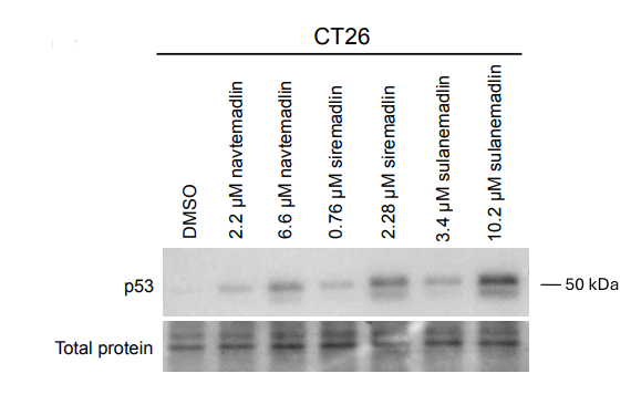
Levels of p53 protein detected by Western blotting in CT26.WT and E B16-F10 cells treated with two different concentrations of Sulanemadlin, Siremadlin (0.76 μM) and Navtemadlin for 6 hours.
-
Cancer Res Commun
Mutant Isocitrate Dehydrogenase 1 Sensitizes Intrahepatic Cholangiocarcinoma Cells to MDM2 Inhibitors. [Abstract]2026 Mar 1;6(3):616-629. PMID: 41747217
Siremadlin succinate purchased from MedChemExpress. Usage Cited in: Cancer Res Commun. 2026 Mar 1;6(3):616-629. [Abstract]
Cell viability after Siremadlin ( HDM201) treatment for 3 doubling times in iCCA cell lines. Data was normalized to the corresponding DMSO control.
Siremadlin succinate purchased from MedChemExpress. Usage Cited in: Cancer Res Commun. 2026 Mar 1;6(3):616-629. [Abstract]
MDM2, TP53, and MIC1 protein expression in two IDH1mut and three IDH1wt iCCA cell lines after DMSO or MDM2 inhibitors treatment for 1 doubling time. MIC1 was used as an indicator of wtTP53 activation. GAPDH was used as an internal control.
-
Carcinogenesis
BRAF V600E-induced distinct DNA damage response defines the therapeutic potential of p53 activation for TP53 wild-type colorectal cancer. [Abstract]2024 Nov 22;45(11):857-867. PMID: 38868979 -
-
bioRxiv
Uncovering senescent fibroblast heterogeneity connects DNA damage response to idiopathic pulmonary fibrosis. [Abstract]2025 Nov 30:2025.11.26.690792. PMID: 41394576 -

Protocols
-
Flow cytometric DNA-content cell-cycle staining
Flow cytometric DNA-content cell-cycle staining measures the fluorescence intensity of DNA-bound fluorochromes in single cells or nuclei to estimate DNA content distributions, allowing assignment of populations to G0/G1, S, and G2/M phases by DNA histogram deconvolution. Propidium iodide (PI) intercalates into DNA, and PI fluorescence is proportional to cellular DNA content when staining is performed under conditions that make DNA accessible and minimize non-DNA signal. Cells with G2/M DNA content are expected to show approximately twice the fluorescence intensity of G0/G1 cells, while S-phase cells occupy intermediate fluorescence values. PI-based DNA-content analysis can also detect cells with fractional DNA content, often reported as sub-G1, when DNA fragmentation and extraction during staining reduce retained DNA signal in apoptotic cells. DAPI is an alternative DNA fluorochrome for univariate DNA-content analysis, while bivariate approaches combining DNA content with proliferation
-
BrdU Incorporation Assay
Bromodeoxyuridine (BrdU) incorporation assay is based on the principle that BrdU, a thymidine analog, is incorporated into newly synthesized DNA during the S phase of the cell cycle, thereby serving as a marker of DNA replication and cellular proliferation. Incorporated BrdU can be detected using anti-BrdU antibodies following DNA denaturation, enabling visualization or quantification of proliferating cells through immunochemical detection methods such as immunofluorescence or immunohistochemistry.
-
Protocol for Cell Cycle
Cell-cycle analysis by flow cytometry measures DNA content in single cells to estimate the fraction of cells in G0/G1, S, and G2/M phases. Propidium iodide intercalates into DNA, and after RNA removal with RNase, fluorescence intensity reflects cellular DNA content: 2N cells are assigned to G0/G1, cells between 2N and 4N to S phase, and 4N cells to G2/M. DNA-content analysis alone cannot reliably separate G0 from G1 or G2 from M. Ki-67 can distinguish quiescent G0 cells from cycling cells, EdU or BrdU incorporation marks active DNA synthesis in S phase, and phospho-histone H3 staining identifies mitotic cells within the 4N population.


Purity & Documentation
References

Calculators
Concentration (start) × Volume (start) = Concentration (final) × Volume (final)


Siremadlin succinate Related Classifications
HY-18658B Related Classifications
 Powered by Bioz