



PKR Antibody (YA124)
(Synonyms: PKR, PRKR, EIF2AK2, Eukaryotic translation initiation factor 2-alpha kinase 2, Interferon-inducible RNA-dependent protein kinase, P1/eIF-2A protein kinase, Protein kinase RNA-activated, Tyrosine-protein kinase EIF2AK2, p68 kinase, eIF-2A protein kinase 2, Protein kinase R)
Based on 1 Customer Validation
PKR Antibody (YA124) is a Rabbit-derived and non-conjugated IgG monoclonal antibody, targeting to PKR.
-
Host:
Rabbit
-
Isotype:
IgG
-
Application:
WB, ICC/IF, IHC-P, IP
-
Reactivity :
Human
-
Formulation:
Supplied in 1*TBS (pH7.4), 0.05% BSA and 40% Glycerol. Preservative: 0.05% Sodium Azide.
-
Conjugation:
Non-conjugated


To place orders, for customer services and technical support, please contact: MedChemExpress USA
Tel: 609-228-6898 E-mail: [email protected] [email protected]





Applications
| Application |
WB

WB: Western Blot
|
ICC/IF
ICC/IF: Immunocytochemistry/
Immunofluorescence |
IHC-P
IHC-P: Immunohistochemistry-Paraffin
|
IP
IP: Immunoprecipitation
|
|---|---|---|---|---|
| Dilution Ratio | 1:1000-1:5000 | 1:50-1:100 | 1:50-1:100 | Use at an assay dependent concentration. |


Product Details
PKR Antibody (YA124) is a Rabbit-derived and non-conjugated IgG monoclonal antibody, targeting to PKR.
-
Host Rabbit

-
Clonality Recombinant,Monoclonal

-
Species ReactivityHuman
-
Observed Molecular WeightObserved band size: 88 kDa
Note: Due to possible protein modifications or aggregation, the molecular weight should be confirmed by actual measurement, and the predicted value is for reference only.

-
Calculated Molecular Weight Predicted band size: 62 kDa

Synthetic peptide corresponding to Human PKR.AA range:500-540.
Endogenous
Protein A affinity purified.
Non-conjugated
Unmodified
IgG


Product Properties
-
Appearance
Solution
-
Formulation
Supplied in 1*TBS (pH7.4), 0.05% BSA and 40% Glycerol. Preservative: 0.05% Sodium Azide.
-
Concentration
Batch-dependent, Please check the COA for the concentration of each lot. Check Lot Concentration
-
Storage & Stability
Stored at -20°C for 1 year. Avoid repeated freeze / thaw cycles.
-
Shipping
Shipping with blue ice.


Verification Images
-
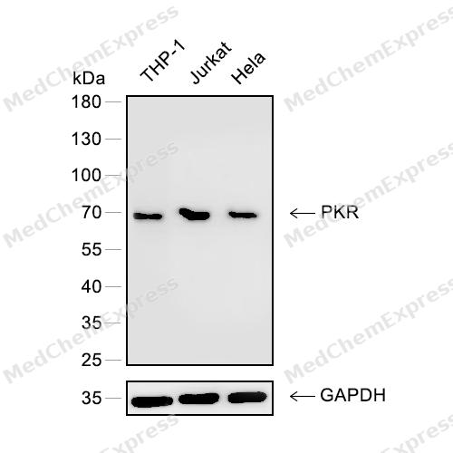 Western blot analysis of extracts from THP-1 (lane 1) and Jurkat (lane 2) and Hela (lane 3) using PKR antibody. Proteins were transferred to a PVDF membrane and blocked with 5% nonfat powdered milk in PBST for 2 hour at room temperature. The primary antibody (1/1000) and loading control antibody (GAPDH, 1/3000) was diluted with 5% nonfat powdered milk in PBST at 4°C overnight. Goat Anti-Rabbit IgG-HRP Secondary Antibody (1/8,000) was incubated for 45min at room temperature.
 -
 Immunohistochemical analysis of paraffin-embedded human ovarian cancer using PKR antibody. The section was pre-treated using heat mediated antigen retrieval with EDTA (pH 9.0) for 14 minutes. The tissues were blocked in 5% BSA for 30 minutes at room temperature, washed with TBST, and then probed with the primary antibody (HY-P80283, 1/100) for 30 minutes at room temperature. The detection was performed using Polymer HRP-conjugated Goat Anti-Mouse/Rabbit lgG(H&L) secondary antibody (HY-P83652). DAB was used as the chromogen. Tissues were counterstained with hematoxylin and mounted with DPX.
 -
 Immunohistochemical analysis of paraffin-embedded human colon cancer using PKR antibody. The section was pre-treated using heat mediated antigen retrieval with EDTA (pH 9.0) for 14 minutes. The tissues were blocked in 5% BSA for 30 minutes at room temperature, washed with TBST, and then probed with the primary antibody (HY-P80283, 1/100) for 30 minutes at room temperature. The detection was performed using Polymer HRP-conjugated Goat Anti-Mouse/Rabbit lgG(H&L) secondary antibody (HY-P83652). DAB was used as the chromogen. Tissues were counterstained with hematoxylin and mounted with DPX.
 -
 Immunohistochemical analysis of paraffin-embedded human prostate cancer using PKR antibody. The section was pre-treated using heat mediated antigen retrieval with EDTA (pH 9.0) for 14 minutes. The tissues were blocked in 5% BSA for 30 minutes at room temperature, washed with TBST, and then probed with the primary antibody (HY-P80283, 1/100) for 30 minutes at room temperature. The detection was performed using Polymer HRP-conjugated Goat Anti-Mouse/Rabbit lgG(H&L) secondary antibody (HY-P83652). DAB was used as the chromogen. Tissues were counterstained with hematoxylin and mounted with DPX.
 -
 Immunohistochemical analysis of paraffin-embedded human breasst cancer using PKR antibody. The section was pre-treated using heat mediated antigen retrieval with EDTA (pH 9.0) for 14 minutes. The tissues were blocked in 5% BSA for 30 minutes at room temperature, washed with TBST, and then probed with the primary antibody (HY-P80283, 1/100) for 30 minutes at room temperature. The detection was performed using Polymer HRP-conjugated Goat Anti-Mouse/Rabbit lgG(H&L) secondary antibody (HY-P83652). DAB was used as the chromogen. Tissues were counterstained with hematoxylin and mounted with DPX.
 -
 Immunohistochemical analysis of paraffin-embedded human tonsil using PKR antibody. The section was pre-treated using heat mediated antigen retrieval with EDTA (pH 9.0) for 14 minutes. The tissues were blocked in 5% BSA for 30 minutes at room temperature, washed with TBST, and then probed with the primary antibody (HY-P80283, 1/100) for 30 minutes at room temperature. The detection was performed using Polymer HRP-conjugated Goat Anti-Mouse/Rabbit lgG(H&L) secondary antibody (HY-P83652). DAB was used as the chromogen. Tissues were counterstained with hematoxylin and mounted with DPX.
 -
 Immunohistochemical analysis of paraffin-embedded human prostate using PKR antibody. The section was pre-treated using heat mediated antigen retrieval with EDTA (pH 9.0) for 14 minutes. The tissues were blocked in 5% BSA for 30 minutes at room temperature, washed with TBST, and then probed with the primary antibody (HY-P80283, 1/100) for 30 minutes at room temperature. The detection was performed using Polymer HRP-conjugated Goat Anti-Mouse/Rabbit lgG(H&L) secondary antibody (HY-P83652). DAB was used as the chromogen. Tissues were counterstained with hematoxylin and mounted with DPX.
 -
 Immunocytochemistry analysis of A549 cells labeling PKR with PKR Antibody (HY-P80283) at 1/50 dilution . Cells were fixed in 4% paraformaldehyde for 15 minutes at room temperature, permeabilized with 0.1% Triton X-100 in PBS for 15 minutes at room temperature, then blocked with quick block buffer for 10 minutes at room temperature. Cells were then incubated with PKR Antibody (HY-P80283) at 1/50 dilution in quick block buffer overnight at 4 ℃. AF488-conjugated Goat Anti-Rabbit IgG H&L(HY-P8002, Green) was used as the secondary antibody at 1/1,000 dilution. PBS instead of the primary antibody was used as the secondary antibody only control. The Nuclear counterstain was DAPI (Blue).
 -
 Immunocytochemistry analysis of A549 cells labeling PKR with PKR Antibody (HY-P80283) at 1/100 dilution . Cells were fixed in 4% paraformaldehyde for 15 minutes at room temperature, permeabilized with 0.1% Triton X-100 in PBS for 15 minutes at room temperature, then blocked with quick block buffer for 10 minutes at room temperature. Cells were then incubated with PKR Antibody (HY-P80283) at 1/100 dilution in quick block buffer overnight at 4 ℃. AF488-conjugated Goat Anti-Rabbit IgG H&L(HY-P8002, Green) was used as the secondary antibody at 1/1,000 dilution. PBS instead of the primary antibody was used as the secondary antibody only control. The Nuclear counterstain was DAPI (Blue).


Background
-
Function
PKR is an IFN-induced dsRNA-dependent serine/threonine-protein kinase that phosphorylates the alpha subunit of eukaryotic translation initiation factor 2 (EIF2S1/eIF-2-alpha) and plays a key role in the innate immune response to viral infection. Inhibits viral replication via the integrated stress response (ISR): EIF2S1/eIF-2-alpha phosphorylation in response to viral infection converts EIF2S1/eIF-2-alpha in a global protein synthesis inhibitor, resulting to a shutdown of cellular and viral protein synthesis, while concomitantly initiating the preferential translation of ISR-specific mRNAs, such as the transcriptional activator ATF4. Exerts its antiviral activity on a wide range of DNA and RNA viruses including hepatitis C virus (HCV), hepatitis B virus (HBV), measles virus (MV) and herpes simplex virus 1 (HHV-1). Also involved in the regulation of signal transduction, apoptosis, cell proliferation and differentiation: phosphorylates other substrates including p53/TP53, PPP2R5A, DHX9, ILF3, IRS1 and the HHV-1 viral protein US11. In addition to serine/threonine-protein kinase activity, also has tyrosine-protein kinase activity and phosphorylates CDK1 at 'Tyr-4' upon DNA damage, facilitating its ubiquitination and proteasomal degradation. Either as an adapter protein and/or via its kinase activity, can regulate various signaling pathways (p38 MAP kinase, NF-kappa-B and insulin signaling pathways) and transcription factors (JUN, STAT1, STAT3, IRF1, ATF3) involved in the expression of genes encoding pro-inflammatory cytokines and IFNs. Activates the NF-kappa-B pathway via interaction with IKBKB and TRAF family of proteins and activates the p38 MAP kinase pathway via interaction with MAP2K6. Can act as both a positive and negative regulator of the insulin signaling pathway (ISP). Negatively regulates ISP by inducing the inhibitory phosphorylation of insulin receptor substrate 1 (IRS1) at 'Ser-312' and positively regulates ISP via phosphorylation of PPP2R5A which activates FOXO1, which in turn up-regulates the expression of insulin receptor substrate 2 (IRS2). Can regulate NLRP3 inflammasome assembly and the activation of NLRP3, NLRP1, AIM2 and NLRC4 inflammasomes. Plays a role in the regulation of the cytoskeleton by binding to gelsolin (GSN), sequestering the protein in an inactive conformation away from actin[1][2][3][4][5][6][7][8][9][10][11][12][13][14][15][16][17][18][19][20][21][22][23][24].
-
Subcellular Localization
Cytoplasm; Nucleus; Cytoplasm, perinuclear region
-
Expression
Tissue_specificity:Compared to non-hematopoietic tissues such as the small intestine, liver, or kidneys, this protein is highly expressed in the thymus, spleen, and bone marrow. In Alzheimer's disease (AD) brain tissue, this protein co-localizes with GSK3β and TAU. In breast and colon cancer, the level of this protein is elevated and associated with tumor progression, invasiveness, or risk of progression.
Induction:By type I interferons -
Isoforms & Post-Translational Modification
P19525 has 2 isomers: P19525-1: 62094 Da (predicted); P19525-2: 57391 Da (predicted).
Autophosphorylated on several Ser, Thr and Tyr residues. Autophosphorylation of Thr-451 is dependent on Thr-446 and is stimulated by dsRNA binding and dimerization. Autophosphorylation apparently leads to the activation of the kinase. Tyrosine autophosphorylation is essential for efficient dsRNA-binding, dimerization, and kinase activation -
Subunit
Homodimer (PubMed:16179258, PubMed:31246429). Interacts with STRBP (By similarity). Interacts with DNAJC3. Forms a complex with FANCA, FANCC, FANCG and HSP70. Interacts with ADAR/ADAR1. Interacts with IRS1 (By similarity). The inactive form interacts with NCK1 and GSN. Interacts (via the kinase catalytic domain) with STAT3 (via SH2 domain), TRAF2 (C-terminus), TRAF5 (C-terminus) and TRAF6 (C-terminus). Interacts with MAP2K6, IKBKB/IKKB, NPM1, TARBP2, NLRP1, NLRP3, NLRC4 and AIM2. Interacts (via DRBM 1 domain) with DUS2L (via DRBM domain). Interacts with DHX9 (via N-terminus) and this interaction is dependent upon activation of the kinase. Interacts with EIF2S1/EIF-2ALPHA; this interaction induces a conformational change in EIF2S1 and its phosphorylation by EIF2AK2 (PubMed:16179258)
-
SwissProt ID
-
Synonyms
PKR, PRKR, EIF2AK2, Eukaryotic translation initiation factor 2-alpha kinase 2, Interferon-inducible RNA-dependent protein kinase, P1/eIF-2A protein kinase, Protein kinase RNA-activated, Tyrosine-protein kinase EIF2AK2, p68 kinase, eIF-2A protein kinase 2, Protein kinase R
-
Research Field
Signal Transduction


Documentation
-
Data Sheet (269 KB)
-
SDS (252 KB)
- English - EN (252 KB)
- Français - FR (252 KB)
- Deutsch - DE (252 KB)
- Norwegian - NO (252 KB)
- Español - ES (252 KB)
- Swedish - SV (252 KB)
- Italian - IT (252 KB)
- Korean - KR (252 KB)
- Portuguese - PT (252 KB)
-
User Guide for Antibodies (1077 KB)
References
[1]. Cardin E, et al. Nck-1 interacts with PKR and modulates its activation by dsRNA. Biochem Biophys Res Commun. 2008 Dec 5;377(1):231-5. [Content Brief]
[2]. Kang JI, et al. PKR protein kinase is activated by hepatitis C virus and inhibits viral replication through translational control. Virus Res. 2009 Jun;142(1-2):51-6. [Content Brief]
[3]. Blalock WL, et al. PKR activity is required for acute leukemic cell maintenance and growth: a role for PKR-mediated phosphatase activity to regulate GSK-3 phosphorylation. J Cell Physiol. 2009 Oct;221(1):232-41. [Content Brief]
[4]. Blalock WL, et al. Multiple forms of PKR present in the nuclei of acute leukemia cells represent an active kinase that is responsive to stress. Leukemia. 2011 Feb;25(2):236-45. [Content Brief]
[5]. Harashima A, et al. Phosphorylation of the NFAR proteins by the dsRNA-dependent protein kinase PKR constitutes a novel mechanism of translational regulation and cellular defense. Genes Dev. 2010 Dec 1;24(23):2640-53. [Content Brief]
[6]. Taghavi N, et al. Protein kinase PKR catalytic activity is required for the PKR-dependent activation of mitogen-activated protein kinases and amplification of interferon beta induction following virus infection. Virology. 2012 Jun 5;427(2):208-16. [Content Brief]
[7]. McAllister CS, et al. Protein kinase PKR amplification of interferon β induction occurs through initiation factor eIF-2α-mediated translational control. J Biol Chem. 2012 Oct 19;287(43):36384-92. [Content Brief]
[8]. Okumura F, et al. Activation of double-stranded RNA-activated protein kinase (PKR) by interferon-stimulated gene 15 (ISG15) modification down-regulates protein translation. J Biol Chem. 2013 Jan 25;288(4):2839-47. [Content Brief]
[9]. Cassady KA, et al. The herpes simplex virus type 1 U(S)11 protein interacts with protein kinase R in infected cells and requires a 30-amino-acid sequence adjacent to a kinase substrate domain. J Virol. 2002 Mar;76(5):2029-35. [Content Brief]
[10]. Chang JH, et al. Double-stranded RNA-activated protein kinase inhibits hepatitis C virus replication but may be not essential in interferon treatment. Liver Int. 2010 Feb;30(2):311-8. [Content Brief]
[11]. Lin SS, et al. A role for protein kinase PKR in the mediation of Epstein-Barr virus latent membrane protein-1-induced IL-6 and IL-10 expression. Cytokine. 2010 May;50(2):210-9. [Content Brief]
[12]. Park IH, et al. PKR-dependent mechanisms of interferon-α for inhibiting hepatitis B virus replication. Mol Cells. 2011 Aug;32(2):167-72. [Content Brief]
[13]. Okonski KM, et al. Stress granule formation induced by measles virus is protein kinase PKR dependent and impaired by RNA adenosine deaminase ADAR1. J Virol. 2013 Jan;87(2):756-66. [Content Brief]
[14]. Zhang L, et al. The modulation of hepatitis C virus 1a replication by PKR is dependent on NF-kB mediated interferon beta response in Huh7.5.1 cells. Virology. 2013 Mar 30;438(1):28-36. [Content Brief]
[15]. Sadler AJ, et al. An antiviral response directed by PKR phosphorylation of the RNA helicase A. PLoS Pathog. 2009 Feb;5(2):e1000311. [Content Brief]
[16]. Bennett RL, et al. The RAX/PACT-PKR stress response pathway promotes p53 sumoylation and activation, leading to G₁ arrest. Cell Cycle. 2012 Jan 15;11(2):407-17. [Content Brief]
[17]. Yoon CH, et al. New Cdc2 Tyr 4 phosphorylation by dsRNA-activated protein kinase triggers Cdc2 polyubiquitination and G2 arrest under genotoxic stresses. EMBO Rep. 2010 May;11(5):393-9. [Content Brief]
[18]. Shen S, et al. Cytoplasmic STAT3 represses autophagy by inhibiting PKR activity. Mol Cell. 2012 Dec 14;48(5):667-80. [Content Brief]
[19]. Li Y, et al. Protein kinase regulated by dsRNA downregulates the interferon production in dengue virus- and dsRNA-stimulated human lung epithelial cells. PLoS One. 2013;8(1):e55108. [Content Brief]
[20]. Bonnet MC, et al. PKR stimulates NF-kappaB irrespective of its kinase function by interacting with the IkappaB kinase complex. Mol Cell Biol. 2000 Jul;20(13):4532-42. [Content Brief]
[21]. Gil J, et al. TRAF family proteins link PKR with NF-kappa B activation. Mol Cell Biol. 2004 May;24(10):4502-12. [Content Brief]
[22]. Silva AM, et al. Protein kinase R (PKR) interacts with and activates mitogen-activated protein kinase kinase 6 (MKK6) in response to double-stranded RNA stimulation. J Biol Chem. 2004 Sep 3;279(36):37670-6. [Content Brief]
[23]. Yang X, et al. The double-stranded RNA-dependent protein kinase differentially regulates insulin receptor substrates 1 and 2 in HepG2 cells. Mol Biol Cell. 2010 Oct 1;21(19):3449-58. [Content Brief]
[24]. Lu B, et al. Novel role of PKR in inflammasome activation and HMGB1 release. Nature. 2012 Aug 30;488(7413):670-4. [Content Brief]

 Powered by Bioz
