



Anticancer agent 105
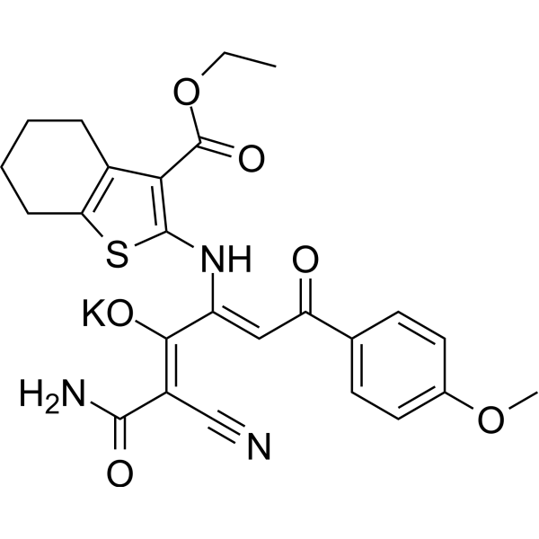
Anticancer agent 105 is a compound based on thienopyrimidine scaffold, with with good safety and anticancer properties. Anticancer agent 105 shows selective toxicity towards melanoma cancer, and induces apoptosis. And Anticancer agent 105 significantly inhibits the metastatic nodules, even in pulmonary metastatic melanoma mouse model.
Nur für Forschungszwecke. Wir verkaufen nicht an Patienten.
- CAS. Nr.: 2450987-57-0
- Formel: C25H24KN3O6S
- Molecular Weight:533.64
-
Speicherung:
Please store the product under the recommended conditions in the Certificate of Analysis.

To place orders, for customer services and technical support, please contact: MedChemExpress USA
Tel: 609-228-6898 E-mail: [email protected] [email protected]


-
 Biologische Aktivität
-
Chemical Information
- Protokoll
- Reinheit & Dokumentation
- Verweise
-
 Help & FAQs




Biologische Aktivität
Beschreibung
Cellular Effect
|
Cell Line

|
Type | Value | Description | References |
|---|---|---|---|---|
| 4T1 | IC50 |
3.8 μM
Compound: 9cb
|
Cytotoxicity against mouse 4T1 cells assessed as cell survival incubated for 24 hrs by alamarblue staining based analysis
Cytotoxicity against mouse 4T1 cells assessed as cell survival incubated for 24 hrs by alamarblue staining based analysis
|
[PMID: 37084598] |
| B16-F10 | IC50 |
2.41 μM
Compound: 9cb
|
Cytotoxicity against mouse B16-F10 cells assessed as cell survival incubated for 24 hrs by alamarblue staining based analysis
Cytotoxicity against mouse B16-F10 cells assessed as cell survival incubated for 24 hrs by alamarblue staining based analysis
|
[PMID: 37084598] |
| CT26 | IC50 |
4.3 μM
Compound: 9cb
|
Cytotoxicity against mouse CT26 cells assessed as cell survival incubated for 24 hrs by alamarblue staining based analysis
Cytotoxicity against mouse CT26 cells assessed as cell survival incubated for 24 hrs by alamarblue staining based analysis
|
[PMID: 37084598] |
| MEF | IC50 |
>100 μM
Compound: 9cb
|
Cytotoxicity against NF2-deficient mouse MEF cells assessed as cell survival incubated for 24 hrs by alamarblue staining based analysis
Cytotoxicity against NF2-deficient mouse MEF cells assessed as cell survival incubated for 24 hrs by alamarblue staining based analysis
|
[PMID: 37084598] |
In Vitro
Anticancer agent 105 (compound 9cb) shows selectivity against cancer cells, while it inhibits survival of cancerous cells (B16–F10 melanoma) and normal non-cancerous fibroblasts (MEF NF2) with IC50s of 2.41 μM, and >100 μM, respectively[1].
Anticancer agent 105 (0.5-10 μM; 24 h) shows toxicity against cancer cells, and (5-20 μM; 48 h) induces apoptosis and necrosis in B16-F10 cells[1].
MedChemExpress (MCE) has not independently confirmed the accuracy of these methods. They are for reference only.
-
Cell Line:B16-F10 melanoma
-
Concentration:0.5 μM, 1 μM, 5 μM, and 10 μM
-
Incubation Time:24 h
-
Result:Exhibited very low toxicity or were non-toxic in the case of MEF NF2 cells in the range of concentration 0.2-55 μM.
-
Cell Line:B16-F10 melanoma
-
Concentration:5 μM, 10 μM, 15 μM, and 20 μM
-
Incubation Time:48 h
-
Result:Induced early apoptosis and resulted nuclear condensation.
In Vivo
MedChemExpress (MCE) has not independently confirmed the accuracy of these methods. They are for reference only.
-
Animal Model:Pulmonary metastatic melanoma mouse model[1]
-
Dosage:I: 9-9.5 mg/kg; II: 42-4.6 mg/kg
-
Administration:IP; once daily every 3rd d for 22 d
-
Result:Decreased the number of metastatic nodules in mouse.


Chemical Information
-
CAS. Nr. 2450987-57-0
-
Molecular Weight 533.64
-
Formel C25H24KN3O6S
-
SMILES
O=C(OCC)C1=C(NC(/C(O[K])=C(C(N)=O)\C#N)=C\C(C2=CC=C(OC)C=C2)=O)SC3=C1CCCC3
-
Versand
Room temperature in continental US; may vary elsewhere.
-
Speicherung
Please store the product under the recommended conditions in the Certificate of Analysis.


Reinheit & Dokumentation
Verweise

Calculators
Konzentration (Stammlösung) × Volumen (Stammlösung) = Konzentration (Ziellösung) × Volumen (Ziellösung)


Anticancer agent 105 Related Classifications
HY-149949 Related Classifications
 Powered by Bioz