



Benzarone

Based on 1 Customer Validation
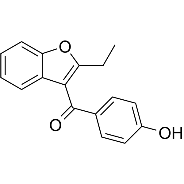
Benzarone is an oral inhibitor of human urate transporter 1 (hURAT1) with an IC50 value of 2.8 μM, and it also acts as an uncoupler of oxidative phosphorylation. Benzarone can cause liver damage and promote cell apoptosis and necrosis. Benzarone can be used to lower serum uric acid levels and for research in vascular diseases.
연구목적의 판매만을 진행합니다. 환자를 대상으로 한 판매는 하지 않습니다.
- Purity : 98.79%
- CAS No.: 1477-19-6
- 화학식: C17H14O3
- 분자량:266.29
-
보관:Powder -20°C, 3 years , 4°C, 2 years ; In solvent -80°C, 6 months , -20°C, 1 month

To place orders, for customer services and technical support, please contact: MedChemExpress USA
Tel: 609-228-6898 E-mail: [email protected] [email protected]


-
 Biological Activity
-
Chemical Information
-
용액&용해도
- Protocol
- 순도&문서
- References
-
 Help & FAQs






Biological Activity
제품 설명
IC50 & Target
IC50: 2.8 μM (hURAT1 in oocyte)[1]
In Vitro
Benzarone (10-100 μM, 1 h) causes a reduction in mitochondrial membrane potential in isolated rat liver cells, decreases the oxidative status of L-glutamate and the respiration control ratio in rat liver mitochondria, and also leads to uncoupling of oxidative phosphorylation and a reduction in β-oxidation[2].
Benzarone (1 nM-100 μM, 1 h) increases the production of reactive oxygen species, causes leakage of cytochrome C from HepG2 cell mitochondria, and induces permeability transition in rat liver mitochondria along with apoptosis and necrosis in rat liver cells[2].
MedChemExpress (MCE) has not independently confirmed the accuracy of these methods. They are for reference only.
-
Cell Line:HepG2
-
Concentration:100 μM
-
Incubation Time:8 h
-
Result:Caused fragmentation and condensation of chromatin, showing that 13% of the cells underwent apoptosis, along with cell death, and a decrease in ATP content.
In Vivo
Benzarone (2 mg/kg, given by gavage, single dose) is excreted in bile in rats, undergoes metabolism through direct conjugation, and exhibits enterohepatic recirculation[3].
MedChemExpress (MCE) has not independently confirmed the accuracy of these methods. They are for reference only.


Chemical Information
-
CAS No. 1477-19-6
-
Appearance Solid
-
분자량 266.29
-
화학식 C17H14O3
-
Color White to light yellow
-
SMILES
CCC1=C(C(C2=CC=C(O)C=C2)=O)C3=CC=CC=C3O1
-
선적
Room temperature in continental US; may vary elsewhere.
-
보관
Powder -20°C 3 years 4°C 2 years In solvent -80°C 6 months -20°C 1 month


용액&용해도
In Vitro:
DMSO : ≥ 100 mg/mL (375.53 mM; Hygroscopic DMSO has a significant impact on the solubility of product, please use newly opened DMSO)
* "≥" means soluble, but saturation unknown.
Please refer to the solubility information to select the appropriate solvent. Once prepared, please aliquot and store the solution to prevent product inactivation from repeated freeze-thaw cycles.
Storage method and period of stock solution: -80°C, 6 months; -20°C, 1 month. When stored at -80°C, please use it within 6 months. When stored at -20°C, please use it within 1 month.
Please refer to the solubility information to select the appropriate solvent. Once prepared, please aliquot and store the solution to prevent product inactivation from repeated freeze-thaw cycles.
Storage method and period of stock solution: -80°C, 6 months; -20°C, 1 month. When stored at -80°C, please use it within 6 months. When stored at -20°C, please use it within 1 month.
Concentration (start) × Volume (start) = Concentration (final) × Volume (final)
In Vivo:
Select the appropriate dissolution method based on your experimental animal and administration route.
- For the following dissolution methods, please ensure to first prepare a clear stock solution using an In Vitro approach and then sequentially add co-solvents:
- To ensure reliable experimental results, the clarified stock solution can be appropriately stored based on storage conditions. As for the working solution for In Vivo experiments, it is recommended to prepare freshly and use it on the same day.
- The percentages shown for the solvents indicate their volumetric ratio in the final prepared solution. If precipitation or phase separation occurs during preparation, heat and/or sonication can be used to aid dissolution.
Add each solvent one by one: 10% DMSO 40% PEG300 5% Tween-80 45% Saline
Solubility: ≥ 2.5 mg/mL (9.39 mM); Clear solution
This protocol yields a clear solution of ≥ 2.5 mg/mL (saturation unknown).
Taking 1 mL working solution as an example, add 100 μL DMSO stock solution (25.0 mg/mL) to 400 μL PEG300, and mix evenly; then add 50 μL Tween-80 and mix evenly; then add 450 μL Saline to adjust the volume to 1 mL.
Preparation of Saline: Dissolve 0.9 g sodium chloride in ddH₂O and dilute to 100 mL to obtain a clear Saline solution.
In Vivo Dissolution Calculator
Please enter the basic information of animal experiments:
-
-
-
-
Recommended: Prepare an additional quantity of animals to account for potential losses during experiments.
Please enter your animal formula composition:
-
%DMSO +
Recommended: Keep the proportion of DMSO in working solution below 2% if your animal is weak.
-
%+
-
+%Tween-80 + +
-
%Saline +
The co-solvents required include: DMSO, . All of co-solvents are available by MedChemExpress (MCE). , Tween 80. All of co-solvents are available by MedChemExpress (MCE).
Working solution concentration: 0.22 mg/mL
Method for preparing stock solution: mg drug dissolved in μL DMSO. Stock solution concentration: mg/mL.
1. Take μL DMSO stock solution;
2. Add μL .
μL , mix evenly;
3. Then add μL Tween 80, mix evenly;
4. Then add μL
Please ensure that the stock solution in the first step is dissolved to a clear state, and add co-solvents in sequence. You can use ultrasonic heating (ultrasonic cleaner, recommended frequency 20-40 kHz), vortexing, etc. to assist dissolution.


순도&문서
-
Data Sheet (277 KB)
-
SDS (392 KB)
- English - EN (392 KB)
- Français - FR (392 KB)
- Deutsch - DE (392 KB)
- Norwegian - NO (392 KB)
- Español - ES (392 KB)
- Swedish - SV (392 KB)
- Italian - IT (392 KB)
- Korean - KR (392 KB)
- Portuguese - PT (392 KB)
-
Handling Instructions (2659 KB)
References
[1]. Wempe MF, et, al. Developing potent human uric acid transporter 1 (hURAT1) inhibitors. J Med Chem. 2011 Apr 28;54(8):2701-13. [Content Brief]
[2]. Priska Kaufmann, et al. Mechanisms of benzarone and benzbromarone-induced hepatic toxicity. Hepatology. 2005 Apr;41(4):925-35. [Content Brief]
[3]. S G Wood, et al. Metabolic fate of the thrombolytic agent benzarone in man: comparison with the rat and dog. [Content Brief]


Complete Stock Solution Preparation Table
Please refer to the solubility information to select the appropriate solvent. Once prepared, please aliquot and store the solution to prevent product inactivation from repeated freeze-thaw cycles.
Storage method and period of stock solution: -80°C, 6 months; -20°C, 1 month. When stored at -80°C, please use it within 6 months. When stored at -20°C, please use it within 1 month.
| Optional Solvent | Concentration Solvent Mass | 1 mg | 5 mg | 10 mg | 25 mg |
|---|---|---|---|---|---|
| DMSO | 1 mM | 3.7553 mL | 18.7765 mL | 37.5530 mL | 93.8826 mL |
| 5 mM | 0.7511 mL | 3.7553 mL | 7.5106 mL | 18.7765 mL | |
| 10 mM | 0.3755 mL | 1.8777 mL | 3.7553 mL | 9.3883 mL | |
| 15 mM | 0.2504 mL | 1.2518 mL | 2.5035 mL | 6.2588 mL | |
| 20 mM | 0.1878 mL | 0.9388 mL | 1.8777 mL | 4.6941 mL | |
| 25 mM | 0.1502 mL | 0.7511 mL | 1.5021 mL | 3.7553 mL | |
| 30 mM | 0.1252 mL | 0.6259 mL | 1.2518 mL | 3.1294 mL | |
| 40 mM | 0.0939 mL | 0.4694 mL | 0.9388 mL | 2.3471 mL | |
| 50 mM | 0.0751 mL | 0.3755 mL | 0.7511 mL | 1.8777 mL | |
| 60 mM | 0.0626 mL | 0.3129 mL | 0.6259 mL | 1.5647 mL | |
| 80 mM | 0.0469 mL | 0.2347 mL | 0.4694 mL | 1.1735 mL | |
| 100 mM | 0.0376 mL | 0.1878 mL | 0.3755 mL | 0.9388 mL |


Benzarone Related Classifications
HY-W011711 Related Classifications
 Powered by Bioz